
A SURVEY OF AUDIO PROCESSING ALGORITHMS FOR DIGITAL
STETHOSCOPES
Fabio de Lima Hedayioglu, Miguel Tavares Coimbra
Instituto de Telecomunicações, Faculdade de Ciências da Universidade do Porto
Rua do Campo Alegre, 1021/1055, 4169 - 007 Porto, Portugal
Sandra da Silva Mattos
Fetal and Pediatric Cardiology Unit (UCMF) at Real Hospital Português de Benecifiencia em Pernambuco
Av. Portugal, 163 - Recife, PE Brazil
Keywords:
Digital stethoscope, Audio processing.
Abstract:
Digital stethoscopes have been drawing the attention of the biomedical engineering community for some
time now, as seen from patent applications and scientific publications. In the future, we expect ’intelligent
stethoscopes’ to assist the clinician in cardiac exam analysis and diagnostic, potentiating functionalities such as
the teaching of auscultation, telemedicine, and personalized healthcare. In this paper we review the most recent
heart sound processing publications, discussing their adequacy for implementation in digital stethoscopes. Our
results show a body of interesting and promising work, although we identify three important limitations of
this research field: lack of a set of universally accepted heart-sound features, badly described experimental
methodologies and absence of a clinical validation step. Correcting these flaws is vital for creating convincing
next-generation ’intelligent’ digital stethoscopes that the medical community can use and trust.
1 INTRODUCTION
Auscultation is one of the oldest, cheapest and most
useful techniques for the diagnosis of heart disease.
Since their invention in 1816, stethoscopes have been
used as part of the initial evaluation of all patients
with suspected heart or lung problems. An experi-
enced physician can diagnose a large number of clin-
ical conditions just from the initial auscultation of the
patient’s chest (Tilkian and Conover, 1984). There
have been several attempts to create electronically en-
hanced stethoscopes, with better sound amplification
and frequency response. However, and according to
Durand (Durand and Pibarot, 1995), their introduc-
tion into clinical practice has been hindered by factors
such as their background noise, unfamiliar sounds
to clinicians due to filtering or fragility and bad er-
gonomic design. Recent advances in electronics and
digital circuits allow us to not only overcome these
problems but also to exploit the benefits of digital sig-
nal processing for signal analysis and visualization.
In this paper we will embrace this novel perspective
and analyze the state-of-the-art in audio processing of
heart sounds that might be adequate for integrating
into this next generation of stethoscopes. A deeper
explanation of digital stethoscopes is given in Section
2, a review of audio processing methods is described
in Section 3, followed by a discussion (Section 4) on
the future of digital stethoscopes and how biomedical
engineers can contribute to the success of this tech-
nology.
2 DIGITAL STETHOSCOPE
It is essential that we define digital stethoscope since
there can be various interpretations from the name
alone. Traditional stethoscopes depend solely on
acoustics to amplify and transmit the heart sounds to
the clinician. The concept of electronic stethoscope
arrives when electronic components were first used to
amplify, filter and transmit the sound (Fig.1) (Durand
and Pibarot, 1995).
There are various examples in literature regard-
425
de Lima Hedayioglu F., Tavares Coimbra M. and da Silva Mattos S. (2009).
A SURVEY OF AUDIO PROCESSING ALGORITHMS FOR DIGITAL STETHOSCOPES.
In Proceedings of the International Conference on Health Informatics, pages 425-429
DOI: 10.5220/0001512104250429
Copyright
c
SciTePress
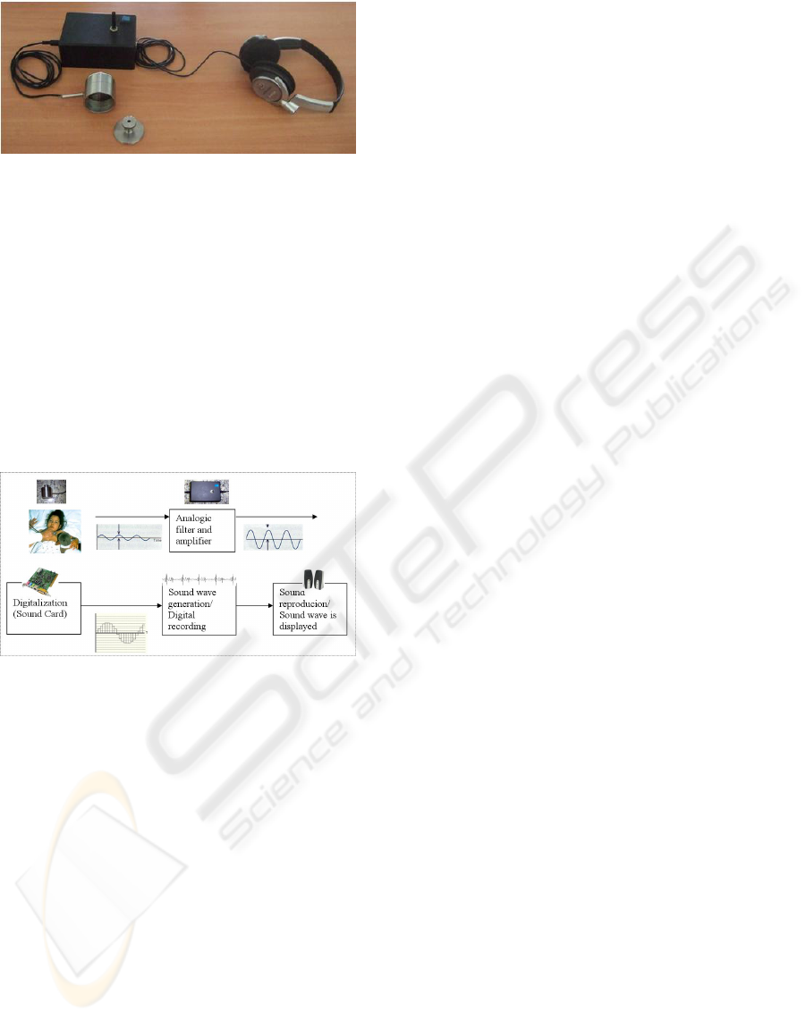
Figure 1: Lab prototype of an electronically enhanced
stethoscope.
ing the development of digital and electronic stetho-
scopes. Bredesen and Schmerler (Bredesen and
Schmerler, 1991) have patented an “intelligent stetho-
scope” designed for performing auscultation and for
automatically diagnosing abnormalities by comparing
digitized sounds to reference templates using a signa-
ture analysis technique. Several other electronically
enhanced and digital stethoscopes have been devel-
oped and described in literature (F. L. Hedayioglu,
2007; M.E. Tavel and Shander, 1994; Durand and
Pibarot, 1995; Brusco and Nazeran, 2005).
Figure 2: Block diagram of a digital stethoscope proto-
type developed by our group and field-tested at Real Hos-
pital Português de Beneficiência em Pernambuco in Recife,
Brazil. Over 100 auscultations were performed during the
clinical validation stage.
Fig. 2 shows a block diagram of a digital stetho-
scope prototype developed by our group. The auscul-
tation quality was considered satisfactory in clinical
trials when compared to auscultation using acoustic
stethoscopes, but clinicians still perceived differences
in audio pitch although this did not affect their abil-
ity to diagnose heart conditions. Our field experience
confirms Durand’s (Durand and Pibarot, 1995) opin-
ion that audio enhancement alone is not enough for
the clinical community to adopt this new technology.
In order to make digital stethoscopes attractive to clin-
ical cardiologists, we clearly need to address the nu-
merous potential improvements provided by a fully
functional, robust digital stethoscope: real-time ac-
quisition, analysis, display and reproduction of heart
sounds and murmurs. Digital stethoscopes must also
open the doors for digital audio archives, simplifying
the acquisition, storage and transmission process of
cardiac exams and murmurs, potentiating functionali-
ties such as the teaching of auscultation, telemedicine,
and personalized healthcare.
3 AUDIO PROCESSING
For the analysis of the state-of-the-art on audio pro-
cessing in cardiology, we have very loosely adopted
some concepts of clinical systematic reviews. A rig-
orous systematic review of such a multi-disciplinary
vast field is quite difficult to implement in practice due
to the large number of papers retrieved by analysis
of both engineering and medical scientific databases.
Our review methodology was as follows:
• Considered that Durand’s (Durand and Pibarot,
1995) excellent review paper fully covers this
topic up to 1995.
• Consulted the IEEE Xplore (ieeexplore.ieee.org)
database with the following query: “(((feature ex-
traction)<in>metadata) <and> ((cardiology)<in>
metadata))”, obtaining 159 results after 1995.
• By title and abstract inspection, we kept only pa-
pers dealing with phonocardiogram data analysis,
reducing this number to 19.
• We analyzed the references from all these papers,
and selected all papers published after 1995 and
with more than 10 citations, obtaining 20 results.
This enabled us to cover additional articles be-
sides the ones published in IEEE journals and
conferences, artificially expanding the scope of
our review to other scientific databases.
• The total number of papers covered by this review
is thus 39 (19+20).
Although we are certain that it is possible to miss
some papers using this methodology, we feel that we
have covered a sufficiently vast and interesting sam-
ple to draw some important conclusions, as described
in Section 4.
3.1 Heart Sound Analysis and Feature
Extraction
The main constituents of a cardiac cycle are the first
heart sound (typically referred to as S1), the systolic
period, the second heart sound (S2) and the diastolic
period. Whenever a clinician is performing an aus-
cultation, he tries to identify these individual compo-
nents, and is trained to analyze related features such
as rhythm, timing instants, intensity of heart sound
HEALTHINF 2009 - International Conference on Health Informatics
426

components, splitting of S2, etc (H. Liang and Har-
timo, 1997b). This analysis allows him to search for
murmurs and sound abnormalities that might corre-
spond to specific cardiac pathologies. From a signal
processing perspective, Heart Sound Analysis (HSA)
is not only interesting by itself (allowing quantita-
tive measures to be displayed automatically in a dig-
ital stethoscope), but is also an essential first step for
the subsequent task of automatic pathology classifica-
tion. In this paper, we will distinguish two sub-tasks
of HSA: Heart Sound Segmentation (HSS) and Aortic
Pulmonary Signal Decomposition (APSD).
3.1.1 Heart Sound Segmentation
In HSS we expect to identify and segment the
four main constituents of a cardiac cycle. This
is typically accomplished by identifying the posi-
tion and duration of S1 and S2, using some sort of
peak-picking methodology on a pre-processed sig-
nal. Liang (H. Liang and Hartimo, 1997a) has used
discrete wavelet decomposition and reconstructed the
signal using only the most relevant frequency bands.
Peak-picking was performed by thresholding the nor-
malized average Shannon energy, and discarding ex-
tra peaks via analysis of the mean and variance of
peak intervals. Finally, they distinguish between S1
and S2 peaks (assuming that the diastolic period is
longer than the systolic one, and that the later is more
constant), and estimate their durations. A classifi-
cation accuracy of 93% was obtained on 515 peri-
ods of PCG signal recordings from 37 digital phono-
cardiographic recordings. The same authors further
improved the statistical significance of their results
by obtaining the same accuracy using 1165 cardiac
periods from 77 recordings (H. Liang and Hartimo,
1997b), and later attempted murmur classification
based on these features and neural network classi-
fiers, obtaining 74% accuracy (Liang and Hartimo,
1998b). Omran (Sherif Omran, 2003) has also studied
this problem using normalized Shannon entropy after
wavelet decomposition of the audio signal, but their
experimental methodology is not so convincing.
3.1.2 Aortic Pulmonary Signal Decomposition
Besides the four main components of the cardiac
cycle, there is a clinical interest in the analysis of
some of its associated sub-components (JingPing Xu,
2000). It has been recognized that S1 may be com-
posed of up to four components produced during ven-
tricular contraction (Durand and Pibarot, 1995), al-
though the complexity of this task has been a very
difficult hurdle for the signal processing community.
The S2 sound is more well known, being composed
of an aortic component (A2), which is produced first
during the closure and vibration of the aortic valve
and surrounding tissues, followed by the pulmonary
component (P2) produced by a similar process asso-
ciated with the pulmonary valve (JingPing Xu, 2000).
Durand (JingPing Xu, 2000) demonstrated that it is
possible to model each component of S2 by a narrow-
band nonlinear chirp signal. Later (JingPing Xu,
2001) he adapted and validated this approach for the
analysis and synthesis of overlapping A2 and P2 com-
ponents of S2. To do so, the time-frequency represen-
tation of the signal is generated and then estimated
and reconstructed using the instantaneous phase and
amplitude of each component (A2 and P2). In this
paper the accuracy evaluation was made by a simu-
lated A2 and P2 components having different overlap-
ping factors. The reported error was between 1% and
6%, proportional to the duration of the overlapping
interval. Nigam (Nigam and Priemer, 2006) also pre-
sented a method for extracting A2 and P2 components
by assuming them as statistically independent. To do
so, four simultaneous auscultations are analyzed us-
ing blind source separation. The main advantage of
this method is the lower dependence on the A2-P2
time interval, although it needs a non-conventional 4-
sensor stethoscope. Leung (T. S. Leung, 1998) also
analyzed the splitting of S2 using time-frequency de-
composition.
3.2 Automatic Pathology Classification
The vast majority of papers we have found regarding
audio processing algorithms, adequate for the integra-
tion into a digital stethoscope, concern the detection
of specific heart pathologies. This highlights the in-
terest of the scientific community on this topic but, as
our analysis shows, there are still some major flaws
in most of them such as the absence of a clinical val-
idation step and unconvincing experimental method-
ologies. Most papers use the well-established pattern
recognition approach of feature extraction followed
by a classifier. Due to space limitations, we will de-
scribe the most interesting papers, leaving a more de-
tailed discussion on this topic to Section IV. Bentley
(P. M. Bentley and Grant, 1995) uses Choi-Williams
Distribution (CWD) as features, working with 45 nor-
mal/abnormal valve subjects. Some features were de-
termined via visual inspection, others automatically
from the CWD by simple rule-based classification.
Latter (P. M. Bentley and McDonnell, 1998), the au-
thors show that CWD is a better method to represent
the frequencies in PCG and to get heart sound de-
scriptors, than other time-frequency (T-F) represen-
tations. According to them, a simple description of
A SURVEY OF AUDIO PROCESSING ALGORITHMS FOR DIGITAL STETHOSCOPES
427

the T-F distribution allows an analysis of the heart
valve’s condition. However, they highlight the need
of a more comprehensive evaluation using a larger
population of test patients. Wang (P. Wang and Soh,
2005) proposes a representation of heart sounds that
is robust to noise levels of 20dB, using mel-scaled
wavelet features. However, details regarding the used
dataset are not clear enough for robust conclusions.
Liang (Liang and Hartimo, 1998a) developed an inter-
esting feature vector extraction algorithm where the
systolic signal is decomposed by wavelets into sub-
bands. Then, the best basis set is selected, and the
average feature vector of each heart sound recording
is calculated. Neural Networks (NN) are used for
classifying 20 samples after being trained with 65,
obtaining an accuracy of 85%. NNs are also used
by Abdel-Alim (Onsy Abdel-Alim and El-Hanjouri,
2002) for the automatic diagnostics of heart valves us-
ing wavelets feature vectors and stethoscope location
information. They use two NNs: one for systolic dis-
eases and the other for diastolic diseases. A total of
1200 cases were used: 970 cases for training and 300
for testing. The recognition rate was 95%. Turkoglu
(Turkoglu and Arslan, 2001), Ozgur (Ozgur Say and
Olmez, 2002) and El-Hanjouri (M. El-Hanjouri and
Alim, 2002) also used wavelets as feature vectors for
classification, although they provide too few details
regarding the used data sets. Trimmed mean spectro-
grams are used by Leung (T.S. Leung and Salmon,
2000) to extract features of phonocardiograms. To-
gether with the acoustic intensities in systole and di-
astole, the authors quantified the distinctive character-
istics of different types of murmurs using NNs. One
of the few papers that is conscious about the impor-
tant clinical validation step is from Kail (E Kail and
Balázs, 2004). The authors propose a novel sound
representation (2D and 3D) and feature extraction al-
gorithm using Morlet wavelet scalograms. After man-
ual classification of the resulting graphs performed by
two cardiologists on 773 subjects, they clinically val-
idated the features as useful for sound and murmur
extraction. Sharif (Zaiton Sharif and Salleh, 2000)
also proposes other features for classification systems
based on central finite difference and zero crossing
frequency estimation.
4 DISCUSSION
By covering the most interesting papers on audio-
processing from a digital stethoscope perspective, we
can make some observations regarding the state-of-
the-art on this field. Section 3.1 has shown us that
there are already important results regarding audio
feature extraction. The S1 and S2 sounds can be ro-
bustly segmented and there is promising work regard-
ing the extraction of secondary sounds such as A2 and
P2.
The scenario is not so bright for automatic pathol-
ogy classification (Section 3.2). Reviewing some of
the papers and simply observing the disparity in the
number of publications when compared with the other
challenges, we conclude that there is a strong inter-
esting in this topic. However, in our opinion, there is
still a long way to go before we can have robust au-
tomatic classification systems that can be introduced
in the clinical routine of hospitals. We have identified
three major problems that afflict most of the papers
reviewed:
• Absence of a set of well-accepted features - We
rarely found papers that selected the same features
for pathology classification. Most acknowledge
that the presence of S1 and S2 is important but
there is no consensus of the scientific community
on how these should be used. We have collected
more than 25 different features with minimum
overlap between papers. We clearly need more
studies on the statistical significance and clinical
importance of heart sound features, from an auto-
matic pattern recognition perspective.
• Badly descript data-sets - It is not enough for au-
thors to mention that they have worked with 300
cardiac cycles. Where were these obtained? From
how many patients? In what conditions? Using
which equipment? All these factors are vital in
the analysis of a system’s performance and robust-
ness. Studies need to be much more rigorous on
this topic so their results can be reasonably con-
vincing.
• Absence of clinical validation - Almost no papers
bothered to handle this vital task of all assisted-
diagnostic systems. No medical specialist will
trust any kind of automatic system without it prov-
ing to be robust and accurate in real field testing.
These conditions are very different from a typical
biomedical engineering research lab, which can
drastically affect results.
As a final conclusion, we can say that working
towards next-generation ’intelligent’ digital stetho-
scopes is highly desirable judging from the signifi-
cant number of scientific publications on this topic but
also examining the undeniable benefits that such sys-
tems can provide. There is already solid work regard-
ing audio feature extraction and many unsolved chal-
lenges in this field such as the complex analysis of the
sub-components of S1. Automatic pathology classifi-
cation is still too undeveloped to be of any practical
HEALTHINF 2009 - International Conference on Health Informatics
428

usage and we hope that the valuable lessons learned
from this study can correct previous mistakes and pro-
vide a precious boost to the challenging field of audio
processing for digital stethoscopes.
ACKNOWLEDGEMENTS
This work was supported by the Programme Al-
ban, the European Union Programme of High Level
Scholarships for Latin America, scholarship no.
E07M402298BR.
REFERENCES
Bredesen, M. and Schmerler, E. (1991). Us patent no.
5,010,889: Intelligent stethoscope.
Brusco, M. and Nazeran, H. (2005). Development of an
intelligent pda-based wearable digital phonocardio-
graph. In Proceedings of the 2005 IEEE Engineering
in Medicine and Biology 27th Annual Conference.
Durand, L.-G. and Pibarot, P. (1995). Digital signal pro-
cessing of the phonocardiogram: Review of the most
recent advancements. Critical Reviews in Biomedical
Engineering.
E Kail, S Khoór, B. K. K. F. and Balázs, F. (2004). Internet
digital phonocardiography in clinical settings and in
population screening. Computers in Cardiology.
F. L. Hedayioglu, S. S. Mattos, L. M. M. E. d. L. (2007). De-
velopment of a tele-stethoscope and it’s application in
pediatric cardiology. Indian Journal of Experimental
Biology, 45.
H. Liang, S. L. and Hartimo, I. (1997a). Heart sound
segmentation algorithm based on heart sound envel-
ogram. Computers in Cardiology, 24.
H. Liang, S. L. and Hartimo, I. (1997b). A heart sound
segmentation algorithm using wavelet decomposition
and reconstruction. In 19th International Conference
- IEEE/EMBS, Chicago, IL, USA.
JingPing Xu, L.G. Durand, P. P. (2000). Nonlinear transient
chirp signal modeling of the aortic and pulmonary
components of the secound heart sound. IEEE Trans-
actions on Biomedical Engineering, 47(7).
JingPing Xu, L.G. Durand, P. P. (2001). Extraction of
the aortic and pulmonary components of the sec-
ound heart sound using nonlinear transient chirp sig-
nal model. IEEE Transactions on Biomedical Engi-
neering, 48(3).
Liang, H. and Hartimo, I. (1998a). A feature extraction
algorithm based on wavelet packet decomposition for
heart sound signals. In Proceedings of the IEEE-SP
International Symposium.
Liang, H. and Hartimo, I. (1998b). A heart sound feature
extraction algorithm based on wavelet decomposition
and reconstruction. In Proc. IEEE EMBS, volume 20.
M. El-Hanjouri, W. Alkhaldi, N. H. and Alim, A. (2002).
Heart diseases diagnosis using hmm. In Electrotech-
nical Conference -MELECON.
M.E. Tavel, D. B. and Shander, D. (1994). Enhanced aus-
cultation with a new graphic display system. In Arch.
Intern. Med., volume 154, page 893.
Nigam, V. and Priemer, R. (2006). A procedure to ex-
tract the aortic and the pulmonary sounds from the
phonocardiogram. In Proceedings of the 28th IEEE
EMBS Annual International Conference, New York
City, USA.
Onsy Abdel-Alim, N. H. and El-Hanjouri, M. A. (2002).
Heart diseases diagnosis using heart sounds. In Radio
Science Conference.
Ozgur Say, Z. D. and Olmez, T. (2002). Classification of
heart sounds by using wavelet transform. In Proceed-
ings of the Second Joint EMBS/BMES Conference,
volume 1.
P. M. Bentley, J. T. E. M. and Grant, P. M. (1995). Clas-
sification of native heart valve sounds using the choi-
williams time-frequency distribution. In IEEE-EMBC
and CMBEC.
P. M. Bentley, P. M. G. and McDonnell, J. T. E. (1998).
Time-frequency and time-scale techniques for the
classification of native and bioprosthetic heart valve
sounds. IEEE Transactions on Biomedical Engineer-
ing, 45(1).
P. Wang, Y. K. and Soh, C. B. (2005). Feature extraction
based on mel-scaled wavelet transform for heart sound
analysis. In Engineering in Medicine and Biology So-
ciety, 2005. IEEE-EMBS 2005. 27th Annual Interna-
tional Conference.
Sherif Omran, M. T. (2003). A heart sound segmentation
and feature extraction algorithm using wavelet. In
Proc. of IEEE MWSCAS ’03, volume 1, pages 27–30.
T. S. Leung, P. R. White, J. C. W. B. C. E. B. A. P. S. (1998).
Analysis of the secound heart sound for diagnosis of
paediatric heart disease. In IEE Proceedings - Sci.
Meas. Technol., volume 145.
Tilkian, A. and Conover, M. (1984). Understanding heart
sounds and murmurs with an introduction to lung
sounds. W.B. Saunders Company.
T.S. Leung, P.R. White, W. B. C. E. B. and Salmon, A. P.
(2000). Classification of heart sounds using time-
frequency method and artificial neural networks. In
Proceedings of the 22nd Annual International Con-
ference of the IEEE, volume 2.
Turkoglu, I. and Arslan, A. (2001). An intelligent pat-
tern recognition system based on neural network
and wavelet decomposition for interpretation of heart
sounds. In Proceedings of the 23rd Annual Interna-
tional Conference of the IEEE, volume 2, pages 25–
28.
Zaiton Sharif, Mohd Shamian Zainal, A. Z. S. and Salleh,
S. H. S. (2000). Analysis and classification of heart
sounds and murmurs based on the instantaneous en-
ergy and frequency estimations. In Proceedings of
TENCON, volume 2.
A SURVEY OF AUDIO PROCESSING ALGORITHMS FOR DIGITAL STETHOSCOPES
429
