Algorithmic Surface Extraction from MRI Data
Modelling the Human Vocal Tract
D. Aalto
1,2
, J. Helle
3
, A. Huhtala
4
, A. Kivelä
4
, J. Malinen
4
, J. Saunavaara
5
and T. Ronkka
6
1
Inst. of Behavioural Sciences (SigMe Group), University of Helsinki, Helsinki, Finland
2
Dept. of Signal Processing and Acoustics, Aalto University, P.O. BOX 13000, FI-00076 Aalto, Finland
3
Media Factory, Aalto University, P.O. BOX 31000, FI-00076 Aalto, Finland
4
Dept. of Mathematics and Systems Analysis, Aalto University, P.O. BOX 11100, FI-00076 Aalto, Finland
5
Dept. of Radiology, Medical Imaging Centre of Southwest Finland, University of Turku, Turku, Finland
6
Aalto Design Factory, P.O. BOX 17700, FI-00076 Aalto, Finland
Keywords:
MRI, 3D Image Processing, Automatic Surface Extraction, FEM Meshing, Physical Modelling.
Abstract:
A procedure for the vectorisation and feature extraction of the human vocal tract is proposed. The raw data
is obtained by high resolution 3D MRI. Because the amount of manual work in the data processing has been
minimised, large datasets can be treated. The vectorised data can be used for both numerical as well as physical
modelling of the vocal tract biophysics, including speech and applications in medicine.
1 INTRODUCTION
We present techniques, algorithms, and results for
automatic vectorisation and feature extraction of
grayscale voxel image files, produced by Magnetic
Resonance Imaging (MRI). Our particular interest is
in the extraction of the human vocal tract (VT) geom-
etry. Numerous test subjects and large datasets are of-
ten involved, and manual data processing must there-
fore be minimised. This is the main motivation for de-
veloping custom software for VT extraction. Applica-
tions of anatomically accurate VT models range from
computational modelling (see (Aalto et al., 2011; De-
douch et al., 2002; Hannukainen et al., 2007; Lu et al.,
1993; Lacis, 2012) and the references therein) to fast
prototyping of physical models for measurements that
are impossible to carry out non-invasively using test
subjects (see (Hirtum et al., 2011; Horá
ˇ
cek et al.,
2011; Šidlof et al., 2012; Takemoto et al., 2010)).
There is wide literature in image processing and
feature extraction methods that have been applied in
medical imaging; see, e.g., (Gonzalez and Woods,
2001; Criminisi et al., 2011). Several software so-
lutions for processing of medical images exist such
as the Vascular Modelling Toolkit (Vascular Model-
ing Toolkit, 2012) and MIMICS (Materialise, 2012)
which was used in (Takemoto et al., 2010) for proces-
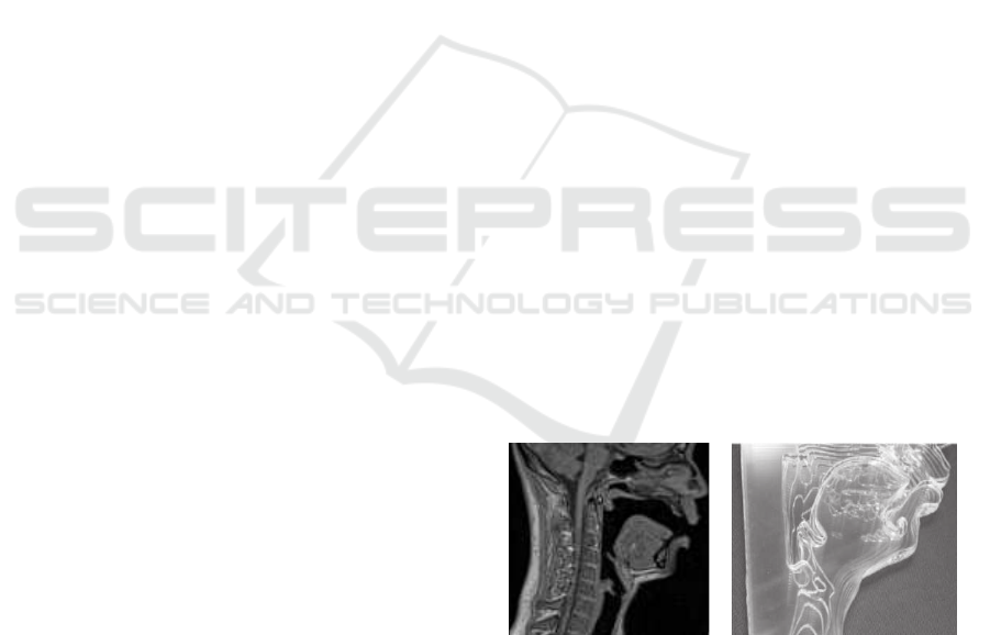
(a) (b)
Figure 1: (a) A mid-sagittal section of a male subject while
pronouncing vowel [œ]. (b) A plastic printout of the same
vowel geometry, consisting of 24 sagittal planes of which
14 is shown here.
sing a small dataset of VT geometries. However,
practical applications require specifically refined cus-
tom procedures that scale with the increasing size of
the dataset.
Compared to cardiovascular modelling and mod-
elling of most internal organs, the extraction of the
VT geometry has complications of its own. Teeth are
missing from raw MRI data because osseous struc-
tures cannot be isolated from air volumes due to their
low hydrogen content. This results in holes and other
artefacts in vectorised images which must be cor-
rected. As discussed below, the highly variable rel-
ative position of the mandible and the maxilla in dif-
ferent VT configurations must be taken into account
257
Aalto D., Helle J., Huhtala A., Kivelä A., Malinen J., Saunavaara J. and Ronkka T..
Algorithmic Surface Extraction from MRI Data - Modelling the Human Vocal Tract.
DOI: 10.5220/0004233302570260
In Proceedings of the International Conference on Biomedical Electronics and Devices (BIODEVICES-2013), pages 257-260
ISBN: 978-989-8565-34-1
Copyright
c
2013 SCITEPRESS (Science and Technology Publications, Lda.)

in automated artefact removal. Separate imaging of
teeth and their alignment with the soft tissue geome-
tries from MRI is a difficult problem which is not dis-
cussed in this article.
2 SURFACE MODELS
In traditional medical imaging, it is sufficient to pro-
duce visualisations for inspection by trained radiolo-
gists. Numerical modelling methods require discrete
representations such as tetrahedral meshes for FEM.
We generate the mesh from a solid triangular repre-
sentation of the desired surface which requires more
data processing than surface extraction for visualiza-
tion purposes only.
We propose a novel algorithm and software for
surface and feature extraction of the 3D VT air vol-
ume Ω. The algorithm comprises the following steps:
1. Pre-processing: The voxel data is smoothed to
remove noise.
2. Initial Surface Extraction: is carried out by ex-
tracting an isosurface corresponding to approxi-
mated air-tissue interface. An isosurface is a level
set corresponding to a constant gray value, and it
consists of triangular elements.
3. Producing the Artefact Prior Model: “Unde-
sired artefacts”, such as vertebrae and maxillae,
are manually identified from the initial surface to
produce two prior models of the artefacts, one for
each maxilla.
4. Removing Artefacts: Artefact models are
aligned with the initial surface using algorithms
provided by PCL (Rusu and Cousins, 2011).
Based on the location information thus obtained,
the undesired artefacts are masked from the origi-
nal voxel data.
5. Final Surface Extraction: An improved isosur-
face Γ is extracted from the artefact free, masked
voxel data.
6. Locating Boundaries: The glottis, the velar port,
and the mouth opening positions are located from
Γ. These openings are covered, and they can be
joined with Γ to produce a triangulated model for
the full boundary ∂Ω.
The raw data consists of MRI sequences that are
stored as a DICOM files that comprising of 44 sagittal
plane images: each image contains 128 ×128 pixels
that are of size d = 1.8mm
1
. These planar images are
aligned and stacked using the location data produced
1
The pixel number is always the same when using this
Figure 2: Shaded visualisation of the initial (left) and the
final isosurfaces. The face is not part of the final computa-
tional geometry boundary ∂Ω.
by the MRI machine. The stacked images form a 3D
matrix of voxel data that represents the VT through
grayscale values. The measurement setup for obtain-
ing this data has been documented in (Aalto et al.,
2011, Section 2.3).
The isosurface extraction requires a threshold gray
value that is determined from 2D bitmaps by estimat-
ing the VT boundary intersection heuristically. Since
the air-tissue interface has a steep gray value gradi-
ent in the MRI voxel data, the extracted surface is not
sensitive to small variations in the threshold value.
2.0.1 Artefact Modelling and Mesh Quality
The initial surface has a lot of artefacts as can be seen
in Fig. 2. The alignment of the artefact models is car-
ried out using the Point Cloud Library (PCL) (Rusu
and Cousins, 2011). The information provided by
PCL gives, in particular, the relative positions of the
mandible and the maxilla which amounts to most of
the positional variation between different VT config-
urations of the same test subject.
The production of the prior artefact models re-
mains the most laborious piece of manual work where
tools such as MeshLab (MeshLab, 2012) or Blender
(Blender, 2012) can used. It takes about one hour to
model the artefacts for one test subject but then the
same artefact model can be used for all VT configura-
tions, albeit only from the same test subject.
As the final result, an artefact free surface model
of the VT geometry is obtained. One such geome-
try is shown in Fig. 2. The side lengths of the sur-
face triangles are bounded above by d
√
3 where d is
the voxel resolution of the MRI data. As reported in
(Aalto et al., 2012, Section 4), the geometric error of
the surface mesh is of order 0.5mm except in those
parts of the model where MRI transparent teeth or the
possibly open velar port cause crude error.
sequence but the pixel size varies according to the physical
dimension of the test subject.
BIODEVICES2013-InternationalConferenceonBiomedicalElectronicsandDevices
258

3 CENTRELINE EXTRACTION
Many simplified computational physics models in
tubular anatomic regions (such as flow mechanics in
vascular structures and the acoustics of the VT) refer
to centrelines and intersectional areas instead of the
full 3D geometry. Our interest in centrelines and area
functions of the VT stems from the generalised Web-
ster’s horn model (see, e.g., (Lukkari and Malinen,
2011), (Story et al., 1996)) which is a low frequency
1D approximation of the 3D wave equation on a tubu-
lar domain. For a discussion on hemodynamics mod-
els, see (Antiga, 2003) and the references therein.
Since centrelines are not uniquely defined on ge-
ometric grounds except in very simple cases, we first
produce a candidate centreline for Ω. However, the
“true” centreline depends on the application and the
model of interest in a possibly intractable way. So as
to Webster’s model, we improve the model accuracy
by choosing optimally from a family of centrelines
that are normal to a fixed set of planar intersections as
can be seen in Fig. 3.
Voronoi diagrams are used for the centreline ex-
traction in Vascular Modelling Toolkit. Our approach
is based on numerically solving the steady state heat
equation with unit source ∆u = 1 in Ω where u = 0 on
VT walls, and
∂u
∂ν
= 0 at mouth and glottis. In our set-
ting, this can be done without much extra effort since
FEM discretisation of Ω has already been produced
for modelling VT acoustics, and a similar approach
has been studied in (Vesom et al., 2008). The solu-
tion of the steady state heat equation can be obtained
in linear time respect to the number n of nodes in the
discretisation. The candidate centreline is, by defini-
tion, the ridge in the solution u. Finding the ridge is a
pathfinding problem that can be solved in O(nlogn)
time.
4 PHYSICAL MODELS
We have produced pilot plastic models of the VT in
1:1 scale. We use these models for acoustic and flow
measurements in order to augment and validate the
numerical results from mathematical models.
We have experimented with hard plastic print-
outs only. Trials were performed using a 3D printer
3DTouch by Bits from Bytes, Ltd. The printing time
for the full VT in 1:1 scale in PLA plastic was in ex-
cess of 12 hours when using a layer height of 0.25mm.
The long printing time combined with the tendency
of PVA to clog the extruder nozzle resulted in a dis-
appointing print quality and a success rate of less
than 25%. Stereolithography as in (Takemoto et al.,
γ(s)
ˆ
γ(s)
{Γ(s)}
Figure 3: A subset of {Γ(s)} and two centrelines γ(s) and
ˆ
γ(s) corresponding to the chosen set of area functions.
2010) or selective laser sintering is expected to pro-
duce higher quality models with better success rate
than fused deposition methods.
The prototype model shown in Fig. 1(b) was pro-
duced by cutting sagittal intersection contours from
3 mm thick acrylic plate. We used Legend 36ext by
Epilog Laser (http://www.epiloglaser.com) which is a
CO
2
laser cutter with P = 60W and two degrees of
freedom. The cutting of all 24 sheets took under 2
hours.
The cutting angle of the sheets is always 90
◦
in
the model of Fig. 1(b), and this results in significant
“stepping” of the VT boundary surface. The stepping
can be reduced either by using thinner acrylic plate or,
preferably, by varying the cutting angle so as to make
the adjacent sheets fit to each other without steps. The
variable cutting angle requires a CNC mill or a more
advanced cutter.
Optically transparent models for Particle Image
Velocimetry studies as in (Horá
ˇ
cek et al., 2011) can-
not be created simply by stacking transparent acrylic
sheets but moulds for casting such models can.
5 CONCLUSIONS
We have developed algorithms for automatic surface
detection and vectorisation of MR images of the head
and neck area, especially concentrating on the vocal
tract. A very large number of MR images are re-
quired for numerical model validation as well as med-
ical applications. Hence, the amount of manual work
must be minimised without sacrificing the data qual-
ity. Apart from some tasks related to manual arte-
fact detection (see Step 3 in Section 2) and remaining
open problems with efficient teeth modelling, the data
processing can be carried out by a fully computerised
procedure described in this work.
In addition to modelling purposes, physical print-
outs of vocal tract geometries may have future appli-
cations in reconstructive surgery and tissue engineer-
ing. Tissue grafts are produced by seeding and at-
tachment of human cells into a scaffold. Scaffolds
AlgorithmicSurfaceExtractionfromMRIData-ModellingtheHumanVocalTract
259

must satisfy many material requirements due to biol-
ogy (Sachlos and Czernuszka, 2003) as well as have
the correct geometric shape, too.
ACKNOWLEDGEMENTS
The authors were supported by the Finnish Academy
grant Lastu 135005, European Union grant Sim-
ple4All, Aalto Starting Grant, and Åbo Akademi In-
stitute of Mathematics.
The current version of the software described in
this paper can be obtained from the authors by re-
quest.
REFERENCES
Aalto, D., Aaltonen, O., Happonen, R.-P., Malinen, J.,
Palo, P., Parkkola, R., Saunavaara, J., and Vainio, M.
(2011). Recording speech sound and articulation in
MRI. In Proceedings of BIODEVICES 2011, Rome,
Italy.
Aalto, D., Huhtala, A., Kivelä, A., Malinen, J., Palo, P.,
Saunavaara, J., and Vainio, M. (2012). How far
are vowel formants from computed vocal tract reso-
nances? arXiv:1208.5963, 13 pp.
Antiga, L. (2003). Patient-Specific Modeling of Geometry
and Blood Flow in Large Arteries. PhD thesis, Po-
litecnico di Milano.
Blender (2012). http://www.blender.org. Ac-
cessed Nov. 7th, 2012.
Criminisi, A., Shotton, J., and Konukoglu, E. (2011). Deci-
sion forests for classification, regression, density esti-
mation, manifold learning and semi-supervised learn-
ing. Technical Report MSR-TR-2011-114, Microsoft
Research.
Dedouch, K., Horá
ˇ
cek, J., Vampola, T., and
ˇ
Cerný, L.
(2002). Finite element modelling of a male vocal tract
with consideration of cleft palate. In Forum Acus-
ticum, Sevilla, Spain.
Gonzalez, R. C. and Woods, R. E. (2001). Digital Image
Processing, 2nd Ed. Addison-Wesley Longman Pub-
lishing Co., Inc., Boston, MA.
Hannukainen, A., Lukkari, T., Malinen, J., and Palo, P.
(2007). Vowel formants from the wave equation. J.
Acoust. Soc. Am. Express Letters, 122(1):EL1–EL7.
Hirtum, A. V., Pelorson, X., and Estienne, O. (2011). Ex-
perimental validation of flow models for a rigid vocal
tract replica. J. Acoust. Soc. Am., 130(4):2128–2138.
Horá
ˇ
cek, J., Uruba, V., Radolf, V., Veselý, J., and Bula, V.
(2011). Airflow visualization in a model of human
glottis near the self-oscillating vocal folds model. Ap-
plied and Computational Mechanics, 5:21–28.
Lacis, U. (2012). Modelling air flow in larynx. Master’s
thesis, Umeå University.
Lu, C., Nakai, T., and Suzuki, H. (1993). Finite element
simulation of sound transmission in vocal tract. J.
Acoust. Soc. Jpn. (E), 92:2577 – 2585.
Lukkari, T. and Malinen, J. (2011). Webster’s equation with
curvature and dissipation. arXiv:1204.4075, 22 pp. +
5 pp. appendix.
Materialise (2012). Mimics. http://biomedical. materi-
alise.com/mimics. Accessed Nov. 7th, 2012.
MeshLab (2012). Visual Computing Lab ISTI -
CNR. http://meshlab.sourceforge.net/. Ac-
cessed Nov. 7th, 2012.
Rusu, R. B. and Cousins, S. (2011). 3D is here: Point Cloud
Library (PCL). In IEEE International Conference on
Robotics and Automation (ICRA), Shanghai, China.
Sachlos, E. and Czernuszka, J. T. (2003). Making tissue en-
gineering scaffolds work. Review: the application of
solid freeform fabrication technology to the produc-
tion of tissue engineering scaffolds. Eur Cell Mater,
5:29–39; discussion 39–40.
Story, B., Titze, I., and Hoffman, E. (1996). Vocal area func-
tions from magnetic resonance imaging. J. Acoust.
Soc. Am., 100(1):537–554.
Takemoto, H., Mokhtari, P., and Kitamura, T. (2010).
Acoustic analysis of the vocal tract during vowel pro-
ductions by finite-difference time-domain method. J.
Acoust. Soc. Am., 128(6):3724–3738.
Vascular Modeling Toolkit (2012). http://www.vmtk.org.
Accessed Nov. 7th, 2012.
Vesom, G., Cahill, N. D., Gorelick, L., and Noble, J. A.
(2008). Characterization of anatomical shape based
on random walk hitting times. In In Proceed-
ings of Mathematical Foundations of Computational
Anatomy (MFCA 2008), New York.
Šidlof, P., Horá
ˇ
cek, J., and
ˇ
Ridký, V. (2012). Parallel CFD
simulation of flow in a 3D model of vibrating human
vocal folds. Computers and Fluids. In press.
BIODEVICES2013-InternationalConferenceonBiomedicalElectronicsandDevices
260
