
A Comparative Study of BRISK, ORB and DAISY Features for Breast
Cancer Classification
Ghada Ouddai, Ines Hamdi and Henda Ben Ghezala
RIADI Laboratory, National School of Computer Science (ENSI), University of La Manouba, La Manouba, Tunisia
Keywords:
Histopathological Image Processing, Feature Extraction, Binary Robust Invariant Scalable (BRISK), Oriented
FAST and Rotated BRIEF (ORB), DAISY Descriptor, Bag-of-Features (BoF), Machine Learning.
Abstract:
Medical data analysis is one of the most emergent fields over the past decades. In Digital histopathology,
images are analysed, mainly, to detect disease or tumors and identify their types and grade. One of the most
used practices in this field is the feature extraction. In this paper, we propose the application of BRISK, ORB
and BRISK/DAISY on RGB histological images. The purpose of this work is to recognise the breast tumor
type (benign or malignant). These features extractors are combined with BoF by kmeans and SVM. A limited
amount of images is used during the training of the system. Out of the three methods, Color-BRISK/BoF/SVM
solution gave the best accuracy value (72.5%) while Color-ORB/BoF/SVM was the fastest.
1 INTRODUCTION
Histopathology, which is also known as pathological
histology, is a bio-medical field that offers a useful
techniques for cancer and disease detection and grad-
ing. Histology and histopathology share the same
sample preparation, called histological process, and
the same study tool: the microscope. The difference
between these two sub-fields is the purpose of study:
in histology, samples as analyzed to observe the cells
morphological development, on the other side, a sam-
ple study in histopathology is performed to detect
abnormal tissues and diseases. When examined for
pathological purposes, a histological sample can be
very effective for detection of tumors as well as defin-
ing its nature and grade.
Following the digitalization of medical data, an
emergence of AI tools applications to these latter has
been observed. In digital histopathology, the main
domain data is the histopathological image, which is
generated by scanning a given specimen. Depend-
ing on the tools, stains and staining techniques used
during the histological process, the image processing
method is selected. In fact, in histology, their are vari-
ous tools for sample cutting, preparation and staining.
The three known-to-date staining techniques are: his-
tochemistry (HC), immunohistochemistry (IHC) and
immunofluorescence (IF), for each one there are hun-
dreds of possible stains. The choice of stains depends
on the target cell and study context; each stain or
stains combination allows the emphasis and highlight
of certain morphological parts, the frequently used
ones are Eosin (E), Hematoxylin (H) and their com-
bination (H&E).
Over the past years, there was a huge number
of researches and attempts to create the perfect au-
tonomous Computer-Assisted Diagnosis (CAD) sys-
tem for disease and cancer detection and grading us-
ing histopathological images. In parallel to that, nu-
merous works focused on the images retrieval and/or
registration. Our study of the state-of-the-art works
in histopathological image analysis field, such as pre-
sented in the papers (Azevedo Tosta et al., 2017),
(Das et al., 2020), (Gurcan et al., 2009), (Irshad et al.,
2014), (Komura and Ishikawa, 2018), (Li et al., 2020),
(Ai et al., 2021), showed that the majority of CADs
proposed in this field depends on deep learning meth-
ods. These latter offer great classification results how-
ever there are some limitations to them:
• First, to achieve good result, a considerable num-
ber of labelled data should be used. The more
images is analysed, the good performance is ob-
tained.
• Second, deep learning methods need powerful
computation machines. GPU-based calculation
offers fast CNN and RNN training however, if the
experiments data-set is very large, a memory over-
flow can occur. In other hand, CPU-based calcu-
lation is slow but the memory is unlimited.
964
Ouddai, G., Hamdi, I. and Ben Ghezala, H.
A Comparative Study of BRISK, ORB and DAISY Features for Breast Cancer Classification.
DOI: 10.5220/0011902200003411
In Proceedings of the 12th International Conference on Pattern Recognition Applications and Methods (ICPRAM 2023), pages 964-970
ISBN: 978-989-758-626-2; ISSN: 2184-4313
Copyright
c
2023 by SCITEPRESS – Science and Technology Publications, Lda. Under CC license (CC BY-NC-ND 4.0)

The available labelled histopathological data-sets
are mainly consisted of HC H&E slides. There are
some IHC data-sets but, to the best of our knowl-
edge, there is no dedicated IF images collections.
The majority of these image-bases regroup breast tu-
mor slides either by type (benign, malignant) or by
grade. In this work, to classify breast tumoral cells,
we use and compare three features extractors: Bi-
nary Robust Invariant Scalable (BRISK) (Leuteneg-
ger et al., 2011), Oriented FAST and Rotated BRIEF
(ORB) (El-Hallak and Lovell, 2013) and BRISK-
keypoints/DAISY-descriptors. These methods are
applied to RGB images rather than gray-scale im-
ages; this allows the exploitation of color informa-
tion. For each feature extractor, an encoding on Bag-
of-Features (BoF) by kmeans/frequency histogram is
performed before the last step of classification by
SVM (Support Vector Machine). A CPU-based cal-
culation and a limited data-set are used in the experi-
ments. The main purpose of this work is to find which
feature extractor is the fastest and the more accurate
in identifying the tumor type.
This paper is organized as follows: in section
2, we give a quick overview of literature works and
features extraction applications in digital histopathol-
ogy, then, in section 3, we introduce and explain our
approach in details beginning by the pre-processing
method used till the classification system. The pro-
posed approach is evaluated in section 4 where all
the specifications of computation architecture, data-
set and test results are listed. In section 5, we give
a conclusion of this paper and a perspective of future
works.
2 BACKGROUND
2.1 Low-Level Information Detection
In digital histopathology, image processing tech-
niques are used mainly for segmentation or Regions-
of-Interest (ROI) detection. Features extraction is
less used. In (
¨
Ozt
¨
urk and Akdemir, 2018), the au-
thors study the efficiency of the combination of dif-
ferent feature extractors with a variety of classi-
fier. For the texture characteristics, Gray-Level Co-
occurrence Matrix (GLCM), Gray-Level Run Length
Matrix (GLRLM) and Segmentation-based Fractal
Texture Analysis (SFTA) are calculated. For the lumi-
nance features, the authors used Local Binary Pattern
(LBP). The Local Binary Gray Level Co-occurrence
Matrix (LBGLCM) is also calculated for common
texture/luminance features. The evaluation of these
methods was performed by the authors using some
common classifiers such as Support Vector Machine
(SVM), K-Nearest Neighbors (KNN), Linear Dis-
criminant Analysis (LDA) and Boosted Trees. The
best performance was achieved by the SFTA/Boosted
Trees system.
Local Binary Pattern (LBP) was used in (Ku-
mar et al., 2018) combined with Bag-of-Visual-Words
(BoVW) and SVM; a comparison with LBP deep
features was established. A variant of LBP, named
mrcLBP which consists of calculating LBP on each
RGB channel separately, was used in (Simon et al.,
2018). Other than that, KAZE features were used in
(Sanchez-Morillo et al., 2018) to classify breast can-
cer H&E-stained images. In (Popovici et al., 2016),
the authors propose to use directly the clustering by
kmeans to construct a local Bag-of-Features (BoF)
from the image, named code blocks. these latter are
jointed to tumor size, grade and gene expression.
Scale-Invariant Feature Transform (SIFT) (Lowe,
1999) was used in (Li et al., 2019b) and (Li et al.,
2019a) alongside other methods to prepare cervical
histopathological images for classification. In (Irshad
et al., 2013), to detect mitosis from H&E stained im-
ages, the authors proposed an system based on SIFT
and texture features to detect the key-points from R
and B channels. In (Bukała et al., 2020), the authors
proposed the exploitation of color information by us-
ing and comparing various Color-SIFT. Similar pro-
cedure was performed in (Ouddai et al., 2023) where
the authors used RGB-SIFT to classify breast cancer
using a small data-set.
2.2 Databases
For CAD, databases are needed to train the ma-
chine/deep learning systems. Digital histopatholog-
ical databases regroup similar images: the study con-
text, size (Whole slide image WSI, patches or reg-
ular sized images) and stains used (histochemistry
(HC), immunohistochemistry (IHC), immunofluores-
cence (IF), Eosin (E), Hematoxylin (H) or their com-
bination (H&E)) must be the same. Databases in gen-
eral need to be labeled by field experts. In the case of
digital histopathology, the size of image, stains used
and studied cells must be provided with the images,
some additional details such as: age, gender, health
situation. . . etc. can be useful in some studies. In Tab.
1, we present some of the existing histopathological
databases.
A Comparative Study of BRISK, ORB and DAISY Features for Breast Cancer Classification
965

Table 1: Examples of Histopathological Image Databases.
Name Type Stains used Cell Dataset size
BreCaHAD (Aksac et al., 2019) Regular images H&E Breast 162
(Wang et al., 2022) WSI H&E ovarian Cancer 288
BreakHis (Spanhol et al., 2016) Regular image H&E Breast 7909 images
Medisp HICL (Glotsos et al., 2008), Regular image H&E/IHC Brain, Breast 3870 images
(Kostopoulos et al., ), Larynx
(Ninos et al., 2016)
Camelyon17 (Litjens et al., 2018) WSI H&E Breast 1339 images
22591
KIMIA Path24 (Shafiei et al., 2021) WSI patches H&E/IHC / (train patches)
1325 (test patches)
3 METHODOLOGY
As stated before, in digital histopathology, color in-
formation is very important; it indicates the histolog-
ical process staining results. The choice of staining
techniques and stains depends on the cells or disease
to detect and study, in fact, for each case exists one or
multiple adequate pigmentation method. The choice
of this latter is an important step in the histological
process.
In this paper, we propose the modification of reg-
ular features descriptors (in our case: BRISK, ORB
and DAISY) to extract key-points from RGB chan-
nel rather than gray-scale single channeled image.
Alongside that, we use an encoding method to re-
group descriptor into clusters of same nature. In the
final step, a supervised classification method is used
to determinate and interpret the input image nature.
The context of our study is the classification of breast
tumor by type (benign/malignant). The main purpose
of this work is to present answers to the following re-
search questions:
• Between color-BRISK descriptor, color-ORB
descriptor and color-BRISK-keypoints/DAISY-
descriptor, which method is the more appropriate
to histopathological images?
• When dealing with a limited database, which
method can offer a better slide classification
scores?
• For each method and for a CPU-based calcula-
tion, what is the maximal execution time to be ex-
pected?
3.1 Pre-Processing
In any computer vision sub-domain, the preparation
of input image for further processing, analysis and
interpretation is really important; the pre-processing
tools and method must be chosen thoroughly. In digi-
tal histopathology in particular, the nature of image is
delicate due to the morphological textures of cells and
tissues. The most frequent noise that can occur dur-
ing the scanning of histological slides are green hues
or shadows and luminance unbalance.
The database selected in our study consists mainly
of HC H&E stained slides. When analysing these
samples, the first global remark is that the majority
of images contain a green shadow; its intensity dif-
fers from image to another. To remedy to this prob-
lem and eliminating the hue without loosing impor-
tant morphological textures, we use a lightweight pre-
processing method; we chose the bias and gain func-
tion (see Eq. 1) to correct the luminance and contrast.
Out put(i, j) = α ∗ Input(i, j) + β (1)
In the definition of the equation above, the param-
eter α and β are fixed in an experimental way: α con-
trols the contrast while β controls the brightness. For
the parameter α, its value must be between 1 and 3;
if α < 0, the result image colors will be compressed.
For the parameter β, its value should range between 0
and 100. The procedure of selection of these latter’s
values depends on the nature of the studied images. In
the case of HC H&E stained slides, we noticed that a
green shadow appears more often than in IHC images.
Our first tentative of adjusting the images quality us-
ing the same parameter’s values on the three chan-
nels was unsatisfactory. This latter led to the fading
of some important morphological and color details.
After numerous experiments, we found that ad-
justing each channel of the RGB image separately is
the best solution to obtain well calibrated contrast and
brightness. For the HC H&E slides, we fix α = 1.2
and β = 25 for the R and B channels, for the G chan-
nel, we fix α = 1.2 and β = −25. In the case of IHC
image, we fix α = 1.1 and β = 10, for the three chan-
nels. The pre-processing results are shown in Fig. 1.
ICPRAM 2023 - 12th International Conference on Pattern Recognition Applications and Methods
966
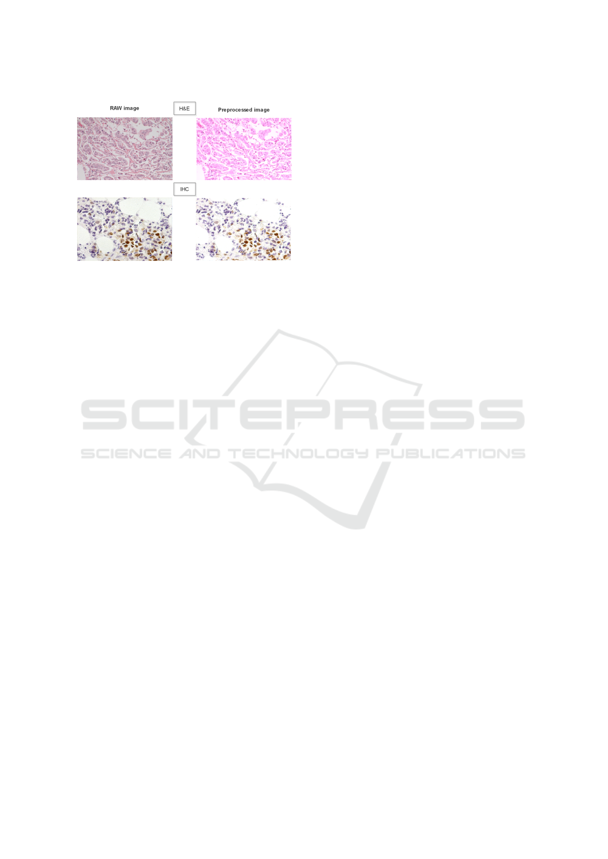
Figure 1: Results of our pre-processing methods – Exam-
ples of H&E and IHC images of Medisp HICL database
((Glotsos et al., 2008), (Kostopoulos et al., ) and (Ninos
et al., 2016)).
3.2 Features Extraction
The main purpose of this work, as stated before, is to
apply, evaluate and compare three methods for fea-
tures extraction. These latter are slightly modified
to operate on RGB image instead of gray-scale im-
ages. With this, we ensure the use of the histolog-
ical staining information as well as the morphologi-
cal textures. In our work, we chose the method: Bi-
nary Robust Invariant Scalable (BRISK) (Leuteneg-
ger et al., 2011), Oriented FAST and Rotated BRIEF
(ORB) and a combination of BRISK key-points and
DAISY descriptors.
3.2.1 Features Extraction by Color-BRISK
The original Binary Robust Invariant Scalable
(BRISK), as presented in (Leutenegger et al., 2011)
is applied to the gray-scale image. This method al-
lows the detection of local key-points and their de-
scriptors construction. The results are rotation, scal-
ing and translation invariant. BRISK follows the same
strategy as SIFT (Lowe, 1999) while being faster.
Gray-scale image is the result generated from a di-
rect transformation of the original image and combi-
nation of its three RGB channels. Some loss of color
information can occur following this conversion. To
make the most use of such details, we propose a mod-
ification to the original BRISK:
• For each RGB channel, apply BRISK and extract
the key-points.
• Generate the descriptor vector by BRISK for each
channel.
• Concatenate the three descriptor vectors into one
vector.
By this methodology, each channel of the RGB
image is considerate by BRISK as a gray-scale im-
age. In the end, and by combining the three descrip-
tors, we are sure to conserve the color information
of each channel. The final vector, resulting from the
concatenation of the three separate vectors, is the new
descriptive representation of the input image.
3.2.2 Features Extraction by Color-ORB
Oriented FAST and Rotated BRIEF (ORB) (El-
Hallak and Lovell, 2013) is a novel approach based on
the original methods Features from Accelerated Seg-
ment Test (FAST) (Rosten et al., 2010) and Binary
Robust Independent Elementary Features (BRIEF)
(Calonder et al., 2010). ORB, as the original works
of FAST, BRIEF and BRISK, is applied to gray-scale
image by calculating the FAST key-points then the
BRIEF descriptors. In this work, we use RGB image
channels separately to compute ORB. The procedure
is the same as Color-BRISK explained above.
3.2.3 Color-BRISK Features and DAISY
Descriptors
In this section, we re-use the Color-BRISK key-
points. These latter are passed as inputs to the Fast
Local Descriptor for Dense Matching (DAISY) (Tola
et al., 2010). We chose this combination to verify if
better results can be obtained by using DAISY de-
scriptor, which is known to be fast and efficient for
Bag-of-Features construction.
3.3 Features Vectors Encoding
In computer vision, descriptors vectors are effective
for image matching, image retrieval or object detec-
tion. For our system, we want to use the descriptors
for classification purposes: rather than classifying di-
rectly the image, low-level features are used instead
as its new representations. Raw descriptor vectors can
not be passed directly to the classification module; an
encoding on Bag-of-Features (BoF) is necessary.
For descriptor encoding on Bag-of-Features
(BoF), we use the method based on kmeans and the
frequencies histogram. This method is proved to be
efficient in the whole image classification task. The
BoFs by kmeans/frequency histogram is performed as
follows:
• After choosing the number of clusters (in our case,
k=5), centroids of each cluster are randomly ini-
tialized by element of the descriptor vectors space.
• For the rest of the descriptor vectors, assign a clus-
ter and recalculate the centroid of the cluster.
A Comparative Study of BRISK, ORB and DAISY Features for Breast Cancer Classification
967

• In the end, the descriptor vector are regrouped into
clusters (in our case, 5 groups)
• For the features dictionary (clusters), a histogram
of frequency is assigned. The latter represents
the apparition number of a given descriptor of the
cluster.
3.4 BoF Classification Using SVM
First introduced in (Cortes and Vapnik, 1995), Sup-
port Vector Machine (SVM) is a widely used su-
pervised classification and regression method. This
method proposed two major contributions to the su-
pervised data classification field. The first aspect of
originality provided by SVM lies in the insurance
of data separability. In fact, the authors state that
if in the original definition space, the data is inter-
leaved or overlapped, a passage to higher dimensional
space secures the obtaining of a linearly separable re-
definitions of the original data. The second goal of
SVM is to find the optimal linear hyperplane which
ensures the classification of the data while optimizing
the margins. In our system, we use the classic binary
SVM. This latter is applied when the experiments data
is contained in twos labelled classes.
4 TESTS AND EVALUATION
4.1 Experimental Setup
The elaboration of our method was performed using a
machine configured as follows: Intel® Core™ I9 10th
Gen up to 5.30GHz CPU, 32 GB of RAM, NVIDIA®
GeForce® GTX 2080 SUPER GPU, 512 GB SSD.
As a software base, we used the Python 3.8 program-
ming language, the library OpenCV 4.4.0 and its ex-
tra modules for the image pre-processing, features ex-
tracting and their encoding on Bag-of-Features. The
training and evaluation of SVM was fulfilled using
TensorFlow CPU 2.4.1.
4.2 Experiments Data
As mentioned before, in the histological process,
there are three possible staining techniques: histo-
chemistry (HC), immunohistochemistry (IHC) and
immunofluorescence (IF), to each method, a multi-
tude of stains can be associated. The most frequently
used ones are: Eosin (E), Hematoxylin (H) and the
combination Eosin-Hematoxylin (H&E). To the bet-
ter of our knowledge, in digital histopathology, there
are no IF databases and a very few IHC databases;
the majority of available data-sets concern HC H&E
breast tumoral slides. Due to this lack of IHC and IF
image bases, we decided to use H&E stained slides
in our experiments. The data-set used is BreakHis
(Spanhol et al., 2016), it offers a collection of H&E
stained SOB slides. The images are categorized,
firstly, following magnification and then following the
breast tumor type (benign or malignant). In each cat-
egory, images are grouped following the cells. (See
Tab. 2 for data-set details and total image number for
each category)
Table 2: BreakHis Dataset Details.
Type cell X40 X100 X200 X400
Benign
A 114 113 111 106
F 253 260 264 237
PT 109 121 108 115
TA 149 150 140 130
Malignant
DC 864 903 896 788
LC 156 170 163 137
MC 205 222 196 169
PC 145 142 135 138
The cells categories of the benign class are:
Adenosis (A), Fibroadenoma (F), Phyllodes Tumor
(PT) and Tubular Adenoma (TA). For the malignant
cells, there categories are: Ductal Carcinoma (DC),
Lobular Carcinoma (LC), Mucinous Carcinoma (MC)
and Papillary Carcinoma (PC). The total image con-
tained in the database is equal to 7909 images, 2480
for benign tumor and 5429 for malignant tumor. The
images size is 700 x 420 pixels.
In our work, the image are resized to 201 x 150
pixels. For the system training step, a total of 380
randomly selected images is used, 180 for the benign
class and 180 for the malignant class. In the validation
step, 120 image were used, 60 for benign and 60 for
malignant; these latter were selected randomly from
the original data-set. The purpose of this is to limit
the training data and observe which method obtains
better results.
4.3 Classification Results
The interpretation of the extracted features must be
given by a trained machine/deep learning system. As
state before, in this paper, we use Support Vector Ma-
chine to classify our Bag-of-Features (BoF). We eval-
uate our system using two criteria: classification ac-
curacy and computation time. The results are shown
in Tab. 3.
ICPRAM 2023 - 12th International Conference on Pattern Recognition Applications and Methods
968

Table 3: Experiments results.
Model Classification accuracy Precision Recall Computation time
RGB-BRISK/BoF/SVM 72.5% 75.47% 66.67% 4 hours 33 minutes
RGB-ORB/BoF/SVM 65% 68.75% 55% 2 hours 50 minutes
RGB-BRISK/DAISY/BoF/SVM 57.5% 57.89% 55% 4 hours 54 minutes
4.4 Results Discussion
The results of our experiments, as shown in Tab. 3,
prove that even if the learning data is limited, an av-
erage and acceptable classification accuracy can be
achieved. From the table, we can retain the follow-
ing:
• The best accuracy value of 72.5% was obtained by
the RGB-BRISK/BoF/SVM system. This system
remains, however, slightly slow.
• The highest value of precision and recall were also
achieved by the RGB-BRISK/BoF/SVM system.
• The fastest system is the one based on RGB-ORB,
however, this latter gave a lower classification per-
formances compared to the RGB-BRISK one.
• The RGB-BRISK key-points/DAISY descriptors
method was the slowest and achieved poor classi-
fication accuracy, precision and recall.
These results were obtained using BoF by kmeans
where K=5, however, we believe that the accuracy
values can increase for a greater k value (10, 20 or
50 and more). In the case of a bigger k value, it is
to be expected that the computation time will drasti-
cally increase. Also, compared to deep learning archi-
tectures, such as ResNet, the RGB-BRISK/BoF/SVM
system remains faster in CPU-based computation. In
the literature works, such as in (Ouddai et al., 2023),
ResNet18 training on similar amount of data took
more than 7 hours. In the case of GPU-based com-
putation, CNN and RNN architectures can be trained
in a significantly shorter time.
5 CONCLUSION AND FUTURE
WORKS
In this work, we applied different features extraction
methods for breast tumoral histological slides classi-
fication. The methods used are: BRISK, ORB and
BRISK/DAISY. An encoding on BoF by kmeans was
performed and the classification was done by SVM.
We proposed the exploitation of color information
by computing features from each RGB channel sep-
arately then fusing the three in one. The obtained
results showed that Color-BRISK gave the best clas-
sification accuracy, Color-ORB was the fastest and
achieved an accuracy of 65% and the combination
Color-BRISK/DAISY gave the worst results in both
computation time and classification accuracy. For fu-
ture works, we intend to exploit other hybrid features
extractors, other than BRISK/DAISY. Another per-
spective is to apply BRISK or ORB to other color-
spaces.
REFERENCES
Ai, S., Li, C., Li, X., Jiang, T., Grzegorzek, M., Sun, C., Ra-
haman, M. M., Zhang, J., Yao, Y., and Li, H. (2021).
A State-of-the-Art Review for Gastric Histopathology
Image Analysis Approaches and Future Development.
BioMed Research International, 2021.
Aksac, A., Demetrick, D. J., Ozyer, T., and Alhajj, R.
(2019). BreCaHAD: A dataset for breast cancer
histopathological annotation and diagnosis. BMC Re-
search Notes, 12(1):10–12.
Azevedo Tosta, T. A., Neves, L. A., and do Nascimento,
M. Z. (2017). Segmentation methods of H&E-stained
histological images of lymphoma: A review. Infor-
matics in Medicine Unlocked, 9(May):35–43.
Bukała, A., Cyganek, B., Koziarski, M., Kwolek, B., Ol-
borski, B., Antosz, Z., Swad
´
zba, J., and Sitkowski,
P. (2020). Classification of histopathological images
using scale-invariant feature transform. VISIGRAPP
2020 - Proceedings of the 15th International Joint
Conference on Computer Vision, Imaging and Com-
puter Graphics Theory and Applications, 5(Visigrapp
2020):506–512.
Calonder, M., Lepetit, V., Strecha, C., and Fua, P. (2010).
BRIEF: Binary robust independent elementary fea-
tures. Lecture Notes in Computer Science (including
subseries Lecture Notes in Artificial Intelligence and
Lecture Notes in Bioinformatics), 6314 LNCS(PART
4):778–792.
Cortes, C. and Vapnik, V. (1995). Support-vector networks.
Machine Learning, 20(3):273–297.
Das, A., Nair, M. S., and Peter, S. D. (2020). Computer-
Aided Histopathological Image Analysis Techniques
for Automated Nuclear Atypia Scoring of Breast Can-
cer: a Review. Journal of Digital Imaging.
El-Hallak, M. and Lovell, D. (2013). ORB an efficient.
Arthritis and Rheumatism, 65(10):2736.
Glotsos, D., Kalatzis, I., Spyridonos, P., Kostopoulos, S.,
Daskalakis, A., Athanasiadis, E., Ravazoula, P., Niki-
foridis, G., and Cavouras, D. (2008). Improving accu-
racy in astrocytomas grading by integrating a robust
least squares mapping driven support vector machine
A Comparative Study of BRISK, ORB and DAISY Features for Breast Cancer Classification
969

classifier into a two level grade classification scheme.
Computer Methods and Programs in Biomedicine,
90(3):251–261.
Gurcan, M. N., Boucheron, L. E., Can, A., Madabhushi, A.,
Rajpoot, N. M., and Yener, B. (2009). Histopatho-
logical Image Analysis: A Review. IEEE Reviews in
Biomedical Engineering, 2:147–171.
Irshad, H., Jalali, S., Roux, L., Racoceanu, D., Naour, G.,
Hwee, L., and Capron, F. (2013). Automated mito-
sis detection using texture, SIFT features and HMAX
biologically inspired approach. Journal of Pathology
Informatics, 4(2):12.
Irshad, H., Veillard, A., Roux, L., and Racoceanu, D.
(2014). Methods for nuclei detection, segmentation,
and classification in digital histopathology: A review-
current status and future potential. IEEE Reviews in
Biomedical Engineering, 7:97–114.
Komura, D. and Ishikawa, S. (2018). Machine Learn-
ing Methods for Histopathological Image Analysis.
Computational and Structural Biotechnology Journal,
16:34–42.
Kostopoulos, S., Glotsos, D., Cavouras, D., Daskalakis,
A., Kalatzis, I., Georgiadis, P., Bougioukos, P., Rava-
zoula, P., and Nikiforidis, G. ANALYTICAL AND
QUANTITATIVE CYTOLOGY AND HISTOLOGY
® ARTICLES Computer-Based Association of the
Texture of Expressed Estrogen Receptor Nuclei with
Histologic Grade Using Immunohistochemically-
Stained Breast Carcinomas. Technical report.
Kumar, M. D., Babaie, M., Zhu, S., Kalra, S., and Tizhoosh,
H. R. (2018). A comparative study of CNN, BoVW
and LBP for classification of histopathological im-
ages. 2017 IEEE Symposium Series on Computational
Intelligence, SSCI 2017 - Proceedings, 2018-Janua:1–
7.
Leutenegger, S., Chli, M., and Siegwart, R. Y. (2011).
BRISK: Binary Robust invariant scalable keypoints.
Proceedings of the IEEE International Conference on
Computer Vision, pages 2548–2555.
Li, C., Chen, H., Li, X., Xu, N., Hu, Z., Xue, D., Qi, S.,
Ma, H., Zhang, L., and Sun, H. (2020). A review for
cervical histopathology image analysis using machine
vision approaches, volume 53.
Li, C., Chen, H., Xue, D., Hu, Z., Zhang, L., He, L., Xu, N.,
Qi, S., Ma, H., and Sun, H. (2019a). Weakly Super-
vised Cervical Histopathological Image Classification
Using Multilayer Hidden Conditional Random Fields.
In Advances in Intelligent Systems and Computing,
volume 1011, pages 209–221. Springer Verlag.
Li, C., Chen, H., Zhang, L., Xu, N., Xue, D., Hu, Z., Ma,
H., and Sun, H. (2019b). Cervical Histopathology
Image Classification Using Multilayer Hidden Condi-
tional Random Fields and Weakly Supervised Learn-
ing. IEEE Access, 7:90378–90397.
Litjens, G., Bandi, P., Bejnordi, B. E., Geessink, O., Balken-
hol, M., Bult, P., Halilovic, A., Hermsen, M., van de
Loo, R., Vogels, R., Manson, Q. F., Stathonikos, N.,
Baidoshvili, A., van Diest, P., Wauters, C., van Dijk,
M., and van der Laak, J. (2018). 1399 H&E-stained
sentinel lymph node sections of breast cancer patients:
The CAMELYON dataset. GigaScience, 7(6):1–8.
Lowe, D. G. (1999). Object recognition from local scale-
invariant features. In Proceedings of the IEEE Inter-
national Conference on Computer Vision, volume 2,
pages 1150–1157. IEEE.
Ninos, K., Kostopoulos, S., Kalatzis, I., Sidiropoulos,
K., Ravazoula, P., Sakellaropoulos, G., Panayiotakis,
G., Economou, G., and Cavouras, D. (2016). Mi-
croscopy image analysis of p63 immunohistochemi-
cally stained laryngeal cancer lesions for predicting
patient 5-year survival. European Archives of Oto-
Rhino-Laryngology, 273(1):159–168.
Ouddai, G., Hamdi, I., and Ben Ghezala, H. (2023). Color-
SIFT Features for Histopathological Image Analysis.
In Lee, R., editor, Software Engineering, Artificial In-
telligence, Networking and Parallel/Distributed Com-
puting. SNPD 2022. Studies in Computational Intelli-
gence. (in press). Springer cham.
¨
Ozt
¨
urk,
˚
A and Akdemir, B. (2018). Application
of Feature Extraction and Classification Methods
for Histopathological Image using GLCM, LBP,
LBGLCM, GLRLM and SFTA. In Procedia Com-
puter Science, volume 132, pages 40–46. Elsevier
B.V.
Popovici, V., Budinsk
´
a, E.,
ˇ
C
´
apkov
´
a, L., Schwarz, D.,
Du
ˇ
sek, L., Feit, J., and Jaggi, R. (2016). Joint analysis
of histopathology image features and gene expression
in breast cancer. BMC Bioinformatics, 17(1):1–9.
Rosten, E., Porter, R., and Drummond, T. (2010). Faster
and better: A machine learning approach to corner de-
tection. IEEE Transactions on Pattern Analysis and
Machine Intelligence, 32(1):105–119.
Sanchez-Morillo, D., Gonz
´
alez, J., Garc
´
ıa-Rojo, M., and
Ortega, J. (2018). Classification of Breast Cancer
Histopathological Images Using KAZE Features. Lec-
ture Notes in Computer Science (including subseries
Lecture Notes in Artificial Intelligence and Lecture
Notes in Bioinformatics), 10814 LNBI:276–286.
Shafiei, S., Babaie, M., Kalra, S., and Tizhoosh, H. R.
(2021). Colored Kimia Path24 Dataset: Configura-
tions and Benchmarks with Deep Embeddings.
Simon, O., Yacoub, R., Jain, S., Tomaszewski, J. E., and
Sarder, P. (2018). Multi-radial LBP Features as a Tool
for Rapid Glomerular Detection and Assessment in
Whole Slide Histopathology Images. Scientific Re-
ports, 8(1):1–11.
Spanhol, F. A., Oliveira, L. S., Petitjean, C., and Heutte,
L. (2016). A Dataset for Breast Cancer Histopatho-
logical Image Classification. IEEE Transactions on
Biomedical Engineering, 63(7):1455–1462.
Tola, E., Lepetit, V., and Fua, P. (2010). DAISY: An effi-
cient dense descriptor applied to wide-baseline stereo.
IEEE Transactions on Pattern Analysis and Machine
Intelligence, 32(5):815–830.
Wang, C. W., Chang, C. C., Khalil, M. A., Lin, Y. J., Liou,
Y. A., Hsu, P. C., Lee, Y. C., Wang, C. H., and Chao,
T. K. (2022). Histopathological whole slide image
dataset for classification of treatment effectiveness to
ovarian cancer. Scientific Data, 9(1):1–5.
ICPRAM 2023 - 12th International Conference on Pattern Recognition Applications and Methods
970
