
Influence of Sport on Autonomic Dysreflexia of a Patient with Spinal
Cord Injury
Laura Bellintani de Freitas
1a
, Rafaella Camilo de Oliveira
1b
, Bruna Valentina Zuchatti
2c
,
Ed Wilson Ferrari Junior
1d
, Orcizo Francisco Silvestre
1e
and Alberto Cliquet Junior
1f
1
Faculty of Medical Sciences, UNICAMP - State University of Campinas, SP, Brazil
2
Faculty of Nursing, UNICAMP - State University of Campinas, SP, Brazil
Keywords: Spinal Cord Injury, Sport, Tetraplegia, Autonomic Dysreflexia.
Abstract: Spinal Cord Injury (SCI) causes loss of motor, sensory and autonomic functions below the injured level. The
increase in the number of cases of SCI, the main cause being motor vehicle accidents, and the social impact
that this condition causes makes the study on this condition very relevant. SCI brings physiological changes
and physical adaptations to the individual, such as cardiovascular problems and increased blood pressure due
to lacking of the sympathetic nervous system, known as autonomic dysreflexia (AD). Topographic
observation of the retina can help to understand the change that occurs in the autonomic response of these
individuals. The objective of the study was to analyze the vasculature changes in SCI with AD in athletes and
non-athletes, through retinal photography of these individuals. Retinal photographs were taken of 40 subjects
participating in the research, and these were divided into 4 groups: non-athletes with AD, athletes with AD,
SCI without AD, individuals without SCI. We can conclude that there is a higher prevalence of vascular
changes in patients with AD, especially in the athlete group.
1 INTRODUCTION
Spinal Cord Injury (SCI) is characterized by
disruption of afferent and efferent nerve
communication between the brain and the peripheral
nervous system, which leads to loss or reduction of
body function below the damaged level. SCI may be
complete or incomplete. Complete injury occurs
when there is no neurological function mediated by
the spinal cord below the level of injury. Incomplete
lesion occurs when there is at least some function
such as an intact sensation or as light distal motor
function (Eckert & Martin, 2017). More than half of
the individuals with SCI have the complete lesion,
which leads to total loss of sensitivity and
movements. The condition is classified as paraplegia
or tetraplegia, which are distinguished by the level of
the spinal cord affected by the injury (Holmes, 2017).
The term tetraplegia refers to the decrease or total
a
https://orcid.org/0000-0003-0213-4013
b
https://orcid.org/0000-0002-2868-6601
c
https://orcid.org/0000-0002-9288-3394
d
https://orcid.org/0000-0002-6870-4528
e
https://orcid.org/0000-0001-8537-4906
f
https://orcid.org/0000-0002-9893-5204
loss of motor and sensory functions of the cervical
segments, while the term paraplegia describes the loss
of motor and sensory functions of the thoracic,
lumbar and sacral segments.
The number of people with spinal cord injury has
grown over time, both in incidence and prevalence.
The main causes of injury are motor vehicle
accidents, followed by falls from heights and
firearms. The increase in the prevalence of
comorbidities is a consequence of the longer survival
of these patients due to the better preparation of pre-
hospital care with polytraumatized patients. The
world average of cases is 15 to 40/million inhabitants.
This high prevalence added to the social impact that
this condition brings to the lives of these people,
mostly young male adults still in the age group of 15-
40 years, (that is, the age of great productive
strength), makes the study necessary and relevant
(Casimiro et al., 2016).
Bellintani de Freitas, L., Camilo de Oliveira, R., Zuchatti, B., Ferrari Junior, E., Silvestre, O. and Cliquet Junior, A.
Influence of Sport on Autonomic Dysreflexia of a Patient with Spinal Cord Injury.
DOI: 10.5220/0012248700003587
In Proceedings of the 11th International Conference on Sport Sciences Research and Technology Support (icSPORTS 2023), pages 221-226
ISBN: 978-989-758-673-6; ISSN: 2184-3201
Copyright © 2023 by SCITEPRESS – Science and Technology Publications, Lda. Under CC license (CC BY-NC-ND 4.0)
221

The autonomic nervous system is responsible for
controlling body homeorhesis. It is divided into
sympathetic and parasympathetic, who act in an
antagonistic and coordinated manners. In order for it
to be activated, actions of neurotransmitters are
necessary. Acetylcholine (Ach) is responsible for the
post-ganglion synapses of the parasympathetic
autonomic nervous system. When we refer to the
sympathetic autonomic nervous system, the
neurotransmitter responsible is norepinephrine, or
adrenaline. The sympathetic nervous system leads to
an increase in heart rate, respiratory rate, mydriasis
and a decrease in gastrointestinal functions. The
parasympathetic works in a contrary way, balancing
the effects.
SCI that occurs at or above T6 neurologic level
results in a syndrome called autonomic dysreflexia
(AD). AD is thus defined as acute hypertension
because sympathetic reflexes below the affected level
are no longer modulated. This syndrome is often
accompanied by baroreceptor-mediated bradycardia,
which leads to a short-term control of blood pressure.
It is precipitated by massive somatic stimulation,
which leads to vasoconstriction. As factors of
precipitation of the syndrome, we can cite the
distension of the bladder by accumulation of urine or
intestinal constipation. Systolic blood pressure can
reach high values, being characterized as
hypertensive crisis, thus being a medical emergency.
Other related symptoms are: headaches, excessive
sweating, hyperemia of the skin, blurred vision and
anxiety (Edahan and Rabcheysky, 2018).
The human retina can characterize diseases of the
eye and other systems such as the cardiovascular. The
retina is a highly vascularized tissue, and its
functioning can be drastically affected in the course
of vascular changes. The retinal vasculature, because
it allows the easy visualization of part of the human
circulation through its microvasculature, is
considered as a way to non-invasive vessel network
access, without the need for procedures and with the
use of technologies of simple complexity to access it.
Thus, retinography can be an instrument for early
identification of cardiovascular changes, one of the
consequences of autonomic dysreflexia in spinal cord
injuries. Quantitative measures of retinal vascular
topography have already been proposed as a way of
analyzing the effects of autonomic dysreflexia and its
relationship with the diagnosis of this condition. AD
can occur several times a day, depending on the
amount of stimuli, which would justify the structural
change of the peripheral vasculature and its
remodelation (Edahan and Rabcheysky, 2018).
Retinal vasculature changes caused by
uncontrolled arterial hypertension have already been
described in the literature. These are: change in the
branching pattern of the retinal vessels, change in
caliber, tortuosity, and branching angle (Dai et al.,
2020). In principle, the patterns of vascular changes
that uncontrolled chronic arterial hypertension can
cause would help define the pattern of autonomic
dysreflexia.
The analysis of the vascular topography of the
retina can help to understand the functioning of the
autonomic nervous system in SCI individuals that
have AD and understand the changes that arise from
repetitive stimuli, such as the practice of Paralympic
sports. In addition, it can facilitate the diagnosis of
this condition.
The diagnosis of the presence of AD is important
to manage the hypertensive crisis that these
individuals present, since it is differs from the
management of hypertensive crises of other causes. It
is based on the active search for precipitating factors
of the episode to eliminate this stimulus such as, the
emptying of the bladder. In addition, postural
maneuvers such as sitting or lifting the patient with a
straight back can help lower blood pressure from a
better hydrostatic distribution of blood in the
extremes (Edahan and Rabcheysky, 2018).
2 OBJECTIVES
This study aimed to analyze the differences in the
patterns of retinal structures of spinal cord injured
patients with autonomic dysreflexia with those
without alterations of the autonomic system. It seeks
to diagnose changes in the retinal arterial pattern of
patients with AD, physical activity practitioners and
non-athletes, through photographic analysis of the
retina.
Changes in the parameters of the retinal
vasculature are related to increased systemic blood
pressure. Changes in the autonomic nervous system
of these individuals lead to increased blood pressure.
The detection and quantification of affected
individuals can help to clarify the effect of AD on the
cardiovascular system.
The work counts on the cooperation of the Spinal
Cord Rehabilitation Outpatient Clinic- University
Hospital, the Biomechanics and Rehabilitation of the
Locomotor System Laboratory, Faculty of Medical
Sciences, both at The University of Campinas -
UNICAMP and the GIGANTES Paralympic Rugby
Team, from the city of Campinas, State of São Paulo.
icSPORTS 2023 - 11th International Conference on Sport Sciences Research and Technology Support
222

3 METHODS
40 individuals were recruited (n = 40), 30 individuals
with spinal cord injury and 10 without spinal cord
injury. Group A was composed of 11 SCI subjects
with AD, all non-athletes. Group B was composed of
8 athletes (tetraplegics) who practice Paralympic
Rugby. As control groups, 11 lower level paraplegic
individuals were selected, by definition without
autonomic dysreflexia, comprising Group C. A total
of 10 individuals without SCI comprised Group D.
Participants with previously diagnosed
cardiovascular disease, such as systemic arterial
hypertension, were excluded from the study.
Photographs of the retina were taken from a
portable retinographer of the company Phelcom
(Figure 1), which made it possible to collect the data.
The control groups were also photographed for
comparison
.
Figure 1: Photo of EYER, portable retinographer of the
company Phelcom (Phelcom.com/product/ever).
The caliber of retinal arterial vasculature with
uncontrolled hypertension was not significantly
influenced by systemic blood pressure at the time of
photography (Dai et al., 2020). This conclusion
corroborates the structure developed by this work,
since if the hypertensive peak momentarily altered the
vasculature, retinal photographs could be a
confounding element. Since the photograph is free of
momentary alteration, it became an instrument for
retrograde analysis of the pressure profile of the
analyzed individual. The analysis of retinal
photographs was carried out by two qualified
ophthalmologists, who diagnosed changes in patterns
compared to normality, based on changes previously
described in the literature. When the diagnostic
hypothesis diverged, a third ophthalmologist was
requested for the correct classification of patients.
The description of the changes found by them was
carried out. The Ethics Committee approved the
Project - CAAE number: 31510620.9.0000.5404. All
regulations regarding the ethical use of human
volunteers were followed. Furthermore, all
participants gave their written consent, thus allowing
the use the results obtained for further analysis and
the production of new data.
4 STATISTICAL ANALYSIS
The statistical analysis used the calculation of the
odds ratio for comparison between the 4 Groups (A,
B, C and D) to clarify the relationship between
autonomic dysreflexia and the chance of vasculature
changes. For instance, when comparing group A with
group C, shown on Table 2: (4*8) / (3*7) equals 1.52.
The epidemiological profile of groups A and B,
groups referring to individuals with high spinal cord
injuries and with the presence of autonomic
dysreflexia, was also described from the interview
with the participants (Table 1).
Table 1: Epidemiological profile of patients with autonomic
dysreflexia (athletes and non-athletes).
Injury
level
Age
(years)
Sex Trauma history Injury
time
(years)
C5 38 Female Automobile
Accident
10
C4 34 Male Automobile
Accident
2
T4 23 Male Automobile
Accident
3
C5 19 Male Diving Trauma 3
T3 49 Male Automobile
Accident
15
C5 49 Male Automobile
Accident
18
T4 54 Male Automobile
Accident
23
T4 20 Male Automobile
Accident
2
T4 38 Male Firearm injury 12
C3 45 Male Firearm injury 23
C5 50 Male Fall from Height 18
C6 33 Male Diving Trauma 11
T2 38 Male Automobile
Accident
17
C7 33 Male Automobile
Accident
15
C5 30 Male Automobile
Accident
10
C7 40 Male Diving Trauma 13
C5 34 Male Diving Trauma 16
C6 34 Male Automobile
Accident
15
C5 43 Male Diving Trauma 20
Influence of Sport on Autonomic Dysreflexia of a Patient with Spinal Cord Injury
223
The diagnosis (Table 2) of changes in the arteries
of the retina: groups A, B, C and D were divided into:
with retinal vasculature alterations and without retinal
vasculature alterations
.
Table 2: Presence or absence of retinal changes in the
analyzed groups:
With
Alterations
Without
Alterations
Total
Group A 4 7 11
Group B 4 4 8
Group C 3 8 11
Group D 2 8 10
-
Group A: non-athletes with autonomic dysreflexia
-
Group B: athletes with autonomic dysreflexia
-
Group C: non-athletes without autonomic dysreflexia
-
Group D: individuals without spinal cord injury.
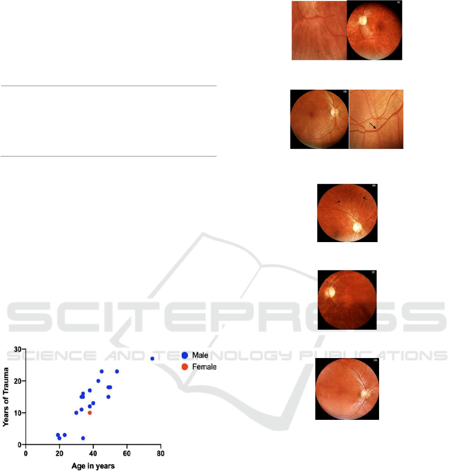
5 RESULTS
According to the epidemiological profile of patients
with autonomic dysreflexia found, we can conclude
that there is a higher prevalence of spinal cord injuries
in males, while automobile accident was the most
prevalent mechanism of trauma. The mean age of the
individuals was 35.2 years old. Mean years of injury
was 12.3 years (Figure 2).
Figure 2: Distribution by sex, age and years of injury.
The retinal changes found and described in spinal
cord injured patients with autonomic dysreflexia
were: arteriolar narrowing, arteriolar tortuosity and
pathological AV crossing. These alterations were also
found in the control groups, but the number of
individuals with alterations in the group with AD was
higher. In addition, cases with two or more
overlapping changes were present in the group with
AD, but this was not observed in the control groups
(Figures 3 to 7).
Figure 3: Arteriolar tortuosities and arteriolar narrowing.
Figure 4: Pathological AV crossing.
Figure 5: Arteriolar narrowing.
Figure 6: Retina without vascular changes (control group).
Figure 7: Retina without vascular changes (control group).
Considering the groups with AD non-athletes and
athletes together (groups A and B), we concluded that
there is a 2.33 times higher chance of developing
retinal artery changes in these individuals, compared
to individuals without AD (Groups C and D).
Considering only individuals with AD, when we
analyzed only groups A and B, we noticed a chance
of changes of 1.75 times more in Group B (athletes),
compared to Group A (non-athletes).
Comparing only individuals with spinal cord
injury that were non-athletes, we noticed a chance of
1.52 times more changes in Group A (with AD),
compared to Group C (without AD).
icSPORTS 2023 - 11th International Conference on Sport Sciences Research and Technology Support
224

Table 3: statistical analysis of the comparison between
groups A, B, C and D.
(A + B) × (C + D) A × B A × C
Odds Ratio (OR) 2.33 1.75 1.52
6 DISCUSSION
From our analysis, it was possible to infer that there
is a higher chance of occurrence of changes in retinal
arterial vasculature in individuals with AD compared
to those who do not present this comorbidity.
The chance of occurrence increases in individuals
with AD who practice physical exercise, compared to
individuals who do not practice sport.
When we analyze only the groups of non- athletes
with SCI, we see that the chance of occurrence is still
greater than 1, thus meaning an increase in
occurrence, but it is still lower than that present in the
athletes.
The results obtained lead to the discussion that it
is possible to have a higher occurrence of retinal
artery changes in these patients. Sport can influence
the occurrence of these vascular changes, and this
may be due to the higher frequency of stimuli that
these individuals are exposed to, due to regular sports.
This study demonstrated the identifiable patterns
in the retina of individuals with such condition, which
may lead in the future to an early diagnosis from the
analysis of the back of the eye from these individuals.
Patterns of alterations such as arteriolar narrowing,
arteriolar tortuosity and pathological AV crossing
were described. These changes were previously
described also in patients with Chronic Systemic
Arterial Hypertension (Ponto et al., 2017). Since the
changes in these arteries reflect systemic changes, it
can be inferred that these patients are more prone to
vascular diseases due to the presence of autonomic
dysreflexia.
This study can serve as a warning about the
increased risk of cardiovascular diseases that
autonomic dysreflexia could bring, presenting as
changes in the retinal vasculature. These can be
considered as target organ lesions, and thus retinal
photography can be a screening method for
comorbidity, enabling the individual to detect early
possible complications of cardiovascular disease that,
as in chronic arterial hypertension, can manifest as
ischemic or hemorrhagic stroke, coronary artery
disease, peripheral vascular disease, and kidney
disease (Oparil et al., 2018).
7 CONCLUSION
The current study was limited by the small number of
participants, especially considering the group of
athletes. More studies are needed, with larger
numbers of individuals, to prove the association that
this work aimed to demonstrate. In the future, early
diagnosis of AD through ophthalmoscopy in these
patients may be beneficial for early detection of the
condition and better monitoring and progression of
comorbidity.
Sport in this study was considered as a risk factor,
but we did not aim to discourage its practice. It has
already proven to be beneficial for this portion of the
population, both in terms of quality of life and health.
The practice of sport leads to a lower risk of
developing shoulder injuries, especially on the
acromioclavicular joint (Medina et al., 2015). In
addition, it is undeniable that sport serves as a form
of psychological support and social interaction for
this population. Our main objective is to stimulate the
regular follow-up of these patients, athletes or not, for
early detection and control of future changes, since all
individuals with AD are exposed to greater risks than
individuals without AD.
Since the results are similar to those found in the
analysis of retinal patterns of chronic hypertensive
patients - pathological AV crossing, tortuosities,
arteriolar narrowing, we can conclude that although
autonomic dysreflexia crises are an acute condition,
the condition can be considered as chronic. This is
explained by the characteristic of AD of high degree
of daily recurrence, justifying that acute pressure
peaks behave as chronic throughout the life of these
patients. Monitoring blood pressure during
paralympic sports involving high lesioned spinal cord
parathletes, tetraplegics in particular, is highly
recommended.
ACKNOWLEDGMENTS
The State of São Paulo Foundation for Research-
FAPESP, The National Council for Scientific and
Technological Development-CNPq (Brasilia) and the
Ministry of Education - CAPES
REFERENCES
Eckert, M. J., & Martin, M. J. (2017). Trauma: spinal cord
injury. Surgical Clinics, 97(5), 1031-1045.
Influence of Sport on Autonomic Dysreflexia of a Patient with Spinal Cord Injury
225

Holmes, D. (2017). Spinal-cord injury: spurring regrowth.
Nature, 552(7684), S49.
Casimiro, F. D. G., Oliveira, G. F. D., Tenório, P. H. D. M.,
Gagliardi, I. D. C., Zoppi Filho, A., & Cliquet Junior,
A. (2016). Clinical and radiographic evaluation of
elbows from spinal cord injuried patients. Acta
ortopedica brasileira, 24, 77-80.
Eldahan, K. C., & Rabchevsky, A. G. (2018). Autonomic
dysreflexia after spinal cord injury: Systemic
pathophysiology and methods of management.
Autonomic Neuroscience, 209, 59-70.
Dai G, Dai, G., He, W., Xu, L., Pazo, E. E., Lin, T., Liu, S.,
& Zhang, C. (2020). Exploring the effect of
hypertension on retinal microvasculature using deep
learning on East Asian population. PloS one, 15(3),
e0230111.
Ponto, K. A., Werner, D. J., Wiedemer, L., Laubert-Reh,
D., Schuster, A. K., Nickels, S., ... & Mirshahi, A.
(2017). Retinal vessel metrics: normative data and their
use in systemic hypertension: results from the
Gutenberg Health Study. Journal of hypertension,
35(8), 1635-1645.
Oparil, S., Acelajado, M. C., Bakris, G. L., Berlowitz, D.
R., Cífková, R., Dominiczak, A. F., Grassi, G., Jordan,
J., Poulter, N. R., Rodgers, A., & Whelton, P. K. (2018).
Hypertension. Nature reviews. Disease primers, 4,
18014
Medina, G. I. S., Jesus, C. L. M., Ferreira, D. M., Pacheco,
E. M. B., Beraldo, G. L., de Franca Urquiza, F., &
Cliquet, A. (2015). Is sport practice a risk factor for
shoulder injuries in tetraplegic individuals?. Spinal
Cord, 53(6), 461-466.
icSPORTS 2023 - 11th International Conference on Sport Sciences Research and Technology Support
226
