
E-MEDI: A WEB-BASED E-TRAINING PLATFORM
FOR BREAST IMAGING
I. Pratikakis, V. Virvilis, D. Kosmopoulos, S. Perantonis
IIT / NCSR “Demokritos”, Athens, Greece
A. Damianakis, D. Tsatsos
Conceptum SA,12 Heyden str., 10434 Athens, Greece
Keywords: e-Training, Breast imaging, web-based learning.
Abstract: This paper concerns a Web-based e-Training platform that is dedicated to multimodal breast imaging. The
assets of this platform are summarised in the following : (i) the efficient representation of the curriculum
flow that will permit efficient training; (ii) efficient tagging of multimodal content appropriate for the
completion of realistic cases and (iii) ubiquitous accessibility and platform independence via a web-based
approach.
1 INTRODUCTION
The essence of a medical doctor’s learning
curriculum is the acquaintance with as many
individual clinical cases as possible since the rate of
success in diagnosis and treatment is directly
proportional to the amount of this accumulated
experience. This learning principle is directly applied
to the field of radiology, where intensive training
with medical images is required. Nowadays, the
training in radiology at a European level is quite
diverse both in curricula and quality due to the lack
of appropriate content (tutorials and case-based
learning material) as well as the lack of qualified
trainers.
Furthermore, there is an increasing demand for
recruiting personnel specialized in radiography
which is proportional to the increase of produced
imaging volumes. In Sweden, imaging volumes are
increasing by 2% to 5% per year. The U.K. and
Canada are both seeing demand for imaging growing
by 5% (ESS, 2000). The demand isn't likely to fall. It
is obvious that there is a gap which may be largely
attributed to the deficiencies of the classic training
methods described in the following.
The number and complexity of the radiographic
imaging modalities have increased dramatically over
time. The last decades have brought about
tremendous innovation in the field with magnetic
resonance imaging, mammography, electron beam
computing tomography, positron emission
tomography just to name a few. While the
multimodal imagery helps the doctor in the diagnosis
it also represents an administrative burden that
renders the work of radiologists more complex,
while the technical and IT skill demands become
increasing and the underpinning knowledge base also
expanding. The range of tasks radiologists have to
perform has increased too. These rapid developments
not only require a well-prepared work force but also
rapidly adapting training programs.
The importance of critical thinking and
professional judgement in professional practice is
obvious. Especially the medical doctors that work in
geographically isolated regions are not able to
contact experienced professionals and lack in
experience due to the fact that the cases they handle
are very few compared to the ones handled in urban
hospitals. The traditional training programmes are
not applicable in these cases due to distance. The
principle of distance learning has been the
motivation factor for the increasing numbers of web
environments dedicated to learning / training
purposes, eg. (WebCT) (Blackboard) (TopClass)
(LotusLS).
In this paper, we present a Web-based training
environment that will facilitate accessibility and
support platform independence as well as will help to
bridge the gap between the availability of highly
338
Pratikakis I., Virvilis V., Kosmopoulos D., Perantonis S., Damianakis A. and Tsatsos D. (2007).
E-MEDI: A WEB-BASED E-TRAINING PLATFORM FOR BREAST IMAGING.
In Proceedings of the Third International Conference on Web Information Systems and Technologies - Society, e-Business and e-Government /
e-Learning, pages 338-345
DOI: 10.5220/0001281003380345
Copyright
c
SciTePress

trained/qualified radiology professionals and the
current needs in Europe for medical doctor’s training
and lifelong learning curriculum.
Emphasis will be placed on (i) the efficient
representation of the curriculum flow that will permit
efficient training; (ii) the tagging of multimodal
content appropriate for the completion of realistic
cases; (iii) a web-based platform that permits
ubiquitous accessibility and platform independence.
Furthermore the actual tagging can be seen as a part
of a strict protocol that helps the doctors file their
data sorted in a hierarchical way by case, time and
modality.
In this work, the focus is on breast imaging, but
the results can be easily extended to imaging
modalities of other anatomical objects like brain,
heart, etc.
The paper will be structured as in the following.
At Section 2, we will review existing relevant
environments for which we will address their
advantages and pitfalls. Section 3 is dedicated to the
overall system architecture wherein a detailed
description of the major components will be given.
Finally, at Section 4, concluding remarks will be
drawn.
2 RELATED WORK
In the following, we review many of the existing e-
learning systems related to radiology with particular
emphasis to breast imaging.
In (Costaridou et al., 1998), the potential of
interactive multimedia and Internet technologies is
investigated with respect to the implementation of a
distance learning system in medical imaging. The
system is built according to a client-server
architecture, based on the Internet infrastructure,
composed of server nodes conceptually modelled as
World Wide Web (WWW) sites. Sites are
implemented by integration and customization of
available components. The system evolves around
network-delivered interactive multimedia courses
and network-based tutoring, which constitute its
main learning features. This potential has been
demonstrated by means of an implemented system,
validated with digital image processing content,
specifically image enhancement. Image enhancement
methods are theoretically described and applied on
mammograms. Emphasis is given in the interactive
presentation of the effects of algorithm parameters
on images. The system end-user access depends on
available bandwidth, so high speed access can be
achieved via LAN or local ISDN connections
. In this
system, the content from real clinical cases was not
supported by all appropriate steps for a decision
making from the medical doctor. Furthermore, it
focused on mammograms only, limiting breast
imaging in a single modality.
In the case of the MammoEd project
(http:\\www.mammoed.com) which was developed
by the University of Washington, the aim was to
provide interactive, comprehensive teaching cases
that could be easily accessed from any computer
connected to the Internet and to provide general
breast imaging education resource for radiology
residents, attending physicians, students, clinicians,
technologists, and patients. It was developed using
teaching cases from daily clinical practice organized
into a computerized database. The screen-film
images were scanned. The student was prompted to
click on the pertinent findings or to answer questions
regarding the images. Each click rendered more
images and questions with discussion of the correct
and incorrect answers and management issues. Links
were embedded to relate teaching files and
references. Although, MammoEd system deals with
clinical cases from the daily clinical practice
supported by corresponding images that the trainee
can manipulate with minimal interactivity, it does
not support multimodal content and does not permit
the trainee to exploit all steps required for the
diagnosis of particular cases.
In January 2004 the Bavarian statutory Health
Care Administration started recertification program
for quality assurance and quality improvement in
mammography reading (Riesmeir, 2004). The
participating physicians are required to read 50 cases
randomly selected from a larger collection. The
mammography films were digitized using a high-
quality CCD scanner (570 dpi, 4096 shades of grey)
to be viewed on an appropriate display workstation.
In addition to the workstation software, a ‘home
edition’ operating on a standard PC window was
developed, allowing physicians to practice the
procedure at home and get used to working with the
software. Based on this ‘home edition’ of the
software, a CD-ROM with 35 cases for training was
composed and distributed. Future releases of the
home edition with more training cases will be in
DVDs. A shortcoming of this effort is the lack of
distance-based learning making the corresponding
material restricted to being consulted at a particular
place and consequently at particular time periods.
EURORAD (www.eurorad.org) is an e-learning
initiative of the European Association of Radiology
(EAR) that was officially launched at ECR 2001. It
is the first and still the largest peer-reviewed
Radiology teaching files database on the Internet,
and offers free access to a wealth of medical
information and imaging data, whose accuracy and
quality have been validated by some of the most
E-MEDI: A WEB-BASED E-TRAINING PLATFORM FOR BREAST IMAGING
339
experienced Radiologists in Europe. In this
approach, although we have a web-based learning
environment, it cannot permit any interactive
activities with the content itself.
In conclusion, it is clear from the above
overview that a more comprehensive e-learning
system for training is needed that firstly, will
encapsulate all available breast imaging modalities
(mammography, ultrasound, MRI), because
contemporary medical students are taught to
diagnose from a variety of modalities instead of
individual image sources. This ensures decrease of
uncertainties in difficult cases. The structure of such
a multimodal e-learning system should be adaptable
and customizable for any anatomical part (in contrast
to a system that would involve different single
modalities per body organ. Secondly, it is required
that training should be based upon a system that will
address all corresponding cases using real clinical
examples constructed in a manner that will respect
the procedure flow used in a daily clinical practice.
Finally, it is imperative that such a system should be
available for an ubiquitous trainee (anywhere,
anytime, from any computer system), enabling
accessibility, sharing and interoperability. The
proposed e-MedI framework strives toward fulfilling
the above requirements, for which a detailed
description is given in the following Sections.
3 E-TRAINING FRAMEWORK
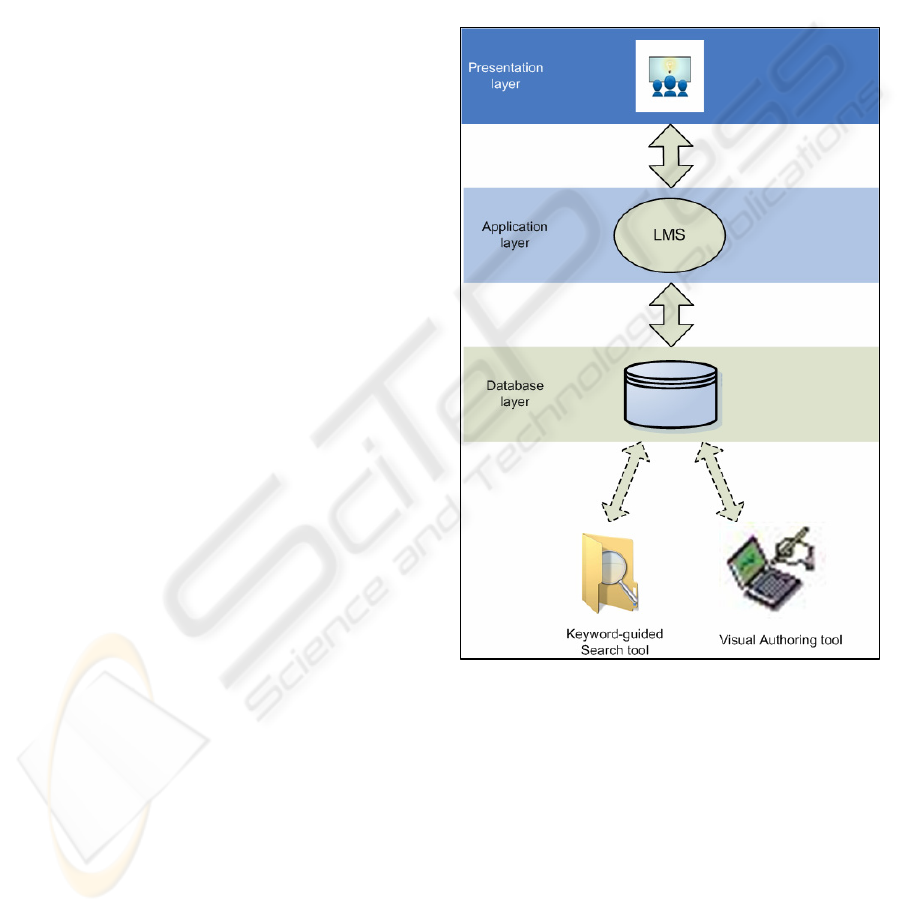
The e-MedI architecture as it is shown in Figure 1 is
based on a client-server 3-tier architecture that
consists of the following core elements: (i) Learning
Management System (LMS); (ii) Visual Authoring
Tool; (iii) Trainee’s interactive e-training
environment and (iv) Keyword-guided Clinical Case
search tool.
3.1 LMS
The LMS is by definition a complex administrative
system used to deliver electronic content in the form
of lessons and to organize people who are attend
these lessons. In the proposed framework, we have
used a proprietary LMS. Specifically, the Lotus LMS
was chosen for a multitude of reasons such as
portability, compatibility, standard compliance and
feature completeness. Lotus LMS is written in Java,
which ensures, up to a certain degree, multiplatform
portability. Lotus provides LMS for several popular
and enterprise class platforms, thus actively
supporting the generic aspect of the learning
platform. Furthermore, LMS supports several
databases, both free and commercially available, as
its storage backend.
For a public access system such as a virtual
school authentication is an essential feature.
However, modern computing often requires
authentication more than one time. This situation
quickly becomes cumbersome encouraging people to
by pass security using weak passwords, sharing
accounts etc. Lotus LMS supports Single Sign On
(SSO) which enables the user to be authenticated
only once in order to access any service offered.
Figure 1: e-MedI e-training platform architecture.
LMS scales its support from simple seminars
lessons up to curriculum lessons sets. Although e-
MedI does use currently only a small percentage of
the full LMS feature set, this setup allows future
expansion.
Finally, Lotus LMS does support SCORM
(Sharable Content Object Reference Model) (LTSC),
the widely accepted standard for e-lessons, thus
making possible to take advantage of the multitude
of SCORM tools available in the market and in the
Free Software world, and avoid a potential future
vendor lock-in.
WEBIST 2007 - International Conference on Web Information Systems and Technologies
340
3.2 Visual Authoring Tool
The Visual Authoring Tool is written in Java in order
to maximize portability and to take advantage of
future and current technologies like Java Web Start
(JWS) which drive the remote execution
technologies. The Authoring Tool is a custom
software made to facilitate the creation of Test cases
for breast imaging, but it can be adapted to other
types of pathology.
The main motivation factor that guided the
Authoring Tool development has been that the
Doctor's (teacher's) time is extremely valuable. With
the Authoring Tool, an experienced author is allowed
to construct a simple test case in about a time period
that ranges from 3 minutes, for a simple case, to 10
minutes for the most complex one.
In order to achieve this level of productivity and
user friendliness the specifications of the Authoring
Tool were crafted by an iterative process. The key
point was that we collected a series of feedback from
a team of doctors from different countries, namely
Ireland, Belgium and Cyprus. In this way, experience
was gathered from users that are familiar from
different medical systems and procedures. This
knowledge is embodied into the Authoring Tool.
The Authoring Tool creates Test Cases that
consist of consecutive stages. Each stage is
associated with one or more visual content object
(2D, 3D images and 2D+t sequences) . Each visual
content object is the acquisition output of one of the
available modalities (Mammogram, Ultrasound,
MRI).
At program startup, the Authoring Tool presents
to the author the opportunity to select and continue
editing an already existing case. If the author selects
a new case then he is prompted to select available
visual content objects from each modality. The
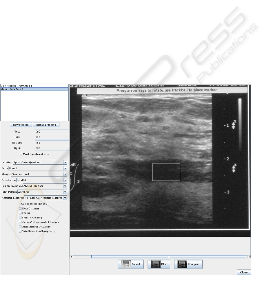
author is required to locate and describe the findings
the student is expected to identify in the image. The
description stage is supported via selection menus
(combo boxes), and boolean check boxes whenever
needed (Figure 2).
When the author has finished with the
description that is related to a particular visual
content object eg. a 2D image, it is prompted for
Figure 2: An instance of the e-MedI Visual Authoring Tool environment.
E-MEDI: A WEB-BASED E-TRAINING PLATFORM FOR BREAST IMAGING
341

another one. When finishes all visual content objects,
it is prompted for a new stage. Stages are data points
in patient's time line. Upon finishing all the desired
stages, the author is prompted to fill a small text field
representing comment's about the specific patient in
free text form, such as age or other important clinical
data.
Since e-MedI is a multilingual system and
despite the fact the Authoring Tool is currently only
in English the resulting Test case is multilingual with
the exception of the free text comment mentioned
above which has to be entered for all currently
supported languages (English, French, Greek).
Finally, it is worth mentioning that the Authoring
Tool gives an essential assistance to the tutors by
supporting image filters such as inversion, blurring
and sharpening that are routinely used by the medical
doctors during an examination.
3.2.1 Training Flow
A novel feature of the e-MedI e-training platform
relies upon the creation of a training flow that
assimilates the real situation in a clinical
environment and guides the interaction between the
trainee and the system during training. The training
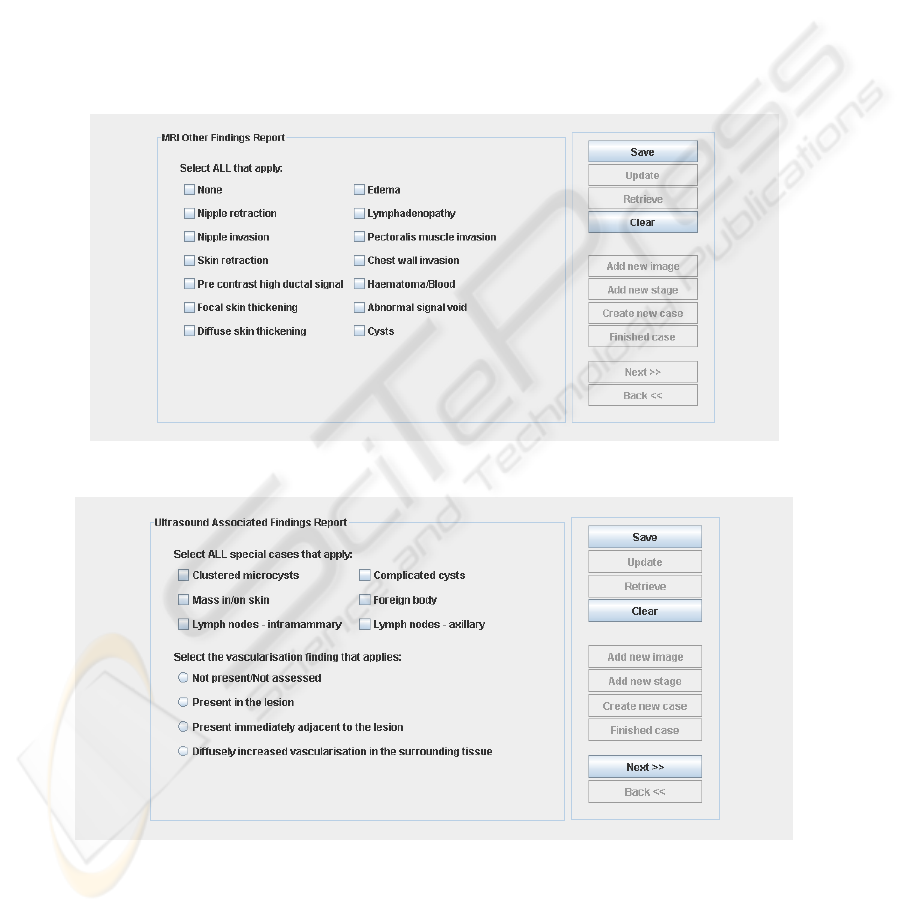
flow depends on the selected modality, as it is
displayed in both the image properties panels and the
associated findings panels. For example in a
Mammogram image it is not possible to detect
‘nipple retraction’, so this specific option does not
exist. This way the Author faces only the relevant
choices, thus accelerating the process of content
creation. Examples of the availability of findings
with respect to the modality can be shown at Figures
6-7.
In the following, the produced XML
document that depicts the training flow is discussed.
After the XML preamble (header) that states that the
encoding is Unicode, each XML file fully describes
a clinical case. For each case, a short free text with
the patient's history is available along with a case
follow up for future reference. The case follow up
can be associated with a small image for a better
overview without actually digging into the case.
The XML format described so far is like this :
<?xml version="1.0" encoding="UTF-8"?>
<case>
<general>
<history>
<caseFollowUp>
<img>
Since each case may have multiple stages these
are described in the data section of the XML file.
<data>
<stage>
<stage>
<stage>
<stage>
<image>
<image>
<image>
...
<action>
<help>
<significance>
<closeCase>
closeCase represents the final diagnosis and the
suggested course of action.
For example:
<closeCase>breastComposition="heterogeneo
usly dense" score="4: suspicious
abnormality" followUp="biopsy"></closeCase>
Each stage may have multiple images and each
image may have more than one finding. In each stage
it is possible for the author to specify further action
(e.g. request ultrasound) and give little guidance to
the trainees in the form of popup windows (<help>
tag)
Each area of the image has only one finding.
<stage>
<image>
<area>
<associatedFindings>
<implant>
<image>
<image>
The Image node has the following tags :
<file>: image filename
<label>: window title
<display>: left, right or both breasts
<modality>: mammo, ultrasound or MRI.
In the area tag the ROI coordinates are stored
along with the finding's type e.g. calcification. The
associated finding tag holds the options that are
displayed in the above figures and they are modality
specific.
Finally, the program gives the opportunity to
classify a finding as implant in order to avoid further
evaluation.
3.3 Interactive e-Training
Environment
The presentation tier is the layer of user interaction.
Its focus is on efficient user interface design and
WEBIST 2007 - International Conference on Web Information Systems and Technologies
342
accessibility throughout the training process. The UI
tier can reside on the user's desktop, on an Intranet,
or on the Internet.
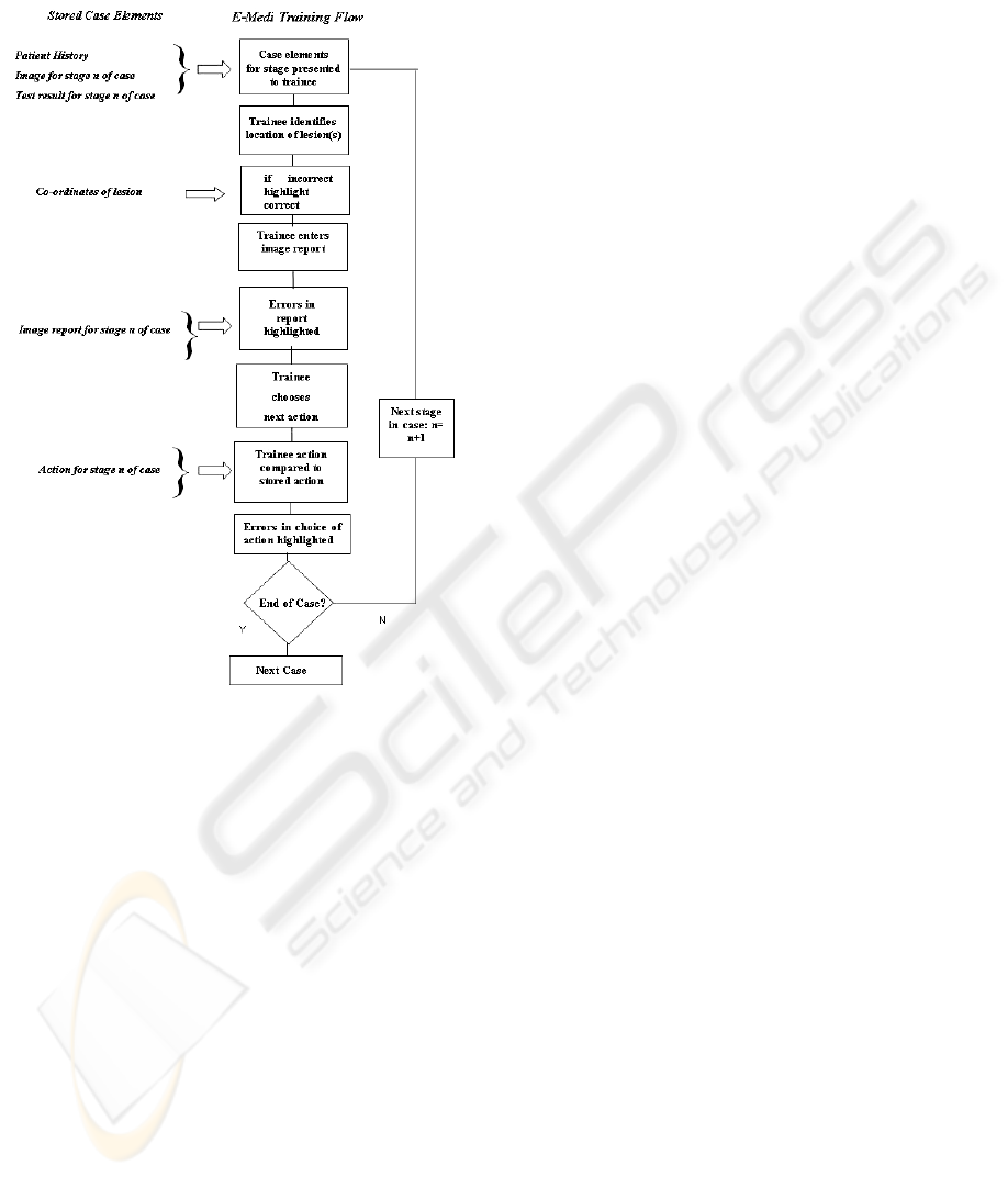
The proposed core aspects of e-MedI as
experienced by the trainee during interaction are
presented in Figure 3. This flowchart shows the core
interaction between the trainee and e-MedI for a
single case. Outside of this core, additional
functionality will exist to manage case selection,
presentation and entry of cases into e-MedI. For
example, additional functionality could be envisaged
to filter cases into categories (eg. ‘easy’ to
‘difficult’), alter the appearance of the user interface,
or provide the trainee with scores and tutorials.
The data are not be revealed in their entirety to
the trainee at the outset but in stages. A stage is the
data that is available to the trainee at any given point
between decision nodes in the decision tree. For
example at the first stage, only the patient data and
clinical breast exam results may be available. On
selecting the correct action for this history, the
images and/or test results associated with the next
stage are displayed. Note that several views (images
taken at different orientations ) will typically exist
for a single examination. Where more than one
image exists for a given examination, images may
need to be presented as thumbnails or equivalent.
The trainee will then click on regions of the image(s)
to identify suspect lesions. The chosen locations will
be crosschecked with the stored lesion coordinates
for that image.
The trainee then enters a report for the current
image using a report data entry table, which displays
a range of standard image features such as “mass”,
“mass margins”, “mass density” etc. Each feature
has standard descriptors. For example the standard
descriptors of the margins of a mass in a
mammogram are “circumscribed’, “microlobulated”,
“obscured”, “indistinct” or “speculated”. Each
descriptor will be associated with a checkbox in the
report entry table. If the trainee believes the margins
of the mass identified are best described by
“circumscribed”, then they will check the box
associated with this descriptor. Errors between the
report entered by the trainee and the stored report
can be highlighted. The trainee must then select the
next action required from a predefined list. For
example possible actions include “carry out
ultrasound” and “carry out spot compression
mammogram”, “score BiRads” or “Biopsy”. Actions
can be selected using an on-screen push button,
dropdown list or similar. Other than scoring BiRads,
any non-imaging action will in general close the
case. An important feature is that several different
image modalities may be available at the same
screen providing the trainee the opportunity to
diagnose simulating the real diagnosis procedures.
This can be shown via a snapshot of the interactive
e-training environment at Figure 4.
3.4 Keyword-based Clinical Case
Search Tool
The e-MedI database holds all available visual
content along with their corresponding descriptors.
The keyword-based clinical case search tool allows
searching through keywords with the aim to assist
the user in finding visual content instances eg. 2D
images that have specific features in order to further
improve training in a particular case diagnosis. For
example if the user has in a mammogram that may
have a ‘ruptured implant’ and wants to verify the
diagnosis, he/she can use this feature as keyword to
retrieve all relevant visual content.
Searching is realised by querying with different
granularity. An example is shown at Figure 5. First,
the user of the system selects what type of image
wants to search eg. Mammogram, Ultrasound or
MRI. After that, based on the selected modality, the
user selects among all the modality-specific
descriptors those that are needed for the particular
Figure 3: Core interaction between the trainee and e-MedI
for a single case.
E-MEDI: A WEB-BASED E-TRAINING PLATFORM FOR BREAST IMAGING
343
image the user searches for. Some descriptors are
further refined by clicking on the button refine next
to it.
4 CONCLUDING REMARKS
The proposed platform for e-training on breast
imaging draws its importance from the underlying
principle that a fruitful training process has to
assimilate and subsequently follow a training flow
which corresponds to a valid step-by-step process
followed in a real clinical routine setting. This
principle is realised via e-MedI through mainly an
authoring tool that supports the creation of the
realistic flow as well as an interactive environment
that is both user friendly and enables the
acquaintance of the trainee with the training flow
process. e-MedI can support an ubiquitous trainee
due to its web-based design providing the students
useable skills that they can directly apply in the real
world. In our immediate future plans, we will apply
to corresponding authorities all over Europe that
support Continuous Medical Education certificates to
consider e-MedI as the means of accreditation on
breast imaging.
Figure 4: A snapshot of the interactive e-training environment.
Figure 5: Example of a refined query.
WEBIST 2007 - International Conference on Web Information Systems and Technologies
344
ACKNOWLEDGEMENTS
This research was supported by the EC Leonardo da
Vinci programme “E-MedI-Virtual School on
Medical Imaging and E-Learning Framewrok”
under contract 2004-EL/2004/B/F/PP-148269.
REFERENCES
Costaridou, L., et al., 1998. Distance Learning in
Mammographic Digital Image Processing, The British
Journal of Radiology, 71(842):167-74.
Riesmeir, J., 2004. Experiences with a workstation
Prototype for softcopy Reading within the Bavarian
Mammography Re-certificating Program, Acad Radiol,
11: 407-418
LTSC, Learning Technologies Standardization Committee
( http://ltsc.ieee.org )
WebCT, ( http://www.webct.com )
Blackboard, ( http://www.blackboard.com )
TopClass, ( http://www.wbtsystems.com/ )
Lotus LS, ( http://www.lotus.com/home.nsf )
ESS, 2000. Employers skill survey. Case study: Health and
social care, Department of education and employment,
Nottingham, UK.
Diagnostic imaging, http://www.diagnosticimaging.com
Figure 6: Valid findings for MRI.
Figure 7: Valid findings for Ultrasound.
E-MEDI: A WEB-BASED E-TRAINING PLATFORM FOR BREAST IMAGING
345
