HUMAN VISUAL PERCEPTION, GESTALT PRINCIPLES AND
DUALITY REGION-CONTOUR
Application to Computer Image Analysis of Human Cornea Endothelium
Yann Gavet, Jean-Charles Pinoli
Centre Ing´enierie et Sant´e & Laboratoire LPMG, UMR CNRS 5148
´
Ecole Nationale Sup´erieure des Mines, Saint-Etienne, France
Gilles Thuret, Philippe Gain
Facult´e de M´edecine, Saint-Etienne, France
Keywords:
Pattern recognition, segmentation, grouping, cognitive and biologically inspired computer vision, mathemat-
ical morphology, medical image analysis, computational geometry, Gestalt Theory, Contour closure, Mosaic
reconstruction, image analysis.
Abstract:
The human visual system is far more efficient than a computer to analyze images, especially when noise or
poor acquisition process make the analysis impossible by lack of information. To mimic the human visual
system, we develop algorithms based on the gestalt theory principles: proximity and good continuation. We
also introduce the notion of mosaic that we reconstruct with those principles. Mosaics can be defined as
geometry figures (squares, triangles), or issued from a contour detection system or a skeletonization process.
The application presented here is the detection of cornea endothelial cells. They present a very geometric
structure that give enough information for a non expert to be able to perform the same analysis as the ophthal-
mologist, that mainly consists on counting the cells and evaluating the cell density.
1 INTRODUCTION
Image processing specialists have often the same
problem. People come with their images of their
own domain of excellence, for example in the field
of ophthalmology, which is the application frame of
the present paper. They ask for counting the cells in
the corneal endothelium (fig. 1), adding it might not
be difficult because a child could perform it.
If it were so simple, it would be a long time we
should have had to work on another subject. To char-
acterize the quality of the cornea, ophthalmologists
first evaluate the cell density and then the shape ir-
regularities of the cells. One method consists on de-
tecting the contours of the cells and then compute the
parameters, like cell density or hexagonality (Gain
et al., 2002; Debayle et al., 2006). Softwares using
the first detection method (Gain et al., 2002) are cur-
rently used in biomedical laboratories, but the special-
ists are still not satisfied because they have to spend a
few minutes checking and correcting the cell borders,
a work that a child could do (fig. 2).
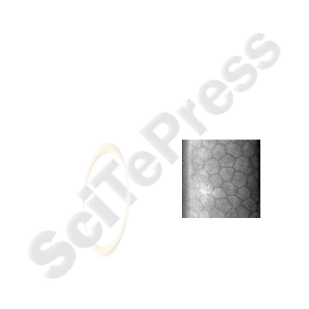
Figure 1: 215 × 215 pixels (0.036mm
2
) image of a hu-
man corneal endothelium acquired by specular optical mi-
croscopy. The endothelial cells are theoretically hexago-
nally shaped. The borders appear as dark contour lines on
this image. But sometimes (for example at the bottom left
of the image) the borders are nearly of the same intensity
level as the inside of the cells, so the segmentation of these
cells cannot be only based on the intensity information.
1.1 Theoretical Human Vision
Processing
When there is not enough information about contours
in an image, no algorithm can really detect anything.
221
Gavet Y., Pinoli J., Thuret G. and Gain P. (2007).
HUMAN VISUAL PERCEPTION, GESTALT PRINCIPLES AND DUALITY REGION-CONTOUR - Application to Computer Image Analysis of Human
Cornea Endothelium.
In Proceedings of the Second International Conference on Computer Vision Theory and Applications - IU/MTSV, pages 221-224
Copyright
c
SciTePress
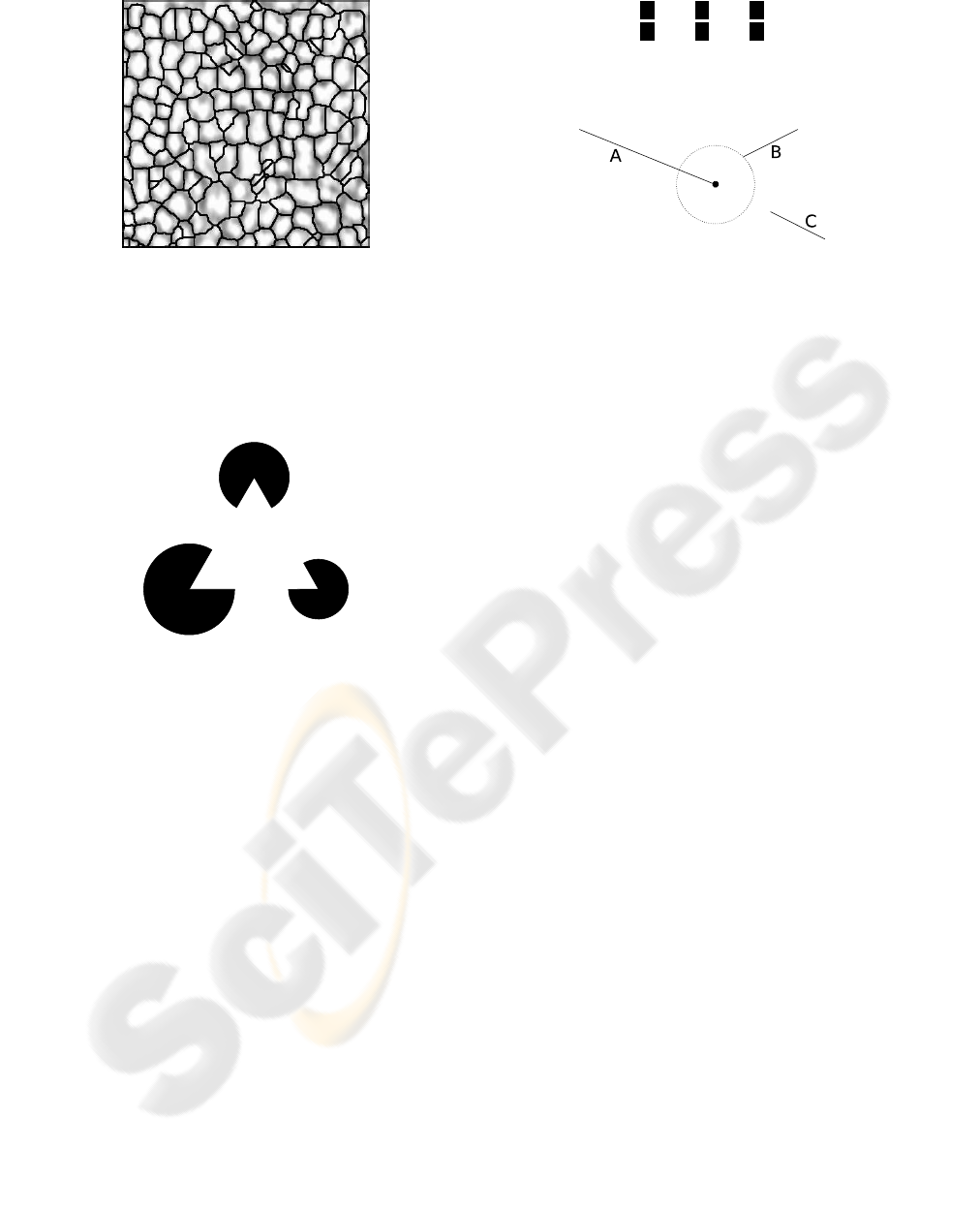
Figure 2: 400 × 400 pixels (0.4mm
2
) image of a hu-
man corneal endothelium acquired by standard optical mi-
croscopy. A software used by ophthalmologists has per-
formed a detection of contours in this image. The detected
cell map appears globally relevant, but some misdetection
can be clearly observed at several locations, (Debayle et al.,
2006).
Figure 3: A Kanizsa illusory triangle. Though not drawn,
the triangle is perfectly seen, or more precisely percepted.
But the fact is that the we see and infer informa-
tions, as illusory or subjective contours, that are not
really present in this image (Koffka, 1935) as for the
Kanisza triangle (Kanizsa, 1980) in the fig. 3.
This is not because we are intelligent, it has been
proved that many animals could see illusory contours
(barn owls (Nieder, 2002), bees, etc.). Then, how
does the human visual system do to distinguish the
cells? Is there an efficient method that could be im-
plemented in a computer to efficiently detect them?
The Gestalt Theory (Wertheimer, 1938) shows sim-
ple visual effects that mainly consist on grouping
some stimuli together according to certain principles
(proximity, good continuation, symmetry, etc.). Al-
though very attractive, they are not proved (they are
asserted by Wertheimer and the gestaltists as axioms),
but recent works show that these laws are biologically
or/and cognitively relevant (Kov´acs and Julesz, 1993;
Mullen et al., 2000). More, they appear really simple
and close to a mathematical or algorithm description,
but in fact, they are not. Those are reasons to develop
Figure 4: Grouping of elementary rectangles here is done
by proximity.
Figure 5: Each segment is grouped to the closest one
following the proximity principle. The circle shows the
Euclidean distance value from the extremity (marked point)
of the segment to the closest extremity. The segments A and
B are thus grouped by proximity.
computer programs to test the effects of the gestalt
principles.
2 ALGORITHMS: COMPUTER
VISION OF MOSAICS
This section introduces some gestalt principles (sub-
section 2.1) used on a real corneal endothelium (sub-
section 2.2). This conducts to define a mosaic as a
network of contours.
2.1 Implementing Gestalt Principles
The most basic objects used to described the gestalt
principles are points or curves. At first sight, a curve
can be simplified as a set of segments and the follow-
ing algorithms use them as inputs to the gestalt prin-
ciples.
Proximity
Experiences done by (Kubovy and Gepshtein, 2000)
prove that proximity plays an important role in group-
ing objects together (fig. 4 and 5).
Good Continuation
Good continuation (fig. 6) is the visual effect noticed
when grouping is done with objects that shows a con-
tinuity of direction.
By using Gabor patches, (Dakin and Hess, 1999)
and (Mullen et al., 2000) prove that good continuation
(in an angular variation of 30
◦
) is important for the
vision: lines have not to be continuously drawn, even
if closed contours are visually more important than
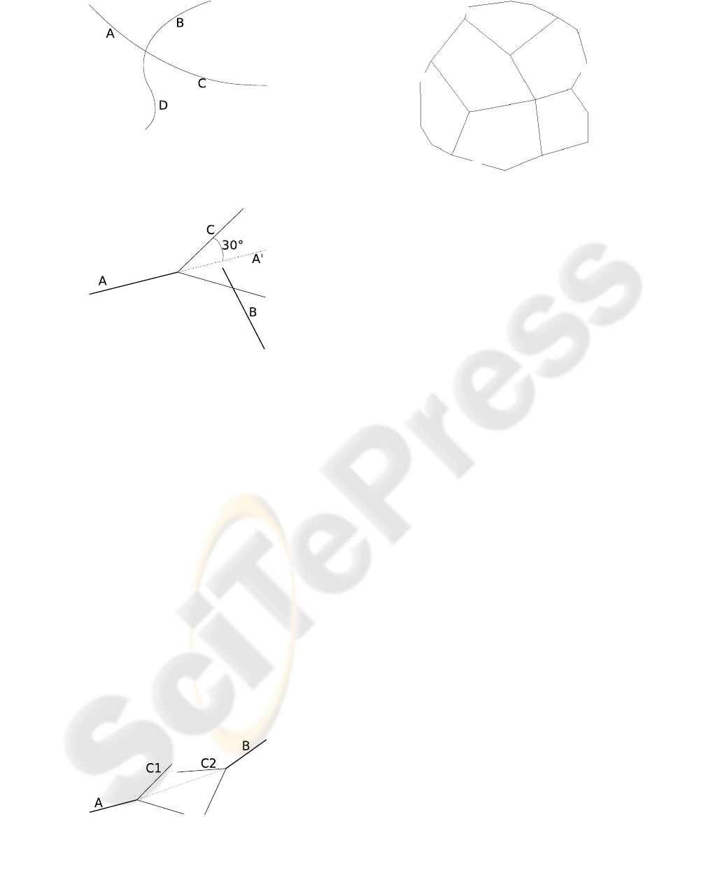
Figure 6: The lines A-C and B-D are grouped by good con-
tinuation. Visually, we have to do an effort to see the groups
A-B and C-D.
Figure 7: The direction (cone C) of continuation is defined
by the segment A and an angle of 30
◦
around the continuing
direction A’. If another extremity is inside the cone C, it
is considered as a weak continuation, as opposed to good
continuation (fig. 8).
incomplete ones (Kov´acs and Julesz, 1993) (see fig. 7
and 8).
Geodesic Proximity and Continuation
The distance transform is an operation that takes an
object and compute the Euclidean distance from this
object to any other point in the image (Rosenfeld and
Pfaltz, 1966): the result is the so-called distance map,
that can be represented as an image. The distance
transform is also used to compute the skeleton (Blum
and Nagel, 1978). The geodesic distance map is the
representation of the geodesic distance from the ob-
ject.
By using the Gestalt theory, the geodesic distance
map can be seen as a mix of proximity and continu-
Figure 8: The good continuation. is considered when the
weak continuity (fig. 7) is effective for every extremity. C1
and C2 englobe respectively the extremities of B and A.
Figure 9: A simple mosaic. Holes are randomly performed
into it and the algorithm tries to close them.
ation. Each extremity of the contour is linked to the
nearest (by the geodesic distance) other extremity.
2.2 From the Corneal Endothelium
According to (Marr, 1983), the first stage of percep-
tion consists in contour detection. This is the way
ophthalmologists analyze the cells in the corneal en-
dothelium.
The contours of the cells can be detected by using
mathematical morphology (Serra, 1988). Our algo-
rithm is inspired from (Chazallon and Pinoli, 1997).
With the skeleton of the borders of the cells (Blum
and Nagel, 1978), the goal is now to reconstruct the
cells with the principles described previously.
This type of image, e.g. the partition of the image
by planar objects, can be shown in different domains,
like materials (grains, dendrites), satellite imagery of
fields or cities, etc. This is why we now introduce an
abstraction of such a network we call a mosaic.
A 2D mosaic is a binary set of curves (we used
segments at first approximation) (fig. 9) in the Euclid-
ean plane space that can be the result of a contour de-
tection or a skeletonization of objects. For testing pur-
poses, we use a hand-drawn mosaic as the reference,
and perform holes on it: one part of the segments are
randomly erased to be then reconstructed (fig. 9).
2.3 Results
Proximity and good continuation perfectly close the
theoretical mosaic (see fig. 9), because they are really
adapted to the holes done into the it and to its segment
structure. About the real cornea (fig. 10), proxim-
ity and good continuation show their limits: the algo-
rithm can link two extremities that are very far from
each other (and draws a segment that crosses the mo-
saic). The geodesic proximity can somehow be con-
sidered as a synthesis of proximity and continuation,
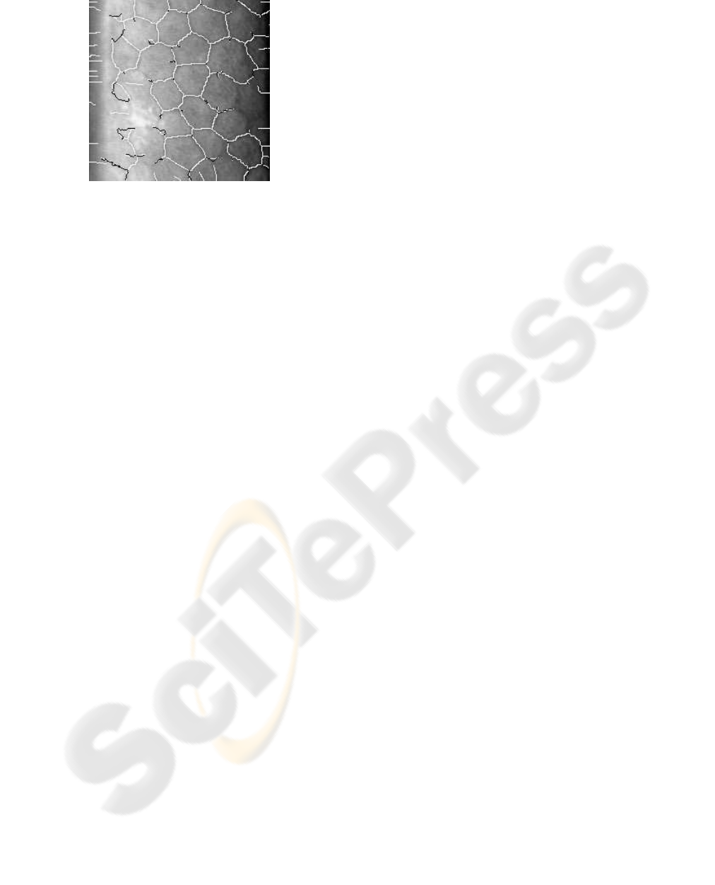
Figure 10: Application of the geodesic distance to recon-
struct the corneal endothelium. This method shows the lim-
its of a pure contour approach. The white pixels represent
the contours detected, and the black pixels represent the clo-
sure computed. The regional information is necessary to
find a good closure.
but is not enough to give what was visually expected.
3 CONCLUSION
We decided to begin with simple principles of vision
and to implement them one by one. Those simple
principles reconstruct well simple mosaics. But real
mosaics like corneal endothelium are complex. The
conclusion of these tests is that when ophthalmolo-
gists count cells on the endothelium, a lot of visual
effects occur at the same time, not only proximity and
continuation (fig. 10). To improve our methods, we
have combined proximity and continuation with a dis-
tance criteria (the geodesic continuation), and the re-
construction is not good yet.
As suggested by Marr, the human visual system
performs a Bottom-up-Top-down dual analysis; we
think that there is more than that: the human visual
system is sensitive to the duality region-contour and
unceasingly reinterpret the informations collected.
PERSPECTIVES
In a near future, we will use regional informations to
choose where to close the cells; and the proximity and
continuation methods will tell us how.
The perspectives for this work are to use regional
descriptors, size and shape parameters and contours
informations at the same time. We will also introduce
comparison methods between the mosaics to evaluate
the reconstruction, and test some other methods with
it. Those will be part of a PhD Thesis (Gavet, 2007).
REFERENCES
Blum, H. and Nagel, R. N. (1978). Shape description using
weighted symmetric axis features. Pattern Recogni-
tion, 10(3):167–180.
Chazallon, L. and Pinoli, J.-C. (1997). An automatic
morphological method for aluminium grain segmen-
tation in complex grey level images. Acta Stereol,
16(2):119–130.
Dakin, S. C. and Hess, R. F. (1999). Contour integration and
scale combination processes in visual edge detection.
Spatial Vision, 12:209–327.
Debayle, J., Gavet, Y., and Pinoli, J.-C. (2006). Image
analysis and recognition, chapter General Adaptive
Neighborhood Image Restoration, Enhancement and
Segmentation, pages 29–40. Lecture Notes in Com-
puter Science 4141. Springer Berlin / Heidelberg.
Gain, P., Thuret, G., Kodjikian, L., Gavet, Y., Turc, P. H.,
Theillere, C., Acquart, S., Petit, J. C. L., Maugery, J.,
and Campos, L. (2002). Automated tri-image analy-
sis of stored corneal endothelium. Br J Ophthalmol,
86(7):801–808.
Gavet, Y. (2007). Human vision principles and completion
of mosaics. application to human corneal endothe-
lium. PhD Thesis. To be published.
Kanizsa, G. (1980). Grammatica del vedere. Saggi su
percezione e Gestalt. Il Mulino, Bologna.
Koffka, K. (1935). Principles of Gestalt Psychology. Har-
court Brace.
Kov´acs, I. and Julesz, B. (1993). A Closed Curve is Much
More than an Incomplete One: Effect of Closure in
Figure-Ground Segmentation. PNAS, 90(16):7495–
7497.
Kubovy, M. and Gepshtein, S. (2000). Perceptual Orga-
nization for Artificial Vision Systems, chapter Gestalt:
from phenomena to laws, pages 41–71. Kluwers Aca-
demic Publishers.
Marr, D. (1983). Vision: A Computational Investigation
into the Human Representation and Processing of Vi-
sual Information. Henry Holt & Company.
Mullen, K. T., Beaudot, W. H. A., and McIlhagga, W. H.
(2000). Contour integration in color vision: a common
process for the blue-yellow, red-green and luminance
mechanisms? Vision Research, 40(6):639–655.
Nieder, A. (2002). Seeing more than meets the eye: process-
ing of illusory contours in animals. Journal of Com-
parative Physiology A: Sensory, Neural, and Behav-
ioral Physiology, 188(4):249–260.
Rosenfeld, A. and Pfaltz, J. L. (1966). Sequential operations
in digital picture processing. J. ACM, 13(4):471–494.
Serra, J. (1988). Image analysis and mathematical mor-
phology: Vol. 2. London: Academic Press.
Wertheimer, M. (1938). A sourcebook of Gestalt Psy-
chology, chapter Laws of Organization in Perceptual
Forms (partial translation), pages 71–88. Hartcourt,
Brace.
