
AN APPLICATION OF A DESCRIPTIVE IMAGE ALGEBRA FOR
DIAGNOSTIC ANALYSIS OF CYTOLOGICAL SPECIMENS
An Algebraic Model and Experimental Study
Igor Gurevich, Irina Koryabkina,Vera Yashina
Dorodnicyn Computing Center, Russian Academy of Sciences, 40 Vavilov str., 119991 Moscow, Russia
Heinrich Niemann
University of Erlangen-Nuernberg, Lehrstuhl fuer Informatik, Martensstr. 3, 91058 Erlangen, Germany
Ovidio Salvetti
Institute of Information Science and Technologies, CNR, Via G. Moruzzi 1, 56124 Pisa, Italy
Keywords: Image mining, image algebras, medical image analysis, feature extraction, pattern recognition in image
understanding, information technologies, automated diagnosis, mathematical models.
Abstract: The paper is devoted to representation of a model of an information technology for automation of diagnostic
analysis of cytological specimens of patient with lymphatic system tumors. The main contribution is
implementation of the model by algebraic means. The theoretical base of the model is the Descriptive
Approach to Image Analysis. The paper demonstrates a practical application of its algebraic tools – it is
shown how to construct a model of a technology for automation of diagnostic analysis of cytological
specimens using Descriptive Image Algebras.
1 INTRODUCTION
The paper is devoted to the development and formal
representation of the descriptive model of the
information technology for automating morphologic
analysis of cytological specimens of patients with
lymphatic system tumors (Gurevich et al., 2003).
The main tasks of the paper are to structure the
information technology and to describe it using
algebraic means provided by the Descriptive
Approach to Image Analysis (Gurevich, 1989). The
developed mathematical model should ensure a
uniform representation of an algorithm for task
solution; this is essential for programming and
useful for comparing different information
technologies designed for solving the same task.
The theoretical base of the model is Descriptive
Approach to Image Analysis (Gurevich, 1989) and
its main tools – Descriptive Image Algebras (DIA)
(Gurevich and Yashina, 2006), Descriptive Image
Modes (DIM) and Generating Descriptive Trees
(GDT) (Gurevich and Yashina, 2005).
DIA is a mathematical language developed for
description, comparison and standardization of
algorithms for image analysis, processing and
recognition. Using image analysis operations as
elements of algebra has made it possible to vary
easily methods for subtask solution, keeping overall
scheme of the technology the same.
Classes of image representation – DIM – are
used for standardization of the data for recognition
algorithms. GDT is an instrument for classification
and representation of all information connected with
image models. GDT is employed to make more
convenient selection and construction of image
models.
The main distinctive feature of the proposed
paper is that the Descriptive Approach tools are
applied for describing algorithms used for applied
task solution. Algebraization of pattern recognition
and image analysis has a long history: Unger,
Sternberg, Serra (Serra, 1982), Zhuravlev
(Zhuravlev, 1998), Grenander (Grenander, 1993),
Ritter (Ritter and Wilson, 2001), but to our
230
Gurevich I., Koryabkina I., Yashina V., Niemann H. and Salvetti O. (2007).
AN APPLICATION OF A DESCRIPTIVE IMAGE ALGEBRA FOR DIAGNOSTIC ANALYSIS OF CYTOLOGICAL SPECIMENS - An Algebraic Model and
Experimental Study.
In Proceedings of the Second International Conference on Computer Vision Theory and Applications, pages 230-237
DOI: 10.5220/0002071502300237
Copyright
c
SciTePress

knowledge algebraic methods in general and
Descriptive Approach methods in particular have
never been employed for solving the task of medical
image analysis and recognition.
In Sections 3, 4 the formal representation of the
descriptive model of the information technology is
presented. In order to make the theoretical basis
clearer Section 2 provides brief introduction into the
essential notions (DIA, DIM, GDT). Section 3
illustrates a simplified model of the image
recognition task based on multi-model image
representation. Section 4 presents a descriptive
model of the information technology developed for
automating morphologic analysis of cytological
specimens of patients with lymphatic system tumors.
The technology has been tested on the specimens
from patients with aggressive lymphoid tumors (de
novo large and mixed cell lymphomas (CL), and
transformed chronic lymphatic leukemia (TCLL)),
as well as innocent tumors (indolent chronic
lymphatic leukemia (CLL)), the results are also
presented in Section 4.
2 ALGEBRAIC TOOLS OF THE
DESCRIPTIVE APPROACH
The main purpose of theoretical apparatus of the
Descriptive Approach to Image Analysis is
structuring of the variety of methods, operations and
representations. The final goal of the Descriptive
Approach is automated image mining: a) automated
selection of techniques and algorithms for image
recognition, estimation, and understanding; b)
automated testing of the raw data quality and
suitability for solving the image recognition
problem.
2.1 Descriptive Image Models
DIM are mathematical objects – classes of image
formal description – providing representation of
information carried by an image in a form
acceptable for a recognition algorithm. There are 4
classes of DIM: P-models (Parametric Models), G-
models (Generating Models), T-models
(Transformation or Procedure Models) and I-models
(initial images as they are). Now we introduce two
of them.
Definition 1: P-model is a description of an
image by numerical features.
An example of P-model is an image
representation by numerical feature vector. An
image feature is a result of calculation of some
function f on an image during or as a result of its
processing. Let I be an initial image, vector
F=(f
1
,f
2
,…,f
n
) be a feature vector (the values of
features are calculated on an image). Thus, a model
M
P
(I)=( f
1
(I),f
2
(I),…,f
n
(I)) is a parametrical models
of an image I.
Definition 2: T-model is an image representation
as a sequence of transforms converting one or
several initial images into a given one.
Let
{
}
n
i
I
1
be a set of initial images (fragments of
an image I) used for creating a T-model. Solution of
an image recognition problem often requires
enhancing quality of an image before calculating
feature values. For instance, it can be contrast
enhancement, denoising, histogram equalization, etc.
Let
{
}
m
j
t
1
be transforms which should be applied to
an initial images in sequential or parallel modes to
get some formal description allowable by a pattern
recognition algorithm for further processing. The
transforms could be the predetermined one (to turn
an initial image 90 degrees) or the transforms with
some stopping criterion (to increase an image
contrast till the maximum in brightness histogram
would become equal to some value N). Then
M
T
=
{
}
{
}
)(
1
1
n
j
m
i
It is a T-model of an initial image.
2.2 Descriptive Image Algebras
DIA is a new type of image algebra. Its main
purpose is to provide a new mathematical language
for representation, comparison, testing and
standardization of algorithms for image analysis,
recognition, and processing.
Definition 3: An Algebra is called DIA if its
basic operands are image models or operations on
images, or both the models and operations.
Let us introduce DIA with one ring, which will
be used further for describing an algorithmic scheme
of a recognition task. For each DIA both the
operands and operations are described.
DIA 1 is a set of color images. The operands:
The set U of
{
}
I is the set of images I={{(r(x,y),
g(x,y), b(x,y)), r(x,y), g(x,y), b(x,y) ∈ [0..M-1]},
(x,y) ∈X}, M=256 is the value of maximum
intensity of a color component, n is the number of
initial images, X is the set of pixels. The operations:
The set U of {I} is the DIA with the ring of color
images over the field of real numbers with standard
algebraic operations of addition, multiplication and
multiplication by an element from the field of real
numbers.
AN APPLICATION OF A DESCRIPTIVE IMAGE ALGEBRA FOR DIAGNOSTIC ANALYSIS OF CYTOLOGICAL
SPECIMENS - An Algebraic Model and Experimental Study
231
DIA 2 is a set of gray scale images. The
operands: Elements of DIA2 are images J=
{{gray(x,y)}
(x,y) ∈X
, (x,y)∈[0,...,M-1]}. The
operations: a set V of {J} is the DIA with ring of
color images over the field of real numbers with
standard algebraic operations of addition,
multiplication and multiplication by an element from
the field of real numbers.
DIA 3 is a set F of operations f(U→V)
converting elements from the set of color images
into elements of the set of gray scale images. The
operands: Elements of DIA2 are operations
f(U→V)∈F. Such transforms can be used for
elimination luminance and color differences of
images. The operations: Operations of addition,
multiplication and multiplication by an element from
the field of real numbers are introduced on the set of
functions f as sequential operations of obtaining gray
scale images and their addition, multiplication and
multiplication by an element from the field of real
numbers correspondingly.
DIA 4 is a set G of operations g(V→P
1
) of
calculation of a gray scale image features. The
operands: DIA4 is a ring of functions g(V→P
1
)∈G,
P
1
is a set of P-models. The operations: Operations
of addition, multiplication and multiplication by a
field element are introduced on the set of functions g
as operations of sequential calculation of
corresponding P-models and their addition,
multiplication and multiplication by a field element.
DIA 5 is a set P
1
of P-models. The operands: a
set P
1
of P-models. The operations: a) addition – an
operation of unification of numerical image
descriptions; b) multiplication of 2 P-models – an
operation of obtaining a complement of numerical
image descriptions; c) multiplication by a field
element - operation of multiplication of a number, a
vector, or a matrix by an element of the field. The
addition is applied for constructing joint parametric
image representation. The multiplication is applied
for reducing a set of image features to a set of
significant features. The multiplication by an
element from the field of real numbers is applied for
feature vector normalization.
DIA 6 is a set P
2
of P-models (P
2
includes
feature vectors of the same length). The operands: a
set P
2
of P-models. The operations: Operations of
addition, multiplication and multiplication by a field
element are introduced on the set P
2
as operations of
a vector addition, multiplication and multiplication
by a field element.
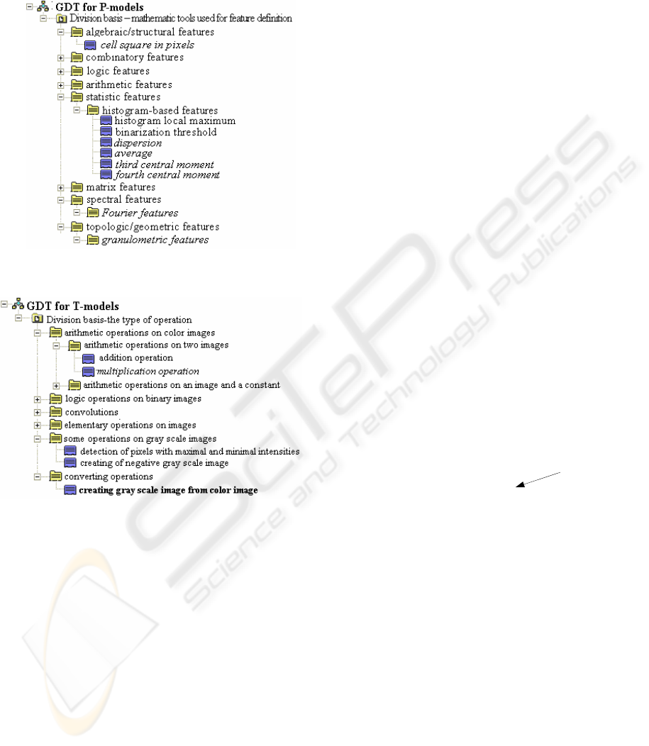
Table 1 shows all DIA with one ring presented
above, which are used for describing the algorithmic
scheme for solving the task of cytological image
recognition.
Table 1: DIAs with one ring used for describing
algorithmic scheme for solving the task of cytological
image recognition.
Ring
elements
Ring operations Purpose
1 color images standard
algebraic
operations
description of
initial images
2 gray scale
images
standard
algebraic
operations
description of
separated
nucleus on
images
3 operations
reducing
color images
to gray scale
images
standard
algebraic
operations
elimination
luminance and
color
differences of
images
4 operations of
image
feature
calculation
standard
algebraic
operations
feature
calculation
5 P-models image algebra
operations
(union,
complement,
multiplication
by real number)
selection of
informative
features
6 P-models standard
algebraic
operations
image
reduction to a
recognizable
form
2.3 Generating Descriptive Trees
GDT (Gurevich and Yashina, 2005) is a
mathematical object for generation multitude of
image models, i.e. it is a tool for creating and
combining image models.
Definition 4: GDT is a tree-like structure
intended for classification and automation of
generating formal image descriptions with the
following properties:
1) each element of the tree (descriptor) reflects
some image property;
2) each GDT combines descriptors of the same
type, i.e. GDT represents single-type properties of
an image (parametric, generic, procedural GDT and
I- GDT);
3) each element of a GDT can be combined with
another one to generate a new partial multi-aspect
model of an image.
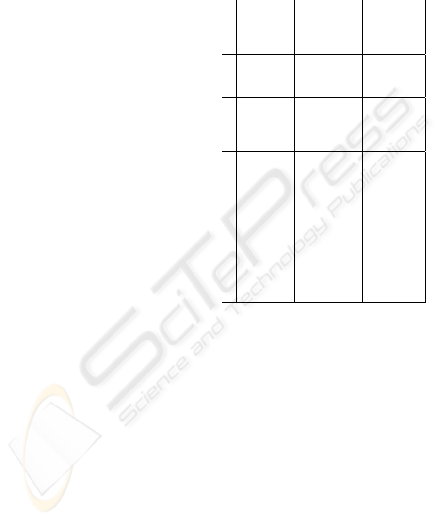
Every type of GDT represents the properties of
the image model class that constitutes its basis. P-
models are based on image features; hence
parametrical GDT is a tree of feature descriptions
VISAPP 2007 - International Conference on Computer Vision Theory and Applications
232
(Figure 1). T-models are based on image
transformations; so procedural GDT is a tree of
operations over images (Figure 2).
Figure 1: GDT for P-models.
Figure 2: GDT for T-models.
3 DESCRIPTIVE MODEL OF AN
IMAGE RECOGNITION
PROBLEM
The Descriptive Approach provides the following
model for an image recognition process (Gurevich,
1989; Gurevich and Yashina, 2005):
{
}
{
}
n
j
rlsn
IPAMI
11111
)}({}{ →→→
(1)
}{
1
n
I is a set of initial images.
i
r
n
KI
1
1
}{ ∪⊂ ,
where
{
}
r
K
1
is a set of classes determined by image
recognition task.
}{
1
s
M is multi-model
representation of an image I
j
. An algorithm
combination
{
}
l
A
1
solves an image recognition
problem, if it puts a set of predicates
n
j
r
IP
11
)}({
into correspondence to the set of initial images,
where predicate P
j
(I
i
)=α
ij
has the values: α
ij
=1, if an
image I
i
belongs to a class K
j
; α
ij
=0, if an image I
i
does not belong to a class K
j
; α
ij
=∆, if an algorithm
combination does not establish membership of an
image I
i
to a class K
j
.
Multi-model representation is generated by the
set of GDT. Different ways for constructing multi-
aspect image representations are used different GDT
types. Image representation becomes multi-model if
it is generated by different types of GDT.
Multi-model image representation
s
M
1
is created
as follows: 1) a set of GDT
{}
n
j
T
1
is generated; 2)
each GDT T
j
generates one or several formal
representations
{
}
j
n
k
M
1
of an image, they precisely
reflect image properties essential for solving the
problem at hand; 3) these representations are united
into one multi-model representation Ψ =
{}
{
}
n
n
k
j
M
1
1
оr several multi-model representations
s
M
1
, which may be used for all, оr for some initial
images presented for recognition.
This scheme takes no account of training sample
recognition. To correct this fact the scheme should
be modified as follows:
{
}
{
}
parametersAMI
l
Training
sm
→⎯⎯⎯→⎯⎯→⎯
1
)2(
1
1
1
}{
{
}
∞
+
∞
+
→⎯⎯⎯⎯→⎯⎯→⎯
111
)3(Re
1
1
1
)}({}{}{
mj
rl
cognition
S
m
IPAMI
(2)
The step of image model (models) construction
is a step of “image reduction to a recognizable form”
(Step 1). Construction of the multi-model
representation is conceptually the same for both
training set and recognition set; however, as it will
be shown below, training and recognition process
can ramify inside Step 1. Step 2 is a training step
and Step 3 is a recognition step.
4 THE MORPHOLOGICAL
ANALYSIS OF THE LYMPHOID
CELL NUCLEUSES
The developed information technology will be
described below and represented by the algorithmic
scheme (2) which is interpreted by means of DIA,
DIM and GDT.
AN APPLICATION OF A DESCRIPTIVE IMAGE ALGEBRA FOR DIAGNOSTIC ANALYSIS OF CYTOLOGICAL
SPECIMENS - An Algebraic Model and Experimental Study
233
4.1 Initial Data
A database (DB) of specimens of lymphatic tissue
imprints was created to select and describe
diagnostically important features of lymphocyte
nuclei images. DB contains 1830 specimens of 43
patients. DB contains both specimen images and the
contours of diagnostically important lymphocyte cell
nucleus indicated by experts.
Table 2: Database filling.
Diagnosis Patient
number
Image
number
Nucl
ei
number
CL
18 986 1639
TCLL
12 536 1025
CLL
13 308 2497
Total:
43 1830 5161
Footprints of lymphoid tissues were
Romanovski-Giemsa stained and photographed with
digital camera mounted on Leica DMRB microscope
using PlanApo 100/1.3 objective. The equivalent
size of a pixel was 0,0036 mcm
2
. 24-bit color images
were stored in TIFF format.
Initial images
{
}
n
I
1
are described by DIA1
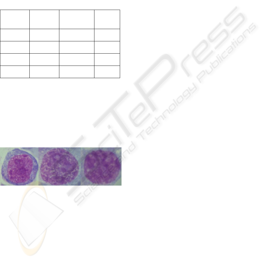
(n=1830). Figure 1 gives specimen nucleus of
patients with CL, TCLL and CLL diagnosis.
Figure 3: Specimen nucleus of patients with CL, TCLL
and CLL diagnosis accordingly (from left to right).
4.2 Reducing an Image to a
Recognizable Form
All initial images are divided into two groups:
training image set
{
}
]2/[
1
n
I and recognition image
set
{
}
n
n
I
1]2/[ +
. Below the steps 1.1-1.5 (that form
together step 1 “Reducing an image to a
recognizable form”) are described as follows:
description, model construction, step operands, step
operations. It will be highlighted where processing
of training and recognition sets differs.
Step 1.1 (equation 3): segmentation of
diagnostically important nucleus on images. The
contours of indicated by experts lymphocyte cell
nucleus were defined on images of cytological
specimens. The applied algorithm of threshold
segmentation was supplemented by morphological
processing of derivable nuclei images in order to
obtain a corresponding mask. The mask
multiplication by initial image gives indicated nuclei
image (m is the number of segmented nucleus on all
images).
{}
1.1 1
1() 1
1
1
{(,)}
,{ }
1
1
Step DIA
Tm
ij j
DIA
DIA
n
m
MI B
IB
ij
−
⎯⎯⎯⎯→
(3)
Model construction: The image T-models
m
jji
Tm
j
BIMI
1)(11
1
)},({}{ ≡ are constructed for
both the initial images
{
}
n
i
I
1
and binary masks
{
}
m
j
B
1
(n=1830, m=5161, index i(j) corresponds to
the mask number j). The number of binary masks
equals to the number of separated nucleus on initial
images. At Fig. 2 operation of this type is marked by
italics - multiplication operation.
Step operands: initial images and binary masks
represented as color
images
⎩
⎨
⎧
=∈
=∈
=
1),(,),(),1,1,1(
,0),(,),(),0,0,0(
yxvalueXyx
yxvalueXyx
I .
Step operation: Operation of segmenting cell
nucleuses on the initial image is operation of
multiplication of 2 elements of DIA1 (an initial
image is multiplied by corresponding binary mask).
Step 1.2 (equation 4): reducing color images to
gray scale images. To compensate different
illumination conditions and different colors of stain
specimen images were processed before feature
calculation.
Model construction: The image T-models
m
j
Tm
j
IMI
1
1
21
2
)}({}{ ≡ are constructed from image
models
m
j
I
1
1
}{ . At Fig. 2 operation of this type is
marked by bold - creating gray scale image from
color image.
Step operands: image models
m
j
I
1
1
}{ .
Step operations are described by the elements of
the DIA2. Such representation gives flexibility for
using different kinds of processing operations. Here
the function f(U→V)∈F (DIA 2 element) has a form
3
1
1
2
22.1
1
1
1
)}({}{
DIA
m
j
T
DIAStep
DIA
m
j
IMI ⎯⎯⎯⎯→⎯
−
(4)
VISAPP 2007 - International Conference on Computer Vision Theory and Applications
234

(I={{(r(x,y),g(x,y),b(x,y)),r(x,y),g(x,y),b(x,y)
∈[0..M-1]}
(x,y) ∈X
}): f(I)=J={{gray(x,y)}
(x,y)∈X
,
(x,y)∈[0..M-1]}, where gray(x,y)=g(x,y)
M
R2
, R is an
average brightness of a red component of an initial
RGB-image. The green tone in this case is the most
informative.
Step 1.3 (equation 5): feature calculation on
constructed image models of the training set. To
describe each image 47 features were selected: the
size of nucleus in pixels, 4 statistical features
calculated on the histogram of nucleus intensity, 16
granulometric and 26 Fourier features of nucleus.
(m
1
equals to the number of segmented nucleus on
training set).
5
1
2
1
43.1
2
1
2
11
))}({}{
DIA
m
j
P
DIAStep
DIA
m
j
IMI ⎯⎯⎯⎯→⎯
−
(5)
Model construction: P-model is generated by
features from P-GDT. 47 selected features are
marked in italics on P-GDT (Fig. 1). P-model
)()(
2
11 j
PP
IMjM ≡ is the vector with dimension
47, j=1,...,m
1
.
Step operands: image models
m
j
I
1
2
}{ .
Step operations are described by the elements of
the DIA4. Such representation gives flexibility for
calculation of different features.
The step 1.4 is additional step of image model
reduction. As it will be shown below the recognizing
algorithm was applied to both full model
)(
1
jM
P
(j=m
1
+1,...,m), and reduced model )(
2
jM
P
.
(j=m
1
+1,...,m).
Step 1.4 (equation 6): selection of informative
features. At this step the constructed image
descriptions are investigated for selecting most
informative features. Applying factor analysis to
training image set detects 14 important features
(Vorobjev et al., 2004).
1 1
1.4 5
11 2 11
56
{ ( )} { ( ( ))}
mStepDIA m
PPP
DIA DIA
Mj MM j
−
⎯⎯⎯⎯→
(6)
Model construction: Image representation
)}({
1
jM
P
is reduced to image representation
))(()(
122
jMMjM
PPP
≡ (j=1,...,m
1
). In our case
it is a vector with dimension 14.
Step operands: Image models
1
11
)}({
m
P
jM are
feature vectors with dimension 47. Step operands are
any P-models, represented as feature vectors that
form a part of vector
1
11
)}({
m
P
jM .
Step operations are described by the operations
of the DIA5.
Step 1.5: feature calculation (calculation of
features of full model
)}({
1
jM
P
or reduced model
)(
2
jM
P
for recognition set (j=m
1
+1,…,n)). Note
that multi-model representation of images
)()()(
12
jMjMj
PP
∨≡Ψ was constructed.
4.3 Training and Recognition
Algorithms based on estimate calculations (AEC)
were chosen as recognition algorithms since they
can be conveniently represented by algebraic tools
(Zhuravlev, 1998).
The set of predicate values {0, 1, ∆} derived by
algorithm combination
{
}
l
A
1
(equations 1, 2) does
not allow to construct intentional algebraic
operations. To realize algebra of algorithms the
algebraic approach of Yu.I.Zhuravlev goes from the
set of predicate values to more general set of values
- the field of real numbers. Each AEC is defined as
A = B·C, where B is recognition operator (it
calculates real estimates), and C is decision rule (for
example, threshold rule).
Recognition algorithm В requires the feature
vectors as the initial data. DIA 6 describes these
initial data.
AEC was applied to both full image models
)(
1
jM
P
(j=1,...,m, 47 features) and reduced image
models
)(
2
jM
P
(j=1,...,m, 14 features). Figures 2
and 3 present the descriptive and the structural
scheme of information technology.
The software system «Recognition 1.0»
(Zhuravlev et al., 2005), used for experimental
investigation, includes effective realization of AEC
methods and allows to apply them for practical task
solution. It was experimentally verified that the best
results are achieved by voting using all possible
support sets, while automatic definition of support
set capacity and definition of fixed support set
capacity give lower precision.
Recognition rate for full feature set amounts to
86,75%, while the rates differ for different
recognition classes (see Table 3). High recognition
rates for CLL diagnosis are likely to be connected
with innocent nature of CLL as opposed to LC and
TCLL, which are malignant. Thus cells of CLL
diagnosis have pronounced distinctions from cells of
other diagnosis, and cells of LC and TCLL diagnosis
are more similar to each other.
AN APPLICATION OF A DESCRIPTIVE IMAGE ALGEBRA FOR DIAGNOSTIC ANALYSIS OF CYTOLOGICAL
SPECIMENS - An Algebraic Model and Experimental Study
235
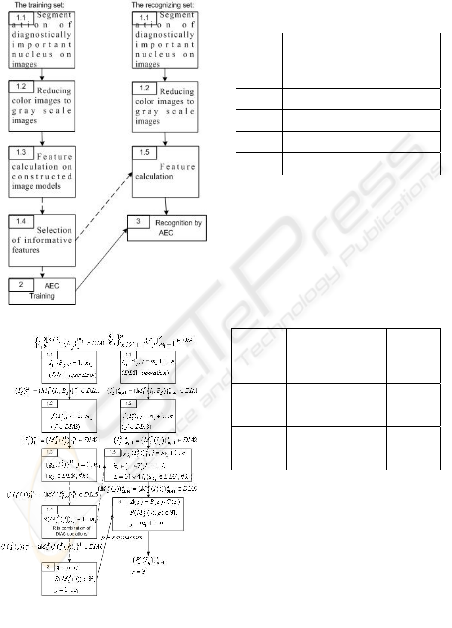
Table 3: The recognition rates for feature description
consisted of 47 features.
Diagnos
is
The
number of
correctly
recognized
cells
Total
number of
cells
The
recogniti
on rate
LC 693 820 84,51%
TCLL 325 513 63,35%
CLL 1221 1248 97,84%
Total
cell set
2239 2581 86,75%
Reducing feature set to 14 features obtained by
factor analysis the recognition rate decrease to
83,18% (see Table 4). This feature set includes
following features: the size of nucleus in pixels,
average by intensity histogram (statistic feature), the
number of elements with typical size in nuclear
(granulometric feature), the number of elements with
minimal size (granulometric feature) and 9 Fourier
features of nucleus.
Table 4: The recognition rates using reduced feature
description consisted of 14 features.
Diagnosis The
number of
correctly
recognize
d cells
Total
number of
cells
The
recognition
rate
LC 626 820 76,34%
TCLL 300 513 58,48%
CLL 1221 1248 97,84%
Full cell
set
2147 2581 83,18%
5 CONCLUSION
The paper demonstrates practical application of
algebraic tools of the Descriptive Approach to Image
Analysis - it is shown how to construct a model of a
technology for automation of diagnostic analysis of
cytological slides of patient with tumors of the
lymphatic system using Descriptive Image Algebras.
The presented model of the information technology
for automation of diagnostic analysis of medical
images will be used for creating software
Figure 4: The descriptive scheme of recognition.
Figure 5: The structural scheme of recognition.
VISAPP 2007 - International Conference on Computer Vision Theory and Applications
236

implementation of the technology, its testing and
performance evaluation.
While the method for solving medical task has
been developed previously, the contribution of this
paper is construction of the model of the information
technology, providing uniform representation for the
technology. So the paper solves dual task: firstly it
presents technology by well structured mathematic
model, and secondly it shows how DIA can be used
in image analysis task.
In the future research the Descriptive Approach
to Image Analysis and its main tools (DIA, DIM,
and GDT) will be applied for constructing models of
information technologies for automation of
diagnostic analysis in other fields of medicine.
ACKNOWLEDGEMENTS
This work was partially supported by the Russian
Foundation for Basic Research, Grants No. 05-01-
00784, 06-01-81009, by the project “Development
and Implementation Knowledge Base for Supporting
of Semantic Image Analysis” (the Program of the
Presidium of the Russian Academy of Sciences
“Fundamental Problems of Computer Science and
Information Technologies”, by the project
“Descriptive Algebras with One Ring Over Image
Models” (the Program of the Basic Research
“Algebraic and Combinatorial Techniques of
Mathematical Cybernetics” of the Department of
Mathematical Sciences of the Russian Academy of
Sciences).
REFERENCES
Grenander, U., 1993. General Pattern Theory. A
Mathematical Study of Regular Structures, Clarendon
Press, Oxford.
Gurevich, I.B., 1989. The Descriptive Framework for an
Image Recognition Problem. In Proceedings, 6
th
Scandinavian Conference on Image Analysis, 1, 220-
227. Pattern Recognition Society of Finland.
Gurevich, I., Harazishvili, D., Jernova, I., et al., 2003.
Information Technology for the Morphological
Analysis of the Lymphoid Cell Nuclei. In
Proceedings, The 13
th
Scandinavian Conference on
Image Analysis, LNCS 2749, 541-548.
Gurevich, I.B., Yashina, V.V., 2005. Generating
Descriptive Trees. In Proceedings, 10
th
International
Fall Workshop on Vision, Modeling, and
Visualization, 367-374. Infix.
Gurevich, I.B., Yashina, V.V., 2006. Operations of
Descriptive Image Algebras with One Ring. In Pattern
Recognition and Image Analysis: Advances in
Mathematical Theory and Application, 16(3), 298-
328. MAIK "Nauka/Interperiodica", Moscow.
Ritter, G.X., Wilson, J.N., 2001. Handbook of Computer
Vision Algorithms in Image Algebra, CRC Press Inc.
2
nd
edition.
Serra, J., 1982. Image Analysis and Mathematical
Morphology, Academic Press. London.
Vorobjev, I., Gurevich, I., et al., 2004. The elements of
information technology for cytological specimen
image analysis: taxonomy and factor analysis. In
Proceedings of the 7th International conference
Pattern Recognition and Image Analysis: New
Information Technologies, 3, 966-969. SPbETU.
Zhuravlev, Yu.I., 1998. An Algebraic Approach to
Recognition and Classification Problems. In Pattern
Recognition and Image Analysis: Advances in
Mathematical Theory and Applications, 8, 59-100.
MAIK "Nauka/Interperiodica", Moscow.
Zhuravlev, Yu.I., Ryazanov, V.V., et al., 2005.
RECOGNITION: A Universal Software System for
Recognition, Data Mining, and Forecasting. In Pattern
Recognition and Image Analysis, 15 (2), 476-478.
MAIK "Nauka/Interperiodica", Moscow.
AN APPLICATION OF A DESCRIPTIVE IMAGE ALGEBRA FOR DIAGNOSTIC ANALYSIS OF CYTOLOGICAL
SPECIMENS - An Algebraic Model and Experimental Study
237
