
AN OBJECTIVE METHOD TO EVALUATE FORCE AND KNEE
JOINT MOMENTS DURING ISOMETRIC EXTENSION
F. Paez
a,b
, C.
Frigo
a,b
, E. Pavan
a,b
, E. Guanziroli
a,b
and S. Frasca
a,b
a
Laboratory of Movement Biomechanics and Motor Control (TBM lab)
b
Department of Bioengineering, Polytechnic of Milan, Milan, Italy
Address: Via Garofalo, 39 Milan Italy
Keywords: Knee extension, moment of knee joint, isometric knee extensor torque.
Abstract: A simple method to evaluate force and moments of knee joint during isometric extension has been
developed and provides to the physicians a fast and objective tool for the evaluation of patients before and
after a surgery or rehabilitative program. The experiment was made on normal young patients. Graphs of
angle-moment were obtained. The patients started from 90° of knee flexion and extended step-by-step the
knee joint until the maximum knee extension was achieved. Force, angle and moment were measured at
each step. In comparison with literature, even if significant differences of technical instrumentation, age and
activity of the patients are present, the maximum moment-angle behaviour during extension is the same but
different magnitude. Future development of this device is to make it easy to use directly in clinical
applications.
1 INTRODUCTION
When a patient with a neuromuscular disease is
subject to an intervention or physical rehabilitation,
it is always necessary to make a physical evaluation
to check the functional state of the muscles and
joints. In the specific case of spastic patients, several
methods exist to see the deficit of active extension
angle (DAE) and the maximum extension force
(MEF) (Rabaiotti, 2004). Also, most mathematical
models describe the forces in the knee under
isometric quadriceps contractions (Huss et al.,2000).
The most common methods of measure used are: A
manual force test, manual dynamometers and
isokinetic dynamometers. It is usually assumed that
the moment measured by the dynamometer is
equivalent to the resultant joint moment
(Adamantios et al.,2004). Some of those methods, as
the manual force test, are subjective and not precise
because it depends on the magnitude of the manual
force the evaluator can exert on the patient. Other
methods require a big instrumentation or are
relatively expensive because of the technology of the
machine, such as the Isokinetic Dynamometer.
The aim of the present work is to create a simple,
portable and economic device for the measure of
forces and moments of knee joint during extension,
and to provide to physicians a fast and objective tool
for the evaluation of their patients before and after a
surgery or rehabilitative programs.
2 MATERIALS
A mechanical device that is attached to the base of
the bed where the patients are lying supine (See Fig.
1).This device includes a force cell connected with a
string to the leg of the patient in order to calculate
the tension force made by the leg of the patient
during the knee extension.
An electrogoniometer made by a precision linear
potentiometer in order to measure the flexion angle
of the knee. A conventional video camera
synchronized with the electrogoniometer and the
force cell in order to acquire the different positions
of the knee during the extension. A Software (MB
Ruler) for bidimensional analysis of images
(distance and angles) in order to calculate the angle
of the force cell with respect to the ground and the
angle of the string with respect to the leg of the
patient during the knee extension. A Software
environment in MATLAB to acquire and synchronize
the data of the angles of the electrogoniometer and
the forces of the cell.
228
Paez F., Frigo C., Pavan E., Guanziroli E. and Frasca S. (2008).
AN OBJECTIVE METHOD TO EVALUATE FORCE AND KNEE JOINT MOMENTS DURING ISOMETRIC EXTENSION.
In Proceedings of the First International Conference on Biomedical Electronics and Devices, pages 228-231
DOI: 10.5220/0001055102280231
Copyright
c
SciTePress
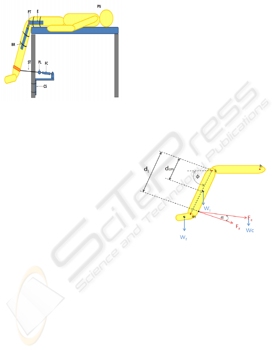
Figure 1: Instrumentation of the patient.
PN: Patient
PT: Precision Linear Potenciometer
E : Elettrogoniometer
ST: String
PL: Pulley
CS: Cell Support and system for attach to bed
BR: Braces for fix the electrogoniometer and the
Cell Support
FC: Force cell
3 METHODS
The mechanical device is attached to the bed and
supports the force cell that is connected by a string
to the ankle of the patient. 26 normal patients
participated in the experiment. (9 boys age 10
+/-
2.24
years and 17 girls age 10.12
+/-
1.87years). The
patient is in supine position with both legs outside
the bed and flexed to 90° (See fig. 1). Considering
that there is a decline of 48
+/-
11% in the mean
dynamic flexion torque by fatigue (Beltman et al.,
2003) and that some differences are caused by the
time-of-day of the exam (Onambele-Pearson et
al.,2007) we recorded only the maximum moment
on the first trial for each angle and made the exam
to each patient at morning. The patient is
instrumented with the electrogoniometer aligned
with the axis of rotation of the knee joint, which is
defined as the midpoint of the segment connecting
the lateral and medial condyles. The knee is flexed
initially at 90° and the patient is ordered to extend
his knee and as a consequence pulling the string, the
force is then recorded by the cell. In a next step the
length of the string is manually increased by an
operator which controls the pulley and consequently
the angle of knee flexion is changed while the force
measurement continues until it arrives to the
maximum extension of the knee which is at 0°. The
conventional video system, synchronized with the
electrogoniometer and the force cell is made in order
to acquire the different positions of the knee during
the extension and be analyzed by the software for
bidimensional images in order to calculate the angle
of the force cell relative to the ground and the angle
of the string with respect to the leg of the patient
during the knee extension.
Anthropometric measurements of the patients
allowed us to calculate mass properties of the leg
and to compensate for gravitational force. A
MATLAB algorithm takes all the data
(electrogoniometer, force cell, inertia properties,
anthropometrical data, force cell-ground angles and
string-leg angles) to calculate the perpendicular
force to the leg during each measure and
consequently the resultant knee torque with planar
analysis. (See Fig. 2).
Figure 2: Free body diagram of the patient shank and force
cell.
dl : Lever arm of force Fr to the knee joint
d
cm: Lever arm of Wl to the knee joint
φ: Angle of knee flexion
α: Angle of resultant force F
r
m: Malleolus joint
k : Knee joint
h: Hip joint
W
f
: Weight of foot
W
l
: Weight
of Leg
W
c
: Weight of force cell
F
c
: Force in extension and measured by the cell
F
r
: Reaction
Force of the cell, perpendicular to
mk segment.
The reaction force F
r was to be assumed
perpendicular to the patient shank mk.
AN OBJECTIVE METHOD TO EVALUATE FORCE AND KNEE JOINT MOMENTS DURING ISOMETRIC
EXTENSION
229
4 RESULTS
4.1 Construction of the Device
A simple method to evaluate force and moments of
knee joint during isometric extension has been
developed. It provides to the physicians a fast and
objective tool for the evaluation of their patients
before and after an orthopaedic surgery or
rehabilitative program.
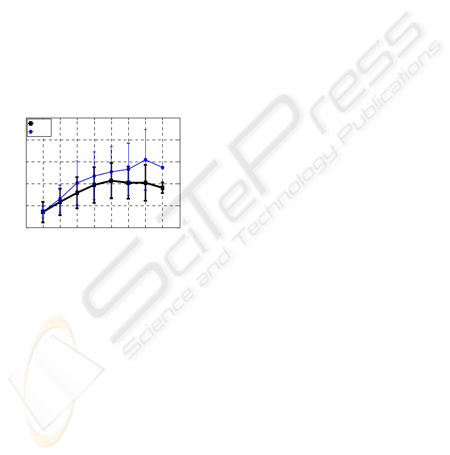
4.2 Extensor Torque in Normal Patients
The obtained information is useful to understand the
isometric extensor torque on normal and
pathological patients. Normalized moment [N*m/kg]
vs. angle [deg] of the normal patients of this study
are reported in figure 3. Graphs are separated in
male (thin line) and female (thick line) subjects.
0 10 20 30 40 50 60 70 80 90
0
0.5
1
1.5
2
2.5
Angle of Knee Flexion [Deg]
Normalized Knee Torque [Nm/kg]
Female
Mal e
Figure 3: Normalized Moment torque vs. flexion angle.
The results demonstrated significant differences
between gender according to Pincivero et al. (2004).
The highest torque was generated at 70° for men and
50° for women. Both curves have a continuous
growing behavior until his maximum value to
decrease until maximum knee flexion as reported in
literature (Beltman et al., 2003; Pincivero et al.,
2004; Welsch et al., 1998; West et al., 2005).
5 DISCUSSION
5.1 Construction of the Device
Future development of this device is to make it
usable in clinical applications. To make the process
faster and more precise, it’s specifically necessary to
eliminate the measures made by the video system
and instead install potentiometers to measure the
shank-string angle and force cell-ground angle.
5.2 Extensor Torque in Normal
Patients
If we make a comparison with literature, even if
significant differences of technical instrumentation,
age and activity of the patients are present, the
results have the same behavior but are different in
magnitude: We made the experiment with a self
constructed device on 9 occasionally active boys age
10
+/-
2.24 years and 17 occasionally active girls age
10.12
+/-
1.87 years, while Pincivero et al. (2004)
experimented with a Biodex Isokinetic
Dynamometer on 14 men age 25
+/-
4years and 14
women 23
+/-
4 years all physically active, as they
reported performing various types of routine
exercises. Beltman et al. (2003) doesn’t report the
data normalized, (only the torque in Nm) but the
behaviour of the curve is similar and he used an
Isokinetic dynamometer (Lido Active, Loredon
Biomedical, Davis) on 7 recreationally active male
subjects age 27
+/-
8 years. Welsh et al. (1998)
experimented with 39 active men age 29.7
+/-
12.6
years and 38 active women age 27.2
+/-
11.3 years
with an isometric knee flexion extension strength
testing device; so we can conclude that differences
in age, activity and instrumentation explains the
higher values of torque of those experiments with
respect to our study.
ACKNOWLEDGEMENTS
The Authors of this study would like to thank the
team of the Istituto Clinico Humanitas, Rozzano
Italy, Prof. Nicola Portinaro MD., Francesco Pelillo
MD. and Federica Spreafico MD. for the
collaboration in the study.
REFERENCES
Adamantios Arampatzis, Kiros Karamanidis, G. De
Monte, Savvas Stafilidis. Gaspar Morey-Klapsing,
Gert-Peter Bruggemann. Differences between
measured and resultant joint moments during
voluntary and artificially elicited isometric knee
extension contractions. Clin. Biomech 19 (2004) 277-
283
Beltman J , Sargeant A., Ball D, Maganaris C. Haan A.
Effect of antagonist muscle fatigue on knee extension
torque. Eur. J. Physiol (2003) 446:735–741
Huss R. A. , Holstein H. , O’Connor J. A mathematical
model of forces in the knee under isometric quadriceps
contractions. Clin. Biomech. 15 (2000) 112-122
BIODEVICES 2008 - International Conference on Biomedical Electronics and Devices
230

Kaufman, K. R. An, K. Chao. A comparison of
intersegmental joint dynamics to isokinetic
dynamometer measurements.J. Biomech. 28, 1243-
1256. (1995)
Onambele-Pearson N.L. Gladys, Pearson Stephen J. Time-
of-day effect on patella tendon stiffness alters vastus
lateralis fascicle length but not the quadriceps force–
angle relationship. Journal of Biomechanics 40 (2007)
1031–1037
Pincivero Danny M., Salfetnikov Yuliya, Campy Robert
M., Coelho Alan J. Angle- and gender-specific
quadriceps femoris muscle recruitment and knee
extensor torque. Journal of Biomechanics 37 (2004)
1689–1697
Rabaiotti G. Rassegna dei principali metodi di misura
della forza muscolare. TdR Servizio di Recupero e
Rieducazione Funzionale - Fondazione Salvatore
Maugeri - Clinica del Lavoro e della Riabilitazione-
IRCCS-Centro M. di Pavia Scienza Riabilitativa. 7-
1998
Welsch M. A., Williams P. A., Pollock M.L., Graves J. E.,
Foster D.N., Futon M.N. Quantification of full range
of Motion Unilateral and Bilateral Knee Flexion and
Extension Torque Ratios. Arch Phys Med Rehabil.
Vol. 79, August 1998
West Sacha J, Smith E Lynne E, Estelle V. Timothy D L.
Noakes E, St Clair Gibson Alan. Submaximal force
production during perceptually guided isometric
Exercise Eur J Appl. Physiol. (2005) 95: 537–542
AN OBJECTIVE METHOD TO EVALUATE FORCE AND KNEE JOINT MOMENTS DURING ISOMETRIC
EXTENSION
231
