
AUTOMATIC DETECTION OF IN VITRO CAPILLARY TUBE
NETWORK IN A MATRIGEL ANALYSIS
Eric Brassart
1
, Cyril Drocourt
1
, Jacques Rochette
2
, Michel Slama
3
and Carole Amant
2
1
LTI, Univerty of Picardie Jules Verne and IUT Amiens, France
2
DMAG, EA 3901, Amiens, France;Univerty of Picardie Jules Verne, Amiens, France and CHU
3
INSERM, ERI-12, Amiens, France;Univerty of Picardie Jules Verne, Amiens, France and CHU
Keywords: Angiogenesis, Image analysis, Capillary tube network.
Abstract: Angiogenesis, the formation of new capillary
blood vessels from pre-existing vessel, has become an
important area of scientific research. Numerous in vivo and in vitro angiogenesis assays have been
developed in order to test molecules designed to cure deregulated angiogenesis. But unlike most animal
models, most in vitro angiogenesis models are not yet automatically analysed and conclusion and data
quantification depend on the observer’s analysis. In our study, we will develop a new automatic in vitro
matrigel angiogenesis analysis allowing tube length and the number of tubes per cell islets as well as cell
islet and tubule mapping to be determined, percentage of vascularisation area, the determination of ratio of
tubule length per number of cells in cell islet and, ratio length/width per tubule determination. This new
method will also take image noise into account. Our method uses classical imaging quantification. For the
first image processing we used image segmentation (Sobel type edge detection) and artefact erasing
(morphologic operator). Subsequent image processing used Snakes: Active contour models in order to
precisely detect cells or cell islets. We suggest that this new automated image analysis method for
quantification of in vitro angiogenesis will give the researcher vascular specific quantified data that will
help in the comparison of samples.
1 INTRODUCTION
Angiogenesis, a complex process whereby new
blood vessels form from pre-existing vasculature in
response to proangiogenic factors, is an essential
physiological process required for growth and
development (Folkman J. 1971 and 1992).
Angiogenesis represents the unique process by
which evolution tissue may be supplied in essential
elements provided by blood. Angiogenesis is
therefore involved in major physiological processes
including embryonic development, female
reproduction, wound healing and collateral
generation in the myocardium. Dysregulated
angiogenesis plays a critical role in various
pathological mechanisms such as solid tumour
formation, metastasis, childhood haemangioma,
diabetic retinopathy, macular degeneration, psoriasis
and in inflammation-related diseases such as
rheumatoid arthritis, osteoarthritis and ulcerative
colitis.
2 PRIOR AND RELATED WORK
In this way, drug design in order to cure
dysregulated angiogenesis is evident. Many in vivo
and in vitro angiogenesis model have been
described. But unlike most animal models in which
blood flow doppler analysis allows vascularisation
quantification (Couffinhal T, 1999), most in vitro
angiogenesis models are not yet automatically
analysed and conclusion and quantification depend
on observer analysis (Vincent L., 2003). most in
vitro angiogenesis cannot be automatically
quantified and require observer participation. The
determination of the effect of drugs on vasculature
development requires the comparison of samples
and the use of data analysis standardization. In this
484
Brassart E., Drocourt C., Rochette J., Slama M. and Amant C. (2008).
AUTOMATIC DETECTION OF IN VITRO CAPILLARY TUBE NETWORK IN A MATRIGEL ANALYSIS.
In Proceedings of the First International Conference on Bio-inspired Systems and Signal Processing, pages 484-489
Copyright
c
SciTePress
study, we focused on an in vitro endothelial cell
differentiation matrigel assay automated image
analysis methods for the quantification of
angiogenesis. Most of the time, tube length and the
number of tubes per cell islet are the only data in
publication that can be found, and are quantified by
the observer himself. Few publications have
described an automatic image analysis approach.
One of these publications, (Niemisto A., 2005)
describes an automatic image analysis method for
quantification of in vitro matrigel angiogenesis. But
in our study we will develop a new automatic in
vitro matrigel angiogenesis analysis allowing in
addition cell islet and tubule mapping, percentage of
vascularisation area determination, ratio of tubule
length per number of cell in cell islet determination,
and ratio length/width per tubule determination. In
this study we will develop a new method in order to
take image noise into account (particles, air bubbles
included in the matrigel).
3 IMAGE ANALYSIS
3.1 Introduction
According to Nicolas Ayache, the problems
encountered in the analysis of medical imagery can
be separated into several categories:
− Restoration: this step consists of recreating an
improved image, in which several faults
connected with the physical acquisition process
have been eliminated (noise reduction, ...).
− Segmentation: separation consists of extracting
points, lines or regions which are then used as
data in complimentary work such as
realignment, measurement, analysis of
movement, visualisation etc.
− Realignment: this a problem common to many
tasks concerning the analysis of medical
imagery, and is necessary to compare the
images acquires from one single patient, or with
varying modalities.
− Morphometry: this consists of studying the
geometry of the forms, in particular the
calculation of average forms and the variations
around theses forms.
These treatments occur at different time and in
different order. The reference image we were using
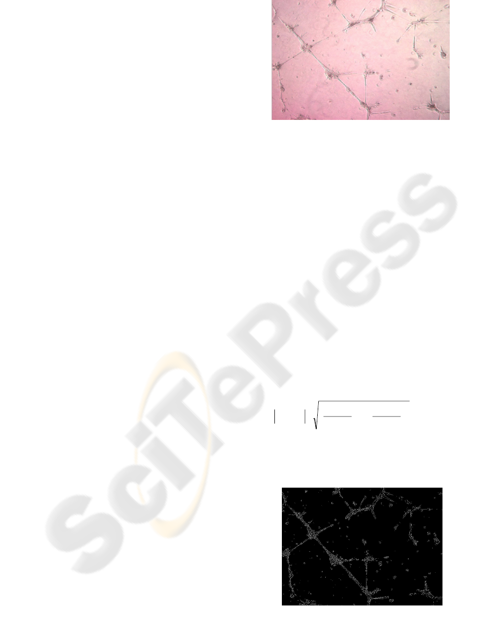
in this article is in figure 1.
Figure 1: The reference image, to which all the developed
processing will be applied in this article.
3.2 Segmentation
After having tested several methods of binarisation
(Fisher, Otsu,) (Antti Niemistö 2005) we determined
that this type of simple processing was not suitable,
principally because of its sensitivity to the variation
in luminosity within the image. Indeed, projections
of light on to tissues are not homogenous, and often
darker zones appear at the edges of the images
acquired, leading to poor separation of classes in
OTSU's formulation. We therefore chose to pre-
process our images in several successive stages,
allowing us to isolate only the cells and the
background. These steps, undertaken one
independently of the other correspond to traditional
processing in digital imagery, but bring about an
efficient solution:
− a detection of the contours by means of the use
of a gradient operator(
∇
G
), and more
specifically the norm of this operator.
2
2
y
)y,x(I
x
)y,x(I
)y,x(I
⎟
⎟
⎠
⎞
⎜
⎜
⎝
⎛
∂
∂
+
⎟
⎠
⎞
⎜
⎝
⎛
∂
∂
=∇
G
with its
discreet estimation coming down to the calculation
of two convolutions in the x and y directions. The
operator we preferred is that of Sobel (Sobel, I
1973); see Figure 2.
Figure 2 : Detection of the contours with Sobel's operator.
AUTOMATIC DETECTION OF IN VITRO CAPILLARY TUBE NETWORK IN A MATRIGEL ANALYSIS
485
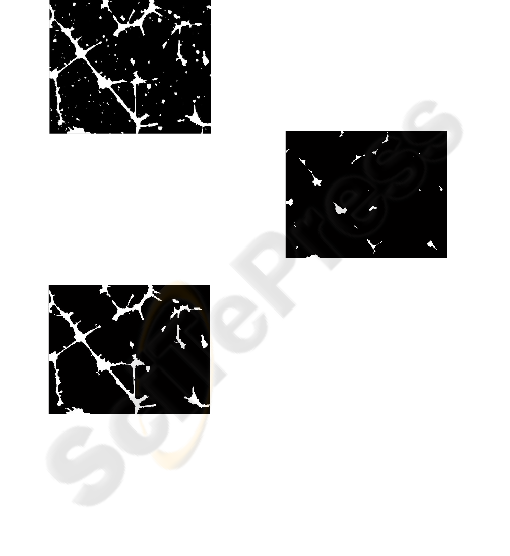
− Closure of the objects in the image which
allows the joining of neighbouring pixels to
close the contours and the unconnected
surfaces. This allows us to make the "textured"
surfaces homogenised and to create a complete,
uniform object. (Figure 3),
Figure 3 : Convolution and closure of the image
− Elimination of objects which are too small
(Restoration phase). The aim here is to eradicate
objects whose size does not satisfy the criterion
of the average size of all the images composing
the image. In the majority of the images
contained in our library, this step permits us to
attribute a sufficiently precise localisation of the
network, without necessarily being able to
identify the cells (or mass of cells) of the
connecting tubes.
Figure 4 : Isolated network after noises elimination and
the isolated elements.
The second phase of this study consists of extracting
the different elements characteristic of what we will
call the cells (or the mass of cells) from the image,
and the tubes joining the cells, when they exist. The
idea developed in this paper is firstly to isolate
everyone which resembles cells, and then to try,
from these latter, to establish the connections (tubes)
which, after all, characterise the mesh of our
network. The different stages put into place are the
elimination of the various noises in the image,
(reflection from bubbles of air in the network,
particles, non-consideration of isolated cells; the
elimination of tubes. From the resulting image, with
the remaining lines, we are specifically looking for
the exact contour of the cells or mass of cells. To do
this, we used an algorithm based on the active
contours, for which the initialisation of the starting
points is done automatically.
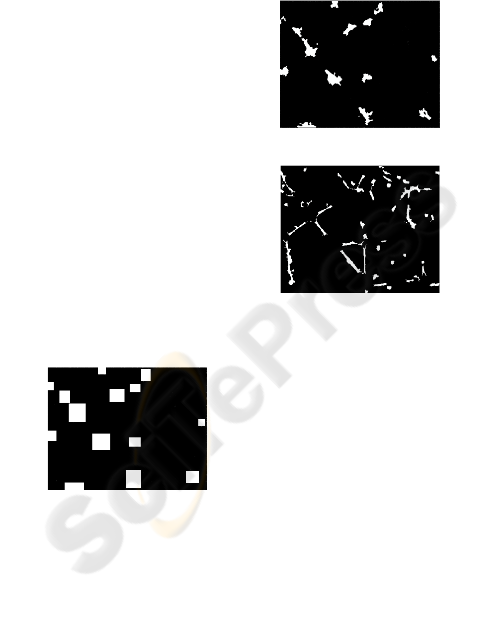
Erosion of the picture descended of the previous
stage permits to suppress the information of type
tubes and to only keep information of type cells.
This stage remains the most appreciable part of our
algorithm because it is from this one that the set of
cells will be initialized. (Figure 5),
Figure 5 : Erosion of the image and initialisation of the
starting point characterising the cellular mass.
− Use of snakes
A snake (Kass M 93, Xu C 97) is an elasticised curve
which can be modelled by a parametric shape
normalised as follows:
s
→
v ( s ) = {x (s), y(s)}
Where s is the curvilinear abscissa or the parameter
on the curve ∈ in the spatial domain Ω,
It ensues from the previous definition that a model
of snake is a problem of optimisation of a functional.
− Several resolution approaches exist, let us quote
a variation method which consists of resolving
Euler's
Ω = [0, 1] → R
2
v(s) is the vector of position of the point of contour
of coordinates x (s) and y(s),
v(1) and v(0) are the vectors of position of the
extremities of the contour.
BIOSIGNALS 2008 - International Conference on Bio-inspired Systems and Signal Processing
486
The total energy of the contour which we try to
minimize is represented by the following function
(Kass M 93):
E
ake
=
0
∫
1
E
snake
(v(s)) ds =
sn
0
∫
1
E
int
(v(s)) + E
image
(v(s)) + E
cont
(v(s)) ds
Where E
int
represents the internal energy of the
snake, E
image
is the energy derived of the image
(contours, gradients) and E
cont
represents the energy
of constraints.
Initialisation of the detection process.
One of the major concerns which exist within the
framework of the use of the active contours is the
initialization. Indeed, in the majority of the
applications using this technique, the initialization of
snakes is done manually by asking the user to select
points around the shape to detect what will
constitute the initial contour. In our application each
image zone corresponding to a cell is automatically
framed by the max coordinates resulting from a
labelling procedure The initial points correspond to
the totality of points characterising the perimeter of
each rectangle concerned (figure 6). The number of
iteration points on the snakes is limited to 200, not to
have a too long treatment on images. Of course, if
the snake converges toward a solution before this
maximum number the process stops on the usual
criteria.
Figure 6 : Initialisation of the snake son the zones marked
in white.
The use of the snakes permits to isolate precisely the
surfaces associated with the cells (Figure 6). Finally
the subtraction of the image obtained with that of the
previous step to isolate the tubes (Figure 7).
Figure 7: Initialisation of the snakes on the zones marked
in white.
Figure 8: Detection of the tubes.
By carrying out a subtraction between the binarised
images of the cells and those of the tubes we are
now able to establish the network of cells, i.e. the
cells and the tubes that connect them. To represent
this network, we positioned the centre of theses cells
by calculating the barycentre of each one of them.
The result of this method is detailed in the following
part of this article.
4 RESULTS
We have developed a new software for the
processing of images, able to automatically analyse
angiogenesis images. For this article a collection of
10 images of this type was used to validate the
results obtained. This software was written under
Matlab with the image processing toolbox. All the
images were obtained using a light microscopy. The
concern in these images acquirement is to obtain
images having sufficient contrast to be able to
clearly show the cells and the tubes, and the noise
inherent to these images as at the lowest level as
possible :
• homogenous light to avoid the effects of
poor binarisations,
AUTOMATIC DETECTION OF IN VITRO CAPILLARY TUBE NETWORK IN A MATRIGEL ANALYSIS
487
• particles and bubbles of air leading to
the detection of objects capable of being
assimilated with cells.
We will show in this part the results obtained with
various processing on a reference image, but the
reader will find the complete results obtained from
all the samples used at the following address:
www.iut-amiens.fr/Angio-results.
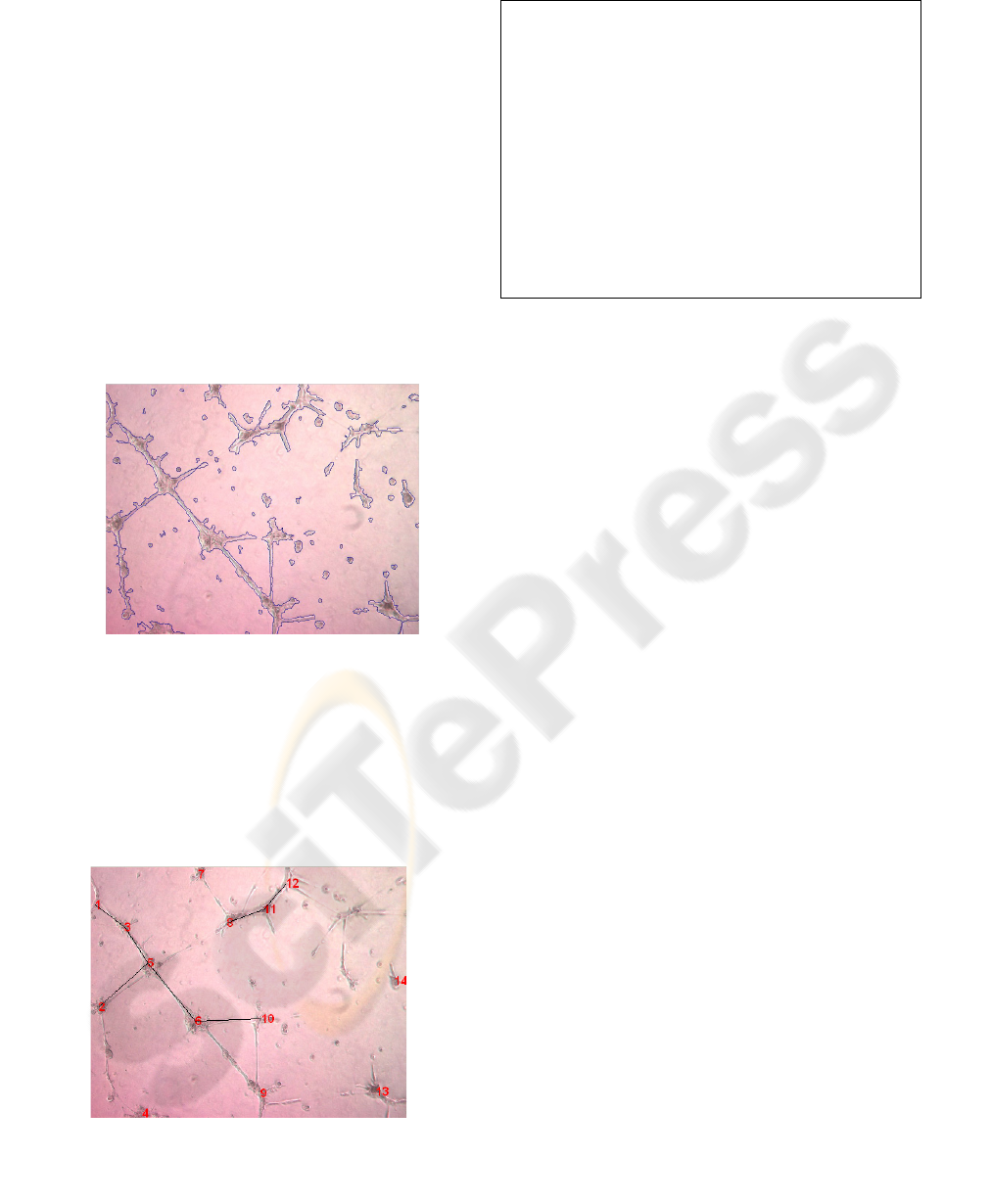
The first result given is to familiarize the practitioner
with the vascularisation surface of the sample that
has been imaged. On the image in figure 8, the
percentage of vascularisation (%Sv) obtained is
10,247%. The values given are determined by the
following ratio of surfaces :
The surface of the pixels within the contours (Sc)
divided by the total pixel surface of the image (St).
Figure 10: percentage of the vascularisation surface: %Sv
= Sc/St.
The results obtained are given in the form of a ratio
that the user of our program may consult following
the processing. On one hand it is visual with the
illustration in figure 9, on the other hand it is
numerical by means of consultation of the statistics
shown in the following table.
Figure 11: Aspect of the network corresponding to the
cellular development.
Report of the detection:
Connection
1 connected with 3
2 connected with 6
3 connected with 6
6 connected with 10
8 connected with 11
11 connected with 12
Number of Cells: 14 :
Number of Tubes: 48
Number of Connections : 6
Mean tube length: 96,923 (in arbitrary units)
Surface of the cells: 4088 (in arbitrary units)
5 CONCLUSIONS
We have developed a new technique of automatic
detection of a vascular network in a matrigel gel.
This technique is based on basic image processing
techniques such as the detection of contours,
morphologic operators combined with more
sophisticated processing such as the use of active
contours. On this latter point we have developed the
original idea of automatic placement of the initial
points on the cells. In literature dealing with this
aspect, there are very few methods avoiding the
placement of these points manually. Our technique
can be used to measure the length and size of tubular
complexes automatically, to localize cell islet and
tubule, to measure the percentage of the
vascularisation area, the ratio of tubule length per
number of cells in a cell islet and the ratio
length/width per tubule. Our software also propose
the structure of the capillary network.
Concerning the software, a certain number of
developments still have to be completed. Indeed,
during the detection of the cells in the image, a
certain number of them are considered as noise or
are ignored since the contrasts are not significant to
allow automatic detection (the light is not adapted).
In order to consider them as an integral part of the
mesh, the practitioner must be able to make them
active by manual intervention, and reintroduce them
in the detection of the capillary network.
6 PERSPECTIVES
This software will help the researcher to quantify
samples and to determine the effect of new anti-
angiogenic or pro-angiogenic agents in deregulated
BIOSIGNALS 2008 - International Conference on Bio-inspired Systems and Signal Processing
488

angiogenesis processing. An other application of
these new in vitro angiogenesis quantification
techniques in ischemic hind limb or ischemic
myocardial cell therapy will be to test the ability of
bone marrow stem cells or endothelial progenitor
cells to differentiate in endothelial cells and to
establish a vasculature shortly before the injection in
the ischemic tissue.
REFERENCES
Antti Niemistö, Valerie Dunmire, Olli Yli-Harja, Wei
Zhang, and Ilya Shmulevich ‘Robust Quantification of
In Vitro Angiogenesis through Image Analysis’ IEEE
Transactions on Medical Imaging, vol. 24, no. 4, April
2005 pp 549- 553.
Couffinhal T, Silver M, Kearney M, Sullivan A,
Witzenbichler B, Magner M, Annex B, Peters K, Isner
JM. “Impaired collateral vessel development
associated with reduced expression of vascular
endothelial growth factor in ApoE-/- mice.”
Circulation. 1999 Jun 22;99(24):3188-98.
Couffinhal T, Silver M, Kearney M, Sullivan A,
Witzenbichler B, Magner M, Annex B, Peters K, Isner
JM. “Impaired collateral vessel development
associated with reduced expression of vascular
endothelial growth factor in ApoE-/- mice.”
Circulation. 1999 Jun 22;99(24):3188-98.
Folkman J., “Tumor angiogenesis : therapeutic
implications.” N. Engl. J. Med. 1971 ; 285 : 1182-6.
Folkman J., Shing Y. “Angiogenesis. J. Biol. Chem. 1992;
267 : 10931-4.
Kass M, Witkin A and Terzopoulos D. "Snakes: Active
contour models". Proc. 1st Int. Conference on
Computer Vision, London, 1987, pp. 259-268.
Sobel, I., Feldman,G., "A 3x3 Isotropic Gradient Operator
for Image Processing", presented at a talk at the
Stanford Artificial Project in 1968, unpublished but
often cited, orig. in Pattern Classification and Scene
Analysis, Duda,R. and Hart,P., John Wiley and
Sons,'73, pp271-2.
Spyridopoulos I, Brogi E, Kearney M, Sullivan AB,
Cetrulo C, Isner JM, Losordo DW. “Vascular
endothelial growth factor inhibits endothelial cell
apoptosis induced by tumor necrosis factor-alpha:
balance between growth and death signals.” J Mol Cell
Cardiol. 1997 May;29(5):1321-30.
Vincent L., Varet J., Pille JY., Bompais H ., Opolon P. ,
Maksimenko A., Malvy C., Mirshahi. M., Lu H.,
Vannier JP., Soria C., Li H. « Efficacy of dendrimer-
mediated angiostatin and TIMP-2 gene delivery on
inhibition of tumor growth and angiogenesis : in vitro
and in vivo studies.” Int. J. Cancer : 2003, 105 : 419-
429.
Williams Donna J. and Shah Mubarak – "A Fast
Algorithm For Active Contours and Curvature
Estimation" – Image Understanding, Vol55, N°1,
January 1992, pp14-26.
Xu C, and Prince J. L. . "Gradient Vector Flow: A New
External Force for Snakes". In IEEE Proc. Conf. on
Comp. Vis. Patt. Recogn. (CVPR' 97).
AUTOMATIC DETECTION OF IN VITRO CAPILLARY TUBE NETWORK IN A MATRIGEL ANALYSIS
489
