
MINIATURIZED ELECTROCHEMICAL SENSING SYSTEMS
FOR IN VITRO AND IN VIVO BIOMEDICAL APPLICATIONS
V. I. Ogurtsov, K. Twomey, N. V. Bakunine, C. Mc Caffrey, J. Doyle, V. Beni and D. W. M. Arrigan
Tyndall National Institute, University College Cork, Ireland
Keywords: Miniaturized, Electrochemical, Sensor, Sensing system, In vitro, In vivo, Potentiostat.
Abstract: Development of miniaturized electrochemical sensing systems for in vitro and in vivo biomedical
applications is discussed. The systems are based on high sensitivity potentiostatic instrumentation, which is
suitable for chemical and biochemical sensors. The in vitro application is an 8 channel hand-held PC-
controlled system with user-friendly interface. This is capable of implementing different electrochemical
potentiodynamic techniques. The in vivo applications are realized using two approaches: a small sized PCB
with commercially available ICs, and a specially developed on-chip system. The performance of the
systems is validated through electrochemical characterization of a microarray sensor.
1 INTRODUCTION
Electrochemical sensors play an important role in
biomedical applications, including clinical disease
diagnostics (Peng et al., 2008), (Seo et al., 2008),
(Ndamanisha and Guo, 2008), (Wang and Ha, 2007),
(McLaughlin et al., 2002), drug testing (Abbaspour
and Mirzajani, 2007), microbiological pollutant
determination (Morales et al., 2007), and detecting
and quantifying DNA and proteins (Shiddiky et al.,
2008). These sensors give rapid and sensitive
measurements, and can detect solid, liquid or
gaseous analytes. If we consider the disease
diagnostic applications, this diversity can be
illustrated with the following examples. Nitric oxide
sensors (Peng et al., 2008) have found application in
the monitoring of neural conditions such as stroke,
heart attack and epilepsy, through the link between
elevated nitric oxide levels and these diseases.
Various glucose sensors are available for in vitro or
continuous in vivo monitoring of blood, urine and
saliva to diagnose diabetes (Seo et al., 2008). There
are oxygen sensors (McLaughlin et al., 2002) for
monitoring blood oxygen levels in pathways to
different organs. Recently sensors for uric acid have
been developed which indicate the presence of gout
and Lesch-Nyhan diseases (Ndamanisha and Guo,
2008). Sensors, which measure pH levels within the
gastrointestinal tract, can detect the presence of
intestinal diseases like gastroesophageal reflux
disease (GERD) (Wang and Ha, 2007). Also,
electrochemical sensors for in vivo drug monitoring
are being explored to monitor drugs administered to
treat different diseases e.g. anti-inflammatory drugs
for the treatment of intestinal diseases (Abbaspour
and Mirzajani, 2007).
In the area of clinical diagnosis, there is a
growing emphasis on early–disease detection.
Through this capability, serious diseases such as
heart disease and cancer can be more successfully
treated. To achieve this challenging goal,
developments in three key areas are essential. Firstly
more sensitive and more diverse sensors should be
created. Sensors with a lower limit of detection, for
example, would be crucial for early diagnostics
(before full-scale disease symptoms develop), and an
expansion of the number of available sensors for
analytes or biomarkers linked to the disease is
important to improve accuracy of disease diagnosis.
Secondly these sensors need to move from their
current use in highly specialized clinical laboratories
to areas of point-of-care (POC) such as at the
patient’s bedside. To enable this move,
developmental work in the area of instrumentation is
necessary, which will provide easy access to these
methodologies for the patient and physician, e.g.
user-friendly portable systems or in vivo systems
such as implantable devices that will allow
continuous supervision of the patient with access
close to the source of disease. This ability would
allow for more accurate monitoring of the disease
and provide early disease diagnosis.
83
I. Ogurtsov V., Twomey K., V. Bakounine N., Mc Caffrey C., Doyle J., Beni V. and W. M. Arrigan D. (2009).
MINIATURIZED ELECTROCHEMICAL SENSING SYSTEMS FOR IN VITRO AND IN VIVO BIOMEDICAL APPLICATIONS.
In Proceedings of the International Conference on Biomedical Electronics and Devices, pages 83-87
DOI: 10.5220/0001546300830087
Copyright
c
SciTePress
There have been notable advances in suitable
sensors for early stage disease detection (Peng et al.,
2008), (Seo et al., 2008), (Ndamanisha and Guo,
2008), (Wang and Ha, 2007), (McLaughlin et al.,
2002), (Abbaspour and Mirzajani, 2007), (Morales
et al., 2007), (Shiddiky et al., 2008), However, there
remains a need to address the developments in
appropriate systems to allow progression of these
sensors outside of the lab to POC and to in vivo
applications. Currently, sensing instrumentation is
mostly bench-top based equipment, which is large
and not adapted to work outside of the lab
environment. Therefore, the development of
miniaturized sensing instrumentation is essential.
In this study, we discuss approaches to develop
miniaturized sensing systems for in vitro and in vivo
applications based on a potentiostatic solution,
which is commonly used with different
electrochemical sensors, and different sensing
methods (amperometry, voltammetry,
impedometry). Although, fundamentally the systems
for in vitro and in vivo applications are built on
similar circuitry, they vary significantly due to
different requirements in size, power, sensitivity,
biocompatibility and functionality. For the in vitro
applications, we have developed a portable, hand-
held, miniaturized, multichannel potentiostatic
system, which is optimized for operation with a
sensor array on chip. The system has the appropriate
accuracy and sensitivity required by biomedical
applications, and would be suited for use in a
hospital or at a GP’s office. For the in vivo
applications, we have developed two variants of the
system that are both miniaturized and can operate
over extended periods on low power, again with the
required accuracy and sensitivity. The first solution
is based on commercially available low-power, low-
noise, micro-sized small outline integrated circuit
(SOIC) components and chips. The second device
presents a specialized on-chip system that is
developed in-house. The in vivo systems have
similar performance specification but the PCB-based
system costs less. Both of these systems are
optimized for low-power operation, making them
applicable for implantable devices, where an
appliance is implanted and remains long term in the
body, operating on a continued basis.
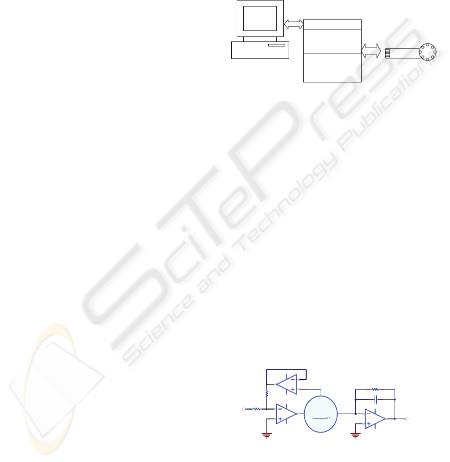
2 SENSING SYSTEM
STRUCTURE
In general, for both system solutions, the
electrochemical potentiostatic sensing system
consists of an electrochemical cell incorporating the
sensor or sensor array, and two main analog
electronic units, the potentiostat and a
transimpedance amplifier, and a microcontroller
unit. These analog units connect to the
microcontroller unit that controls the measurement
process, and provides data acquisition and
connectivity to the personal computer (PC) as shown
in figure 1.
Microcontroller
Multichannel
potentiostat
Transimpedance
amplifier
Microcell
PC
Figure 1: Block diagram of electrochemical sensing
system for in vitro and in vivo applications.
The electrochemical cell is usually a three
electrode structure comprising a counter electrode
(C), reference electrode (R) and working electrode
(W), which is immersed into or covered by sample
solutions to be analyzed. Electrochemical or
biochemical reactions in the cell are detected with
electronics, in particular with a transimpedance
amplifier. The reactions are dependent on the W
potential, therefore, a stable potential at the W
should be provided. The W potential stability is
secured by the R, which supplies a reference
potential independent of the environment, and the
potentiostatic unit, which maintains this W potential,
with respect to the R, to be equal to a stimulation
signal generated by the microcontroller unit. The
shape of the stimulation signal depends on the
sensing methodologies (i.e. DC for amperometry,
staircase for voltametry and so on). A simplified
schematic of the electrochemical potentiostatic
sensing system (without microcontroller unit) is
shown in figure 2
Figure 2: Simplified schematic of the potentiostatic
sensing system.
Implementation of the electrochemical sensing
systems for in vitro and in vivo biomedical
applications is each governed by a different set of
requirements. For in vitro real- time systems, these
2
3
1
Op3
R
W
Vout
2
3
1
Op2
2
3
1
Op1 C
Vin
Current pass
CELL
BIODEVICES 2009 - International Conference on Biomedical Electronics and Devices
84
requirements are high sensitivity, rapid rate of
measurement and corresponding signal processing,
accurate data interpretation, a multi-functional
instrument and simplicity of its operation by the end
user. For in vivo systems the main challenging
requirement besides sensitivity is the size of device,
which must adhere in some cases to millimeter size
scale, and continuous operation over extended
periods of time (i.e. up to one year, or longer for
some implantable devices). Both of these restrictions
impose a serious limitation on the power and size of
the electronic components which can be used for the
system design.
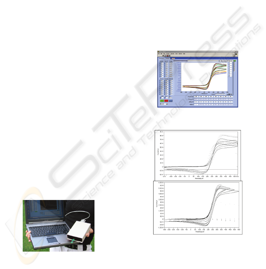
2.1 In vitro System
In keeping with these requirements for the in vitro
applications, a multi-channel potentiostatic system,
optimized for operation with an 8 working electrode
on-chip sensing system, has been developed. The
instrument is based on low-noise, high precision,
pico-ampere input current operational amplifiers
AD8602 (leakage current of 200 fA) and Analog
Devices microconverter, ADuC812, which enables
generation of the stimulation signal (via the in-built
DAC), feeds sensing data into the system (via the in-
built 8 channel ADC), and provides preliminary
signal processing and communication with the PC
(via an RS232 communications port). Running on
the PC is dedicated software with user-friendly
graphical interface, which provides final signal
processing and data interpretation. The prototype of
the system, with overall size 170x110x40 mm and
weight 250 g, is shown in figure 3. The associated
user-friendly interface software (developed in
LabVIEW, figure 4) allows the user to specify the
type of voltammetric technique and its setting (start
and finish potential, scan rate for each channel), and
to visualize obtained experimental data.
Figure 3: The multi-channel potentiostatic system suitable
for in vitro applications.
In order to validate and characterize the system,
a microelectrode array was fabricated using CMOS
techniques. It consisted of an array of gold working
electrodes surrounded by a counter electrode. The
final packaged die connects to a PCB dipstick which
allows for easy connection and testing of the sensor
chip. The solutions with analyte can be placed
directly on the sensor, or the dipstick can be
integrated into an associated fluidic system, or it can
be simply immersed into a container with the sample
to be analysed. The performance of the developed
system can be estimated by example of cyclic
voltammetry applied to the 8 Ws in ferrocene
carboxylic acid solution in simultaneous mode. The
measurements that were carried out over different
scan rates are shown in figure 5a, and the
measurements that were carried out over different
start and finish potentials are shown in figure 5b.
These experiments demonstrate that the system’s
current sensitivity is greater than 1nA, with a noise
level less than 50pA p-p.
Figure 4: PC Interface for the multi-channel potentiostatic
system.
a.
b.
Figure 5: Cyclic voltammograms recorded simultaneously
with the instrument for oxidation of Ferrocene carboxylic
acid at an eight microelectrode array: (a) – with different
scan rates set for each channel (from 50mV/s to 400mV/s),
(b) – with different starting and finishing potentials set for
each channel.
MINIATURIZED ELECTROCHEMICAL SENSING SYSTEMS FOR IN VITRO AND IN VIVO BIOMEDICAL
APPLICATIONS
85
2.2 In vivo System
In keeping with requirements for the in vivo
applications, a potentiostatic system based on a PIC
microcontroller, has been developed. This
microcontroller family contains a number of
microcontroller models including those which are
available in a smaller size, consumes less power and
has more RAM memory than the ADuC, making
them more suitable for this application. The
PIC18F2520 has been chosen which features
6x6mm, 1256 bytes RAM and current consumption
of 2.6mA during normal operation. This is in
contrast to the ADuC812 features of 8x8mm, 256
bytes RAM and 6.2mA during normal operation. In
the programming of the microcontroller, close
attention has been given to reduce the power
consumed during device operation by using
appropriate electronic components with power
switch on/off mode and inbuilt microcontroller
possibilities. There are two realizations of this
system. The first one is a PCB-based device with
separate commercially-available low power and
small size ICs forming a one-channel potentiostatic
system in accordance with figure 1. The size of the
system prototype is 12x24 mm, which is suitable for
a variety of in vivo applications. To validate the
performance of the system, cyclic voltammetry was
carried out in 0.5M sulfuric acid solution at a gold
macroelectrode, figure 6.
Figure 6: Cyclic voltammogram for in vivo PCB-based
system at the macro gold electrode in 0.5M sulphuric acid
solution
The second in vivo system is a specially
developed on-chip system, which is a realization of
the PCB in vivo device in an integrated single die.
The implementation of the developed system on a
silicon substrate was made by using a C5P0 CMOS
5 micron silicon breadboard. There is a digital core
of 1248 gates and an analog oriented section
containing 32 operational amplifier blocks, 1500
units of poly-poly capacitor, approximately 2MΩ of
resistance in 40 sticks of 6 segments each, and a
range of more specialized components. The
schematic of the breadboard operational amplifier
unit and final chip appearance, are shown in figures
7 and 8 correspondingly. The performance of the on-
chip system can be seen from the example of cyclic
voltammetry of a macro gold W in 0.5M sulphuric
acid solution, figure 9, which substantiates its
operability. At present both of the in vivo systems
have similar performance specification but due to
the initial small batch size, the PCB-based system
costs less. The potential advantages of the on-chip
system over PCB based device (a smaller size, a
reduction in noise and power, a decrease of leakage
current, which leads to an improvement of the
system performance) will be realized when complete
integration of sensor and electronics on one chip is
developed.
Figure 7: P-input CMOS OPAMP. Figure 8: Fabricate
d
on-chip potentiosta
t
die, 8x10mm.
Figure 9: Cyclic voltammogram for in vivo on-chip system
at the macro gold electrode in 0.5M sulphuric acid
solution
3 CONCLUSIONS
Miniaturized sensing systems for in vitro and in vivo
biomedical applications suitable for point-of-care
applications have been presented that will facilitate
early-stage disease diagnosis. The systems are based
on potentiostatic instrumentation, which is
commonly employed with different electrochemical
sensors. For in vitro requirements, an 8 channel PC-
controlled system has been developed, capable of
carrying out a number of sensing methods. For the in
vivo applications, two systems have been realized,
both based on the PIC microntroller. The first is a
PCB with separate commercially-available low
BIODEVICES 2009 - International Conference on Biomedical Electronics and Devices
86

power and low size ICs forming a one-channel
potentiostatic system. The second is a specially
developed integrated on-chip system. Future work
will include optimization of the developed systems
for specific biomedical application according to end-
user requirements. Additionally for the on-chip
system the chip will be redesigned to improve its
characteristics and reduce the fabrication cost.
ACKNOWLEDGEMENTS
Financial support of this work by CFTD/04/112,
CFTD /05 /122 Enterprise Ireland and National
Access Programme NAP68 projects is gratefully
acknowledged.
REFERENCES
Peng, Y., Hu, C., Zheng, D., & Hu, S. (2008). A sensitive
nitric oxide microsensor based on PBPB composite
film-modified carbon fiber microelectrode. Sensors
and Actuators B-Chemical, in press.
Seo, H., Park, D., Park, J. (2008). Fabrication and
characterization of platinum black and mesoporous
platinum electrodes for in-vivo and continuously
monitoring electrochemical sensor applications. Thin
Solid Films, 516(16), 5227-5230.
Ndamanisha, J. C., & Guo, L. P. (2008). Electrochemical
determination of uric acid at ordered mesoporous
carbon functionalized with ferrocenecarboxylic acid-
modified electrode. Biosensors & Bioelectronics,
23(11), 1680-1685.
Wang, M., & Ha, Y. (2007). An electrochemical approach
to monitor pH change in agar media during plant
tissue culture. Biosensors & Bioelectronics, 22(11),
2718-2723.
McLaughlin, G. W., Braden, K., Franc, B., & Kovacs, G.
T. A. (2002). Microfabricated solid-state dissolved
oxygen sensor. Sensors and Actuators B-Chemical,
83(1-3), 138-148.
Abbaspour, A., & Mirzajani, R. (2007). Electrochemical
monitoring of piroxicam in different pharmaceutical
forms with multi-walled carbon nanotubes paste
electrode. Journal of Pharmaceutical and Biomedical
Analysis, 44(1), 41-48.
Morales, M. D., Serra, B., de Prada, A. G. V., Reviejo, A.
J., & Pingarron, J. M. (2007). An electrochemical
method for simultaneous detection and identification
of Escherichia coli, Staphylococcus aureus and
Salmonella choleraesuis using a glucose oxidase-
peroxidase composite biosensor. Analyst, 132(6), 572-
578.
Shiddiky, M.J.A., Rahman, M.A., Choel, C.S., Shim, Y.
(2008). Fabrication of disposable sensors for
biomolecule detection using hydrazine electrocatalyst.
Analytical Biochemistry, 379(2), 170-175.
MINIATURIZED ELECTROCHEMICAL SENSING SYSTEMS FOR IN VITRO AND IN VIVO BIOMEDICAL
APPLICATIONS
87
