
DOUBLE PULSE TRANSMISSION - DEAD ZONE DECREASING IN
ULTRASOUND IMAGING
Ihor Trots, Andrzej Nowicki and Marcin Lewandowski
Institute of Fundamental Technological Research, Polish Academy of Sciences, Swietokrzyska 21, Warsaw, Poland
Keywords:
Golay complementary sequences, Double pulse transmission, Dead zone.
Abstract:
This study investigates a new composing method of double transmission of short coded sequences based on
well-known Golay complementary codes, which allows to obtain the higher signal-to-noise ratio (SNR) and
decrease dead zone area. The proposed method can potentially find application in small parts ultrasonography
and play an important role in examination of superficial structures, e.g. in dermatology, ophthalmology, etc.,
where using longer coded sequences leads to increase of a dead zone and single pulse transmission of short
sequences does not assure sufficient SNR.
This paper discusses the results obtained during the examination of four different length pairs of Golay coded
sequences excited at 3.7 MHz: the single 64-bits pair of Golay sequences and combined sequences consisting
of two 8, 16, and 32-bits Golay codes separated in time. The experimental results have shown that double pulse
transmission allows to suppress considerably the noise level, the SNR increases by 5.7 dB in comparison with
the single pulse transmission of Golay sequences of the same length. The presented results of this work
demonstrate the advantage of double pulse transmission method which enhances SNR while maintaining the
dead zone short.
1 INTRODUCTION
Coded ultrasonography has been intensively devel-
oped and studied in the last decade - from para-
metric imaging of bone in the range 0.5 – 2 MHz,
through imaging in classic ultrasonography (3.5 –
10 MHz) up to imaging in micro ultrasonography
(above 20 MHz). The reasons of such interest are
the properties of the coded transmission: increase
of penetration depth, signal-to-noise ratio improve-
ment, exploration of the signal with lower amplitude
and improvement of the axial resolution moving to
the higher frequency range. Nowadays extensively
explored coded sequences are: linearly frequency
modulated signals (chirp) and phase-modulated sig-
nals like Barker codes and Golay complementary se-
quences (side-lobe cancelling codes). Within the last
few years, the increasing interest in visualization of
tissue surface (Altmeyer P., 1992) as well as vessel
wall research using high frequency ultrasound can be
observed among biologists and clinicians. Develop-
ment of high frequency ultrasound is directed to a
new region of application in dermatology (diagnostics
of skin diseases and lesion treatment) (Kielbasa Z.,
2007), (Hildegard M., 2005). The ultrasound diag-
nostics is applied to examine main skin layers: epi-
dermis, cutis vera and hypodermis. It is very impor-
tant in ophthalmology as it allows to examine and to
diagnose the pathological changes of cornea, iris, etc.
The motivation of this work was to find a new
transmission method of coded sequences based on
complementary Golay sequences (CGS) to decrease
a noise level in result RF echo signal and to improve
SNR in ultrasonic imaging. Coded sequences of short
pulses based on CGS separated in time were transmit-
ted in medium and received. This proposed method
can eventually solve the problem connected to dead
zone, because it allows to transmit the shorter codes
which is important in ultrasonography, namely to ex-
amine and diagnose skin layers, cornea, iris, etc. Dou-
ble transmission of shorter codes instead the longer
ones can also allow to obtain the higher SNR.
2 DOUBLE PULSE
TRANSMISSION METHOD
Among the different excitation sequences proposed in
ultrasonography, Golay codes evoke more and more
416
Trots I., Nowicki A. and Lewandowski M. (2009).
DOUBLE PULSE TRANSMISSION - DEAD ZONE DECREASING IN ULTRASOUND IMAGING.
In Proceedings of the International Conference on Bio-inspired Systems and Signal Processing, pages 416-420
DOI: 10.5220/0001777104160420
Copyright
c
SciTePress
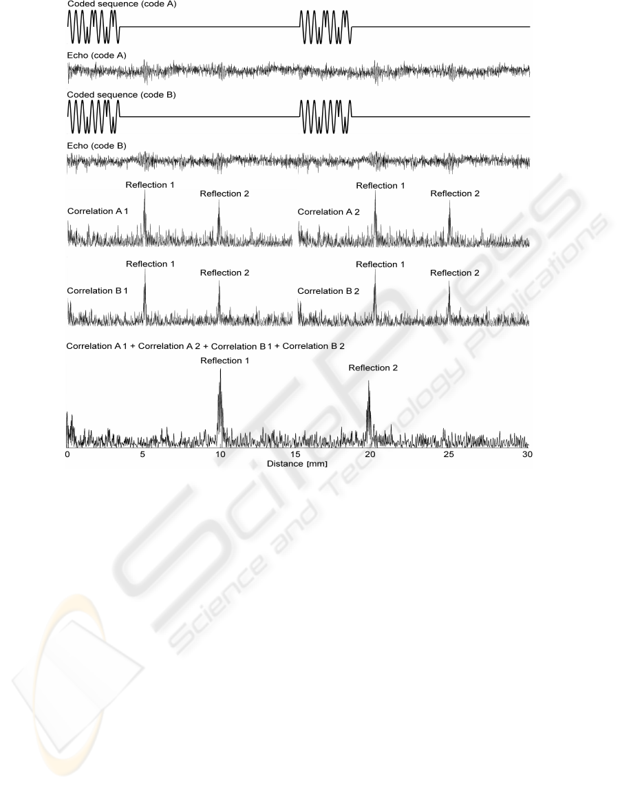
Figure 1: Double pulse transmission method using Golay complementary sequences.
interest in comparison to other signals. The reason
of that lies in the fact that Golay codes, like no other
signals, suppress to zero the amplitude of side-lobes
in ideal case. This type of complementary sequences
was introduced by Golay (Golay, 1961). Practical
implementation of complementary Golay sequences
(CGS) is not widely available in the literature, for the
convenience of the reader a step-by-step principle of
construction and properties of the CGS as well as cor-
relation principle are described in (Trots I., 2004).
Figure 1 shows the double pulse transmission
method using the Golay complementary sequences.
As can be easily seen in practice even for coded trans-
mission the different artifacts are present and because
of that the noise elimination and efficient side-lobe
cancellation cannot be obtained. The noise level can
lead to wrong visualization of the examined organs
and range ambiguity. The nice method which can de-
crease the noise level is to use the longer Golay coded
sequences i.e. 128 or even 512 bit lengths. The usage
of longer coded sequences is welcome in radar tech-
nique or hydrolocation - where information located
closely to transducer is not important. In ultrasonog-
raphy the usage of longer coded sequences is rather
limited since it leads to increase of the dead zone that
is not accepted in some diagnostic applications.
Increase of the echo detection using Golay codes
in comparison to short pulse is evident. As reported
experiments show the two echoes received from re-
flectors distanced one from other by 1 cm. SNR im-
provement in compressed signal in comparison to di-
rect echoes is about 15 dB (Nowicki A., 2004).
The idea of double transmission method is based
on an assumption of mutual noise cancellation, where
noise in the resulted RF signal is averaged by sum-
ming two compressed echo signals obtained by single
transmissions. Such solution allows to improve SNR
maintaining the dead zone area short which can po-
tentially increase the application of ultrasound in der-
matology, ophthalmology, oncology etc.
DOUBLE PULSE TRANSMISSION - DEAD ZONE DECREASING IN ULTRASOUND IMAGING
417
3 EXPERIMENTAL SETUP
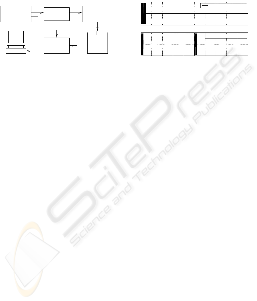
The block diagram of the measurement arrangement
used is shown in Fig. 2.
Pentium
PC
TM
Fantom
HP 8643A
Signal Generator
Synthesized
ENI 3100LA
RF Power Amp.
EPM7064
Bipolar Coder
HP 54810A
Infinium
Oscilloscope
Figure 2: Diagram of experimental setup.
The Golay sequences with different lengths 8, 16,
32 and 64-bits at frequencies 3.7 MHz were synthe-
sized in the following way. The Signal Generator
HP8643A produced a sine wave at 0 dB level at a
given frequency. This signal was fed to the bipo-
lar modulator driven by the -1,1 sequences from the
custom-designed coder. The coder circuitry based
on the programmed logic EPM7064 allowed gener-
ating switched pair of single 64-bits Golay sequences
and combined sequences of other shorter Golay codes
separated in time as well as single coded sequences
transmitted for later comparison. The coded signals
were then amplified via the power RF amplifier ENI
3100LA and the transmitted coded burst excited the
ultrasonic transducer which scanned the tissue phan-
tom model 525 Danish Phantom Design. The uncom-
pressed RF echoes data were acquired using a digi-
tal storage 12-bits oscilloscope Infinium HP 54810A
with a sampling rate of 100 MHz. All processing and
display were done on the computer using Matlab rou-
tines. The processing included amplification, pulse
compression, sum of Golay sequences, envelope de-
tection and the obtained results were in few seconds
displayed on the monitors.
4 RESULTS AND DISCUSSION
Figure 3 shows the comparison of transmitting 32-
bits Golay coded sequences at nominal frequency
3.7 MHz and time duration 8.64 µs and proposed
method of the double transmission of 16-bits Golay
coded sequences with shorter time duration that is
equal 4.32 µs. The start time of the second sequence
depends on penetration depth that is examined. In the
given case, the plot illustrates the examined environ-
ment on penetration depth up to 8 cm. The starting
time of the second sequence can be calculated from:
t = 2d/c = 100 µs (1)
where d is the depth, and c is the speed of the ul-
trasound wave in examined environment and is equal
to 1540 m/s. In the second case the RF echo sig-
0 20 40 60 80 100 120 140 160 180 200
−1
0
1
Amplitude
0 20 40 60 80 100 120 140 160 180 200
−1
0
1
Time [µs]
Amplitude
Traditional transmission
Double transmission
Figure 3: Transmission of the 32-bits Golay sequence with
time duration 8.64 µs (top) and double transmission of the
16-bits Golay sequences with time duration 4.32 µs (bot-
tom).
nals split into two sequences, next compressed and
summed. The amplitude of main-lobe in the resulted
compressed signal will be equal to 64 for both cases.
This is because for single 32-bits coded transmission
two RF echo lines are added, whereas in the case of
double 16-bits coded transmission four RF echo lines
need to be summed.
The tissue phantom model 525 Danish Phan-
tom Design with attenuation of background material
0.5 dB×cm×MHz was used in the experiments. The
pair of the Golay sequences of the different lengths
8, 16, 32, and 64-bits at the frequency 3.7 MHz were
used. The centre RF echo lines obtained from the tis-
sue phantom using the CGS of the different lengths
transmitted by the two methods and calculated SNR
are presented in the Figs. 4 – 7. The target reflections
are the nylon filaments, 0.1 mm in diameter spaced
1 cm one from another.
Figs. 4 – 7 show the advantages of double trans-
mission of Golay coded sequences over single trans-
mission used heretofore. In the double transmission
case the SNR increases by about 5.7 dB in compar-
ison to the single transmission of the same length
coded sequences and by about 4 dB in comparison
to the single transmission of the two times longer se-
quences. According to Trots et al. (Trots I., 2004) in
order to obtain the 4 dB SNR improvement the coded
sequence needs to be about 16 times longer in the sin-
gle transmission method. But using longer coded se-
quences results in increasing of the dead zone area
that increases proportionally to the coded sequences
length and inversely to frequency. Theoretically, the
dead zone area is equal to the half burst pulse time
duration. But in practice the time duration of the
burst pulse is calculated from the beginning to mo-
ment when the power drops to the -3 dB level, so the
BIOSIGNALS 2009 - International Conference on Bio-inspired Systems and Signal Processing
418
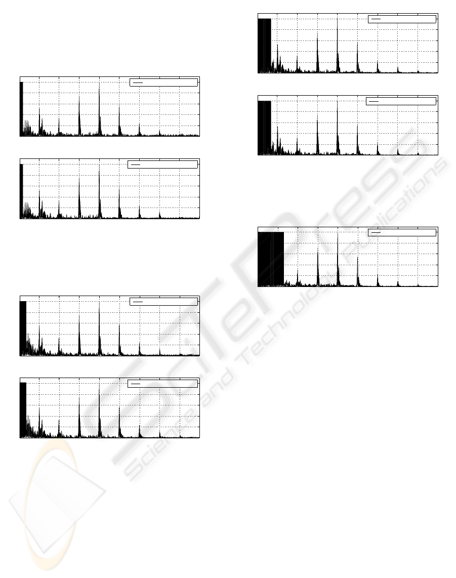
dead zone area is assumed to be equal to burst pulse
time duration. In case of 8-bits Golay sequences the
dead zone area is equal to 1.66 mm (Fig. 4) and in-
creases up to 13.2 mm for 64-bits Golay coded se-
quences (Fig. 7) for the frequency 3.7 MHz.
0 1 2 3 4 5 6 7 8
0.2
0.4
0.6
0.8
1
8−bits Golay coded sequences
Norm. Amplitude
0 1 2 3 4 5 6 7 8
0.4
0.8
1.2
1.6
2
Depth [mm]
Norm. Amplitude
Single transmission
Double transmission
32.3 dB
38.1 dB
Figure 4: The centre RF-lines of the tissue phantom ob-
tained by single transmission (top) and double transmission
(bottom) of 8-bits Golay sequences.
0 1 2 3 4 5 6 7 8
0.2
0.4
0.6
0.8
1
16−bits Golay coded sequences
Norm. Amplitude
0 1 2 3 4 5 6 7 8
0.4
0.8
1.2
1.6
2
Depth [mm]
Norm. Amplitude
Single transmission
Double transmission
34.0 dB
39.5 dB
Figure 5: The centre RF-lines of the tissue phantom ob-
tained by single transmission (top) and double transmission
(bottom) of 16-bits Golay sequences.
The comparison of the obtained 2D ultrasonic im-
ages of a tissue phantom using 8-bits, 16-bis, 32-bits
and 64-bits length Golay sequences transmitted by
different methods is shown in Fig. 8. These record-
ings allow to verify axial resolution and the scan ge-
ometry. It consists of several nylon filaments twists
0.1 mm in diameter positioned every 1 cm ±2% axi-
ally. Additional groups of 11 twisted threads for 6 dB
axial and lateral resolution are placed at the different
depths from top of the phantom. Also some groups of
low contrast cylinders that deviates +3 dB, -3 dB and
-6 dB from the background are placed.
0 1 2 3 4 5 6 7 8
0.2
0.4
0.6
0.8
1
32−bits Golay coded sequences
Norm. Amplitude
0 1 2 3 4 5 6 7 8
0.4
0.8
1.2
1.6
2
Depth [mm]
Norm. Amplitude
Single transmission
Double transmission
35.6 dB
41.3 dB
Figure 6: The centre RF-lines of the tissue phantom ob-
tained by single transmission (top) and double transmission
(bottom) of 32-bits Golay sequences.
0 1 2 3 4 5 6 7 8
0.2
0.4
0.6
0.8
1
64−bits Golay coded sequences
Depth [mm]
Norm. Amplitude
Single transmission
37.1 dB
Figure 7: The centre RF-lines of the tissue phantom ob-
tained by single transmission of 64-bits Golay sequences.
The images in Fig. 8 demonstrate that the ultra-
sound imaging can benefit from double transmission
of Golay sequences yielding a higher SNR and there-
fore a higher contrast resolution, while maintaining
both axial and lateral resolution. The last one de-
pends on transducer acoustic field and is discussed
by NOWICKI et al. (Nowicki A., 2007). Also, it
needs to be noted, that in case of double transmission
method in comparison to single transmission one the
code length is two times shorter.
Obtained 2D ultrasonic images clearly demon-
strate the advantage of double pulse transmission. As
was mentioned above, coded length increase leads to
elongated dead zone area and in the case of 8-bits Go-
lay sequences this area is equal to 1.7 mm while in the
case of 64-bits Golay code the dead zone increases
up to 13.4 mm. The dead zone area on the top of
each 2D ultrasound image is created by high inten-
sity echo amplitude, multiple reflections and is equal
to the transmitted code length. In this area the real
echoes are masked and using the longer codes lead to
limited application in dermatology, where superficial
structures are needed to be readable.
DOUBLE PULSE TRANSMISSION - DEAD ZONE DECREASING IN ULTRASOUND IMAGING
419
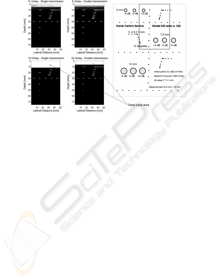
Figure 8: Comparison of 2D ultrasonic images of the tissue phantom obtained using 8-bits, 16-bits, 32-bits and 64-bits length
Golay coded used single transmission and double transmission. Schematic diagram of the examined tissue phantom model
525. The rectangle marks the scanned area.
5 CONCLUSIONS
This paper discusses actual study and development
trend of the coded transmission method in ultrasonog-
raphy. One of the important parameters in ultra-
sound diagnostic is dead zone area that makes the real
echoes lying closely to transducer surface unreadable.
For that reason using of the coded sequences in ultra-
sound imaging is considerably limited.
The proposed work concerns the development
and investigation of a new composing method of
short coded sequences and their transmission based
on well-known Golay complementary codes. This
method allows to increase field of ultrasound diagnos-
tic application where dead zone plays important role,
e.g. dermatology, ophthalmology, etc.
The results obtained show the effectiveness of
double transmission and its resistance to the refrac-
tion, attenuation, and reflection of ultrasound waves.
The SNR gain is evident when applying double pulse
transmission method in comparison to single pulse
transmission. Also, increasing codes length, the SNR
increases and penetration elongates proportionally.
The proposed coded method of double transmis-
sion can be applied also in standard ultrasonography.
Introduction of double coded transmission method in
medical ultrasound equipment can increase the effec-
tiveness and quality of the ultrasound diagnostic.
ACKNOWLEDGEMENTS
This work was supported by the Polish Min-
istry of Science and Higher Education (Grant
N51804432/3434).
REFERENCES
Altmeyer P., El-Gammal S., H. K. (1992). Ultrasound in
Dermatology. Springer-Verlag, Berlin.
Golay, M. (1961). Complementary series. IRE Trans. Inf.
Theory, IT-7:82–87.
Hildegard M., Wendtner S., B. W. (2005). Ultrasound scan-
ning in dermatology. Arch. Dermatol., 141:217–224.
Kielbasa Z., Gawda H., W. B. (2007). Ultrasonography
as a method of diagnosis and treatment monitoring
in dermatology. In Annales Universitates Mariae
Curie-Sklodowska, Lublin-Polonia, volume LXII, 1,
46, pages 242–247.
Nowicki A., Klimonda Z., L. M. L. J. L. P. T. I. (2007).
Direct and post-compressed sound fields for different
coded excitation. Acoustical Imaging, 28:399–407.
Nowicki A., Secomski W., T. I. L. J. (2004). Extending pen-
etration depth using coded ultrasonography. Bulletin
of the Polish Academy of Sciences, Technical Sciences,
52(3):215–220.
Trots I., Nowicki A., S. W. L. J. (2004). Golay se-
quences - side-lobe-canceling codes for ultrasonogra-
phy. Archives of Acoustics, 29(1):87–97.
BIOSIGNALS 2009 - International Conference on Bio-inspired Systems and Signal Processing
420
