ACQUISITION, ANNOTATION AND INTERACTIVE
EXPLORATION OF STEREO IMAGES
WITH VIRTUAL REALITY
Mohammed Haouach*
,
**, Karim Benzeroual*
,
**
Christiane Guinot*
,
** and Gilles Venturini*
* Laboratoire d’Informatique, Université François-Rabelais de Tours, 64 avenue Jean Portalis, 37200 Tours, France
**CE.R.I.E.S., Unité Biométrie et Epidémiologie, 20 Rue Victor Noir, 92521 Neuilly sur Seine, France
Keywords: Stereoscopic acquisition, Camera calibration, Genetic algorithms, 3D visualization, Image annotation,
Hypermedia, Skin relief.
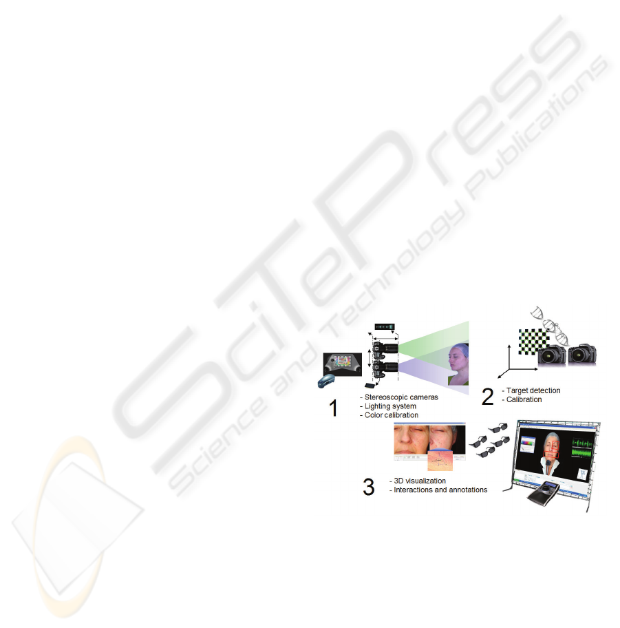
Abstract: We present in the paper a system called Skin3D that integrates all hardware and software to extract
information from 3D images of skin. It is composed of a lighting equipment and acquisition-based
stereoscopic cameras, a camera calibration using genetic algorithms, virtual reality equipment to restore the
images and interact in 3D with them, a set of interactive features to annotate images, annotations and share
these 3D hypermedias. We present a comparative study and an application of Skin3D on faces skin.
1 INTRODUCTION
Relief is a complex and important data for many
domains. In medicine, numerous methods have been
developed in order to acquire relief of various parts
of the human body with the aim of discovering
information and knowledge. In this paper we are
especially interested with the acquisition of a
surface, and more precisely the skin. We have
conceived a complete and operational system called
Skin3D (see an overview in figure 1) which is
compound of three main modules: (1) an acquisition
module that takes stereoscopic photographs of
people with skin problems or specific pathologies,
(2) a camera calibration module that estimates the
cameras parameters which are necessary for
computing 3D information, and (3) a visualization
and exploration module which can be used by
dermatologists to perform 3D measurements, to
create annotations as well as a 3D hypermedia, and
to share the extracted knowledge with others.
In this paper, we will detail respectively each
module in sections 2, 3 and 4, and we present our
motivations and the state of the art for each of them.
In section 5 we described the obtained results on the
precision of camera calibration and a first example
of an annotated 3D hypermedia build on 3D
photographs of faces.
Figure 1: Overview of Skin3D.
2 ACQUISITION AND
CALIBRATION MODULE
2.1 Acquisition of Relief
In the skin domain, two types of relief acquisition
methods can be distinguished, the so-called active
369
Haouach M., Benzeroual K., Guinot C. and Venturini G. (2009).
ACQUISITION, ANNOTATION AND INTERACTIVE EXPLORATION OF STEREO IMAGES WITH VIRTUAL REALITY .
In Proceedings of the International Conference on Health Informatics, pages 369-374
DOI: 10.5220/0001778803690374
Copyright
c
SciTePress

and passive methods (Ben Amor et al. 2005).
Actives methods consist in combining an optical
sensor with a source of light, like for example Laser
scanners, sensors that use structured lights (Salvi,
2004), or profilometry (Rohr and Schrader, 1998).
Passives methods rather use one or more images like
in (Hernandez and Schmitt, 2003) or like
stereophotogrammetry (D'Apuzzo, 2002). In our
application, we have three main constraints: to
acquire the relief in conjunction with the visual
aspect of the skin, to do this with as less constraints
as possible for the experimenter and the subject, to
keep a high visual fidelity to the real skin. These
constraints thus exclude Laser-based systems,
methods that need heavy hardware, or systems that
do not acquire both the relief and texture. Finally,
the last constraint tends to exclude systems that
perform a 3D reconstruction, because this process
alters the visual quality of the acquired relief
compared to a high quality photograph for instance
(Ayoub et al. 1998).
For this module of Skin3D, we have conceived
an acquisition system on the basis of two cameras
assembled together and which are triggered in a
synchronized way. We have designed a specific
lighting system and we have used an optical sensor
to calibrate all graphic devices (cameras, screens,
video projectors, etc).
We have used two Pentax K10 reflex cameras
with a resolution of 10 megapixels and with 50mm
macro objective (ideal for taking pictures of faces).
The stereoscopic support allows us to minimize the
distance between the two cameras (the natural gap
between human eyes is about 6cm) and to maintain
parallelism between cameras (or a small converging
angle of a few degrees only). These conditions are
necessary to ensure a comfortable visualization for
the user during the stereoscopic projection without
important modifications of the original images.
Taking such stereoscopic pictures requires an
adapted lighting system. In our first test we deal
mainly with faces, so we have selected specific
lights to reveal the skin relief while removing all
shadows inherent the shape of the face. This lighting
system is compound of two HMI torches and two
Pentax AF 540 FGZ flashes.
In order to obtain the highest image fidelity both
for the acquisition and visualization, we calibrate all
graphic devices by associating them an ICC profile
(International Color Consortium). This is performed
using an i1 (X-rite) sensor and a standardized color
board.
2.2 Calibration with a Genetic
Algorithm
Camera calibration is a crucial step in stereovision
(Faugeras et al., 1987) because it will determine the
accuracy of the acquired relief. It consists in
estimating the intrinsic and extrinsic parameters of
the cameras (i.e. focal length, distortion,
rotation/translation between the two cameras, etc).
Numerous methods exist in this context (Tsai, 1987)
without a real consensus, even if some algorithms
are relatively common (Zhang, 1998). The type of
methods we have selected consist in taking pictures
of a calibration target with known dimensions, and
then to estimate the parameters that minimize a
target « reconstruction » error. These methods
involve non linear optimization procedures which
may have some problems (stability, initial starting
point). This has lead researchers to make use of
genetic and evolutionary algorithms which are
stochastic procedures with less sensitivity. In this
context, one may cite for instance (Zhang and Ji,
2001) where a single camera is calibrated, (Cerveri
et al. 2001) who use evolution strategies for
stereovision, or (Dipanda et al. 2003) who use one
camera and a Laser.
We have developed a new calibration method,
based on genetic algorithms, and which
distinguishes itself from the others on the following
points: it is specific to stereovision, it uses the notion
of distance between points in its evaluation function
(because we want to make precise measurements), it
can be applied to several models of objectives (“pin-
hole” model but also telecentric model). It proceeds
in the following way (see figure 1, step 2): we take
pictures of a target of known dimensions and with
different orientations, then we detect specific points
(corners) on this target in both left and right images.
The distances between these points are perfectly
known. The objective of our genetic algorithm is to
find the set of parameters that minimizes the
prediction error (i.e. the difference between known
and estimated distances). For this purpose, it uses a
population of individual, where each individual is a
possible set of parameters. At the beginning, the
population is filled with randomly generated
individuals where parameters belong to loosely
defined intervals. Then parents are selected
according to their performance using binary
tournament selection, and we recombine these
individuals using a crossover operator (either a
linear recombination or a discrete uniform
crossover) and using a mutation operator (small
random noise). The evaluation function takes into
HEALTHINF 2009 - International Conference on Health Informatics
370
account the error between the real and estimated
distances, but also other errors computed during the
estimation of points (such as the intersection error,
see for example (Cerveri et al. 2001)).
The parameters that we consider in the
individuals are the 3D transformation between
cameras, the focal lengths, the distortion and the
decentering of the objectives w.r.t. the CCD. After
numerous tests (especially on artificial problems
where the parameters to estimate are known), we
have kept the following parameters: 1000
individuals in the population, stopping of the
algorithm after 30000 generations (one individual is
generated per generation) when the population
improvement is below a given threshold. The
running times for real problems on a standard
computer are about 1 minute.
3 VISUALISATION AND
INTERACTIVE EXPLORATION
3.1 Virtual Reality
This module of skin3D is very important because
once the data are acquired they must be restituted to
the expert with the highest possible fidelity and with
all interactive tools necessary for knowledge
discovery. Using virtual reality is thus necessary in
order to visualize the relief in stereoscopy but also to
let the expert navigate in the 3D image and, for
instance, make annotations. As far as we know
systems for exploring and annotating stereoscopic
images are rare. We may mention (Zhu, 2007) in
ophthalmology but 3D images are basic anaglyphs
with no real interactions.
For the stereoscopic visualization of images, we
have used two types of projection hardware: on the
one hand, standard cathodic screens that
alternatively visualize the left and right images using
active shuttering glasses, and on the other hand, two
video projectors with passive glasses. We have
tested two types of video projectors: the first ones
(F1+ from Projection Design) use a passive
polarization of light (vertical/horizontal
polarizations respectively for each left and right
images), and the others use F2 video projectors with
Infitec filters (equivalent to red/green filters but with
higher quality than basic anaglyphs). These
projectors have allowed us to project skin 3D images
on a 25m2 screen in front of more than 20 people.
As far as navigation is concerned, the user may
move along the 3 axis X, Y et Z. All other moves are
prohibited (like turning around in 3D) because this
would involve a 3D reconstruction that would
decrease the visual and photographic quality of the
pictures. To perform these moves, the expert may
use the mouse or a more specific 3D controller with
6 degrees of freedom (SpacePilot™). Using this
controller is very intuitive, and the user may for
instance navigate in 3D with his left hand and use
the mouse with his right hand for selecting areas.
3.2 Mesuring 3D Information
One may compute the 3D coordinates of a point P
on the skin thanks to the parameters estimated by the
calibration module of Skin3D. Let Pl denote the
projection of P in the left image, and let us suppose
that this projected point was selected by the user. In
order to compute the 3D coordinates of P, one has to
find the point Pr, i.e. the correspondent of Pl in the
right image. This is performed using a pattern
matching algorithm that tries to maximize the
correlation between Pl and Pr. This correlation is
computed using the color values of pixels on two
small images centered respectively on Pl and Pr
(Chambon and Crouzil, 2003). The best candidate is
the point that maximizes the correlation and that
checks other constraints (for instance, the
correspondent of Pr must be, in a symmetrical way,
Pl). Then, using Pl and Pr, the 3D coordinates of P
are known. The expert may thus measure 3D
distances between two selected points. In order to
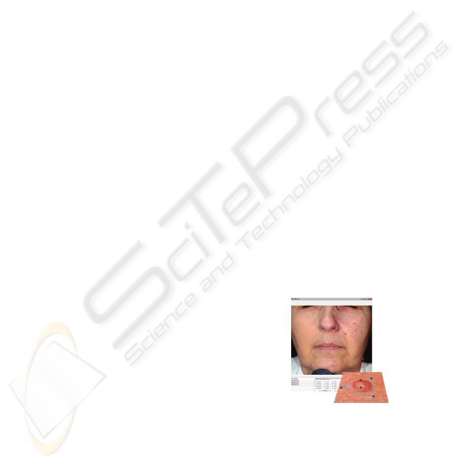
measure depths (or heights), the expert selects 3
points in the image (see figure 2). These 3 points
represents a plane, and the distance between this
plane and a fourth point can be computed, which
results in a height or depth measurement.
Figure 2: Measuring a depth in the stereoscopic images.
3.3 Selecting Point in 3D
The basic pointing device on a standard display is
2D and is not adapted to stereoscopic visualization
because the position of the pointer is relative to the
2D screen and not to the 3D image, which is very
confusing for the user. This is the reason why we
ACQUISITION, ANNOTATION AND INTERACTIVE EXPLORATION OF STEREO IMAGES WITH VIRTUAL
REALITY
371
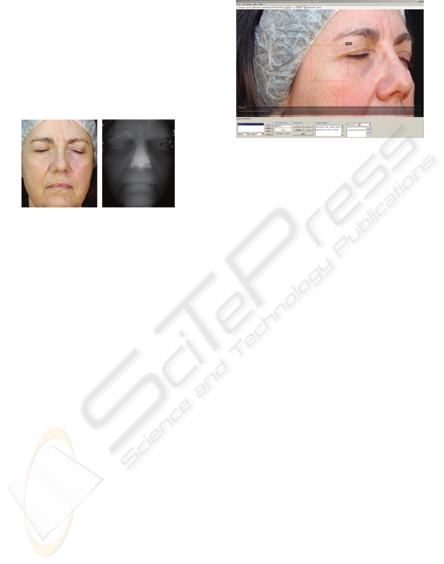
have defined a 3D stereoscopic pointer in Skin3D. In
order to properly position this pointer, one has to
compute, for a subset of pixels in the images, the
correspondence between the left and right images
(see figure 3). We thus obtain a depth map for each
of the selected pixels. This map can be used to
correctly position the pointer in the 3D visualization.
The pointer gives the user the feeling that it is flying
just above the skin and that it follows the variation
of the pointed relief.
Figure 3: Depth map (right) computed from the image
(left) using 1% of the pixels.
3.4 Annotations of Stereoscopic Images
During the exploration of images, the expert may
select some regions of interest in order to annotate
them. Image annotation is currently the object of
many researches, especially for automatic methods.
For interactive or manual methods, one may cite for
instance the work on VirtualLab (Alfonso, 2005)
where microscopic pictures can be annotated, or
(Chalam et al. 2006) where images can be annotated
using a web interface and with several layers that
allows experts to see the evolution of a pathology for
instance. In this last work (ophthalmology), the
authors mention the possibility to view stereoscopic
images but without any details. So as far as we
know, systems for annotating stereoscopic images
are rare.
Skin3D includes the interactive tools necessary
to associate textual or voice annotations to selected
areas (see figure 4). For this purpose, the user selects
a specific area (wrinkle, specific symptoms, etc.)
and may define for this annotation, a title, a text and
a recording of his voice. Furthermore, annotations
have specific parameters: a name, a color, and a
shape. They can be visible or hidden, and specific
pointer events can be associated to them (display of
the title when the pointer surveys the annotation,
automatic zooming with a double click, etc). These
annotations are recorded in an XML file (see next
section).
Figure 4: Example of annotations in Skin3D.
3.5 Interactive Tour, 3D Hypermedia,
Knowledge Sharing in XML
The author of annotations may generate in an
intuitive way an interactive guided tour of the 3D
picture. For this purpose, he may determine an
ordering of the annotations and the corresponding
selected areas. Skin3D may then automatically scan
these annotations in the specified order, with an
adjusted zoom, and with playing the recorded voice.
The expert’s annotations are turned into an
interactive movie. In this way, the expert may
underline some facts and present them first, and then
he may explain their consequences. Skin3D could
thus be used for teaching purposes.
Several image databases exist in dermatology
like DermAtlas (Bernard, 2008) or Dermnet
(Dermnet, 2007). In Dremnet, it is possible to define
links between 2D images. In comparison, Skin3D
manages 3D images, and allows the expert to define
links between annotations. Each annotation may thus
point to several others, either in the same image or in
other images. The semantic encoding of hyperlinks
is « annotation@picture». These links allow the
expert to create a graph of relations between images,
which may represent for instance relations between
pathologies of different subjects.
For each 3D picture, all recorded information
(parameters of cameras, annotations, guided tours,
hyperlinks, etc) is represented in an XML file. These
files can be sent by email in order to obtain
complementary information from other experts. The
images can thus be annotated in a collaborative way.
Finally, as we will describe it in the conclusion, the
XML encoding facilitates the evolution of this
module in Skin3D.
HEALTHINF 2009 - International Conference on Health Informatics
372
4 RESULTS
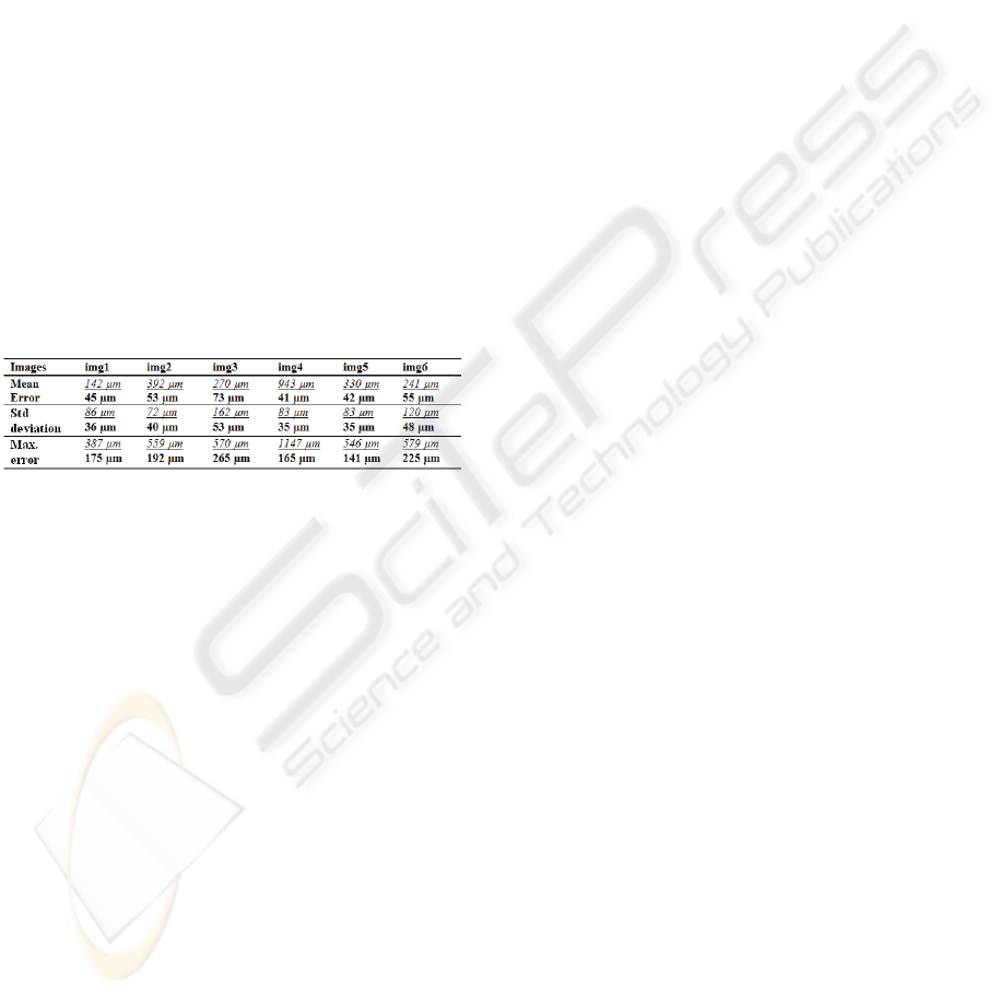
4.1 Experimental Comparison
We have compared our calibration method with a
camera calibration toolbox implemented in MatLab
(Bouguet 2008). For this purpose, we have used
pictures of the calibration target that were taken
during the experiment described in the next section.
6 such pictures have been taken with different target
orientations, and in order to compare the two
approaches, we have used a cross validation
technique: each picture is isolated in turn and is used
as an unseen test case, while the 5 others are used
for learning the parameters. Both methods are
evaluated with the same set of detected points, and
with the same error measure (difference between
real and estimated distances).
Table 1: Evaluation of calibration accuracy using a cross
validation technique over 6 pictures (48 points and 82
distances per picture). In underline and italic are presented
the results of MatLab’s « Camera Calibration ToolBox »,
and in bold the results of Skin3D.
According to the results, our method has the best
accuracy for all images. These results can be
explained by the fact that the two methods are very
different from each others: classical versus genetic
optimization, optimization of parameters for each
image versus all images. These results are thus very
encouraging and we have planned additional
comparative tests.
4.2 Real Study
In order to evaluate our system in a real world
application, we have conducted a study involving 18
women from 20 to 65 years old who presented skin
specificities. For each woman, we have taken 3D
pictures of their face (front and both sides). For
some women who presented specific symptoms, we
have also taken pictures of their hands and of their
back. In order to analyze the pictures, we have
presented them to a panel of international
dermatologists. They have used Skin3D to visualize
the pictures in stereoscopy, to perform 3D
measurements and to annotate the pictures. They
have defined a guided tour. The possibilities offered
by Skin3D (like 3D visualization and annotations)
have improved the diagnostic of different skin
symptoms by making the identification of specific
information easier than in standard photographs.
5 CONCLUSIONS
We have developed the Skin3D system for the
acquisition, visualization and interactive exploration
of stereoscopic pictures in the domain of
dermatology. We have described its 3 main modules.
We have defined a new calibration method which,
after a first experimental comparison, seems to be
efficient and well adapted to our application. We
have proposed the use of specific virtual reality
hardware in order to visualize stereo images and to
navigate through them. A test was performed with
success on a large screen. We have developed
several ways to perform 3D measurements,
annotations and to share the discovered knowledge.
Several perspectives can be derived from this
work. As far as the acquisition module is concerned,
we want to acquire better cameras with a resolution
of 21 Mpixels in order to increase further the
accuracy of the system. We want to perform parallel
computation of the pattern matching algorithm. We
want to improve also the annotation process by
adding specific algorithms for region automatic
detection. We want to add a search engine for
searching specific text in the annotations. Finally,
we could study the use of domain ontology in order
to help the expert for normalizing the annotations.
REFERENCES
Alfonso, B., 2005. Featured Virtual Lab in their NetWatch
section, a publication by the AAAS Vol 308 Science
Magazine.
Ayoub, A.F., P. Siebert, K. F. Moos, D. Wray, C.
Urquhart, T. B. Niblett, 1998. A vision-based three-
dimensional capture system for maxillofacial
assessment and surgical planning. British journal of
oral & maxillofacial surgery, vol. 36, no5, 353-357
Ben Amor, B., M. Ardabilian, L. Chen, 2005. 3D Face
Modeling Based on Structured-light Assisted Stereo
Sensor. Image Analysis and Processing Vol.3617,
842-849
Bernard, A., M. Cohen, U. Christoph et MD. Lehmann,
2008. DermAtlas, Johns Hopkins University.
www.dermatlas.org.
Bouguet,J., 2008, Camera Calibration Toolbox for Matlab,
www.vision.caltech.edu/bouguetj/calib_doc/
Cerveri, P., A. Pedotti et N. A. Borghese, 2001. Combined
Evolution Strategies for Dynamic Calibration of
ACQUISITION, ANNOTATION AND INTERACTIVE EXPLORATION OF STEREO IMAGES WITH VIRTUAL
REALITY
373

Video-Based Measurement Systems. IEEE
Transactions on evolutionary computations, vol.5,
No.3. 271-282
Chalam, KV., P. Jain, VA. Shah, Gaurav Y. Shah, 2006.
Evaluation of web-based annotation of ophthalmic
images for multicentric clinical trials. Indian Journal
of Ophthalmology 54, 126-129.
Chambon, S., A. CROUZIL 2003. Dense matching using
correlation: new measures that are robust near
occlusions. British Machine Vision Conference -
BMVC 2003, vol. 1, 143-152.
D'Apuzzo, N., 2002. Modeling human faces with
multiimage photogrammetry. Three-Dimensional
Image Capture and Applications vol. 4661, 191-197.
Dermnet©, 2007. Interactive Medical Media LLC.
www.dermnet.com.
Dipanda, A., S. Woo, F. Marzani, J.M. Bilbault, 2003. 3-D
shape reconstruction in an active stereo vision system
using genetic algorithms. The journal of pattern
recognition society, Vol.36, 2143-2159.
Hernandez Esteban, C., F. Schmitt, 2003. Silhouette and
Stereo Fusion for 3D Object Modeling; Computer
Vision and Image Understanding Volume 96, Issue 3,
367-392.
Faugeras, O. D., and Toscani, G, 1987. Camera calibration
for 3d computer vision. International Workshop on
Machine Vision and Machine Intelligence, 240–247.
Rohr, M., K. Schrader, 1998. Fast Optical in vivo
Topometry of Human Skin (FOITS) Comparative
Investigations with Laser Profilometry. SÖFW Journal
124, 52 - 59
Salvi, J., J. Pagès, J. Batlle, 2004. Pattern codification
strategies in structured light systems. Pattern
Recognition. Vol. 37, Issue 4, 827-849
Tsai, R. Y., 1987. A versatile camera calibration technique
for high-accuracy 3D machine vision metrology using
off-the-shelf tv cameras and lenses. IEEE Journal of
Robotics and Automation, 3(4), 323-344.
Zhang, Y., Q. Ji, 2001. Camera Calibration With Genetic
Algorithms, IEEE International Conference on
Robotics and Automation. 120-130
Zhang, Z., 1998. A Flexible New Technique for Camera
Calibration. Technical Report MSR-TR-98-71 of
Microsoft Research.
Zhu, Y., 2007. A Java program for stereo retinal image
visualization. Computer Methods and Programs in
Biomedicine, Vol.85, Issue 3, 214-219.
HEALTHINF 2009 - International Conference on Health Informatics
374
