
MAGNETIC RESONANCE IMAGING OF THE VOCAL TRACT
Techniques and Applications
Sandra M. Rua Ventura
Área Científico-pedagógica da Radiologia, School of Allied Health Science – IPP
Rua Valente Perfeito 322, 4400-330 Vila Nova de Gaia, Portugal
Diamantino Rui S. Freitas, João Manuel R. S. Tavares
FEUP – Faculty of Engineering of University of Porto
Rua Dr. Roberto Frias, s/n 4200-465 Porto, Portugal
Keywords: Magnetic Resonance Imaging, Image Processing, 3D modeling, Vocal Tract Study, Speech Production.
Abstract: Magnetic resonance (MR) imaging has been used to analyse and evaluate the vocal tract shape through
different techniques and with promising results in several fields. Our purpose is to demonstrate the
relevance of MR and image processing for the vocal tract study. The extraction of contours of the air
cavities allowed the set-up of a number of 3D reconstruction image stacks by means of the combination of
orthogonally oriented sets of slices for each articulatory gesture, as a new approach to solve the expected
spatial under sampling of the imaging process. In result these models give improved information for the
visualization of morphologic and anatomical aspects and are useful for partial measurements of the vocal
tract shape in different situations. Potential use can be found in Medical and therapeutic applications as well
as in acoustic articulatory speech modelling.
1 INTRODUCTION
Magnetic Resonance (MR) improvements, in the
past decades, allowed vocal tract imaging, making it
currently one of the most promising tools in speech
research.
Speech is the most important instrument of
human communication and interaction.
Nevertheless, the knowledge about its production is
far from being complete or even sufficient to
describe the most relevant acoustic phenomena that
are conditioned at morphological and dynamic
levels. The anatomic and physiologic aspects of the
vocal tract are claimed to be essential for a better
understanding of this process. The quality and
resolution of soft-tissues and the use of non-ionizing
radiation are some of the most important advantages
of MR imaging (Avila-García et al., 2004; Engwall,
2003).
Several approaches have been used up to now for
the study of the vocal tract based on MR images.
Since the first study proposed by Baer et al. (Baer et
al., 1991), many MR techniques have been used
(from static to dynamic studies, and more recently
even done in real-time), starting by studies of vowel
production (Badin et al., 1998; Demolin et al.,
2000), followed by consonant production (Engwall,
2000b; Narayanan et al., 2004), for different
languages such as French (Demolin et al., 1996;
Serrurier & Badin, 2006), German (Behrends et al.,
2001; Mády et al., 2001), and Japonese (Kitamura et
al., 2005; Takemoto et al., 2003).
The work presented in this paper, consisting
basically in the static description of the vocal tract
shape during sustained vowels and consonants and
in the dynamic description of some syllables, is the
first to report the application of MR imaging for the
characterization of European Portuguese (EP). This
study started in 2004, having attained a first series of
results published in 2006 (Rua & Freitas, 2006). Our
approach can be seen as a contribution to the wide
area of articultory speech modeling, since it provides
geometrical data to the acoustic modeling phase or
research.
In the articulatory speech research of EP a few
studies of nasal vowels have been carried through, at
105
Rua Ventura S., S. Freitas D. and R. S. Tavares J. (2009).
MAGNETIC RESONANCE IMAGING OF THE VOCAL TRACT - Techniques and Applications .
In Proceedings of the First International Conference on Computer Imaging Theory and Applications, pages 105-110
DOI: 10.5220/0001792901050110
Copyright
c
SciTePress
the acoustic production and perceptual levels based
on acoustic analysis and electromagnetic
articulography (Teixeira et al., 2001, 2002, 2003).
More recently, another MR study of EP presents
some results relative to oral and nasal vowels
exploring contours extraction from 2D images,
articulatory measures and area functions (Martins et
al., 2008).
In former studies, vocal tract modelling has been
limited to the midsagittal plane (Engwall, 2000a;
Takemoto et al., 2003), but improvement of MR
imaging equipment system capabilities allowed the
expansion into this domain of research and made it
possible to obtain three-dimensional (3D) modelling
(Badin & Serrurier, 2006). The more realistic
models of the vocal tract shape that nowadays are
possible to obtain, are hugely needed in the research
towards improved speech synthesis algorithms and
more efficient speech rehabilitation.
The main purpose of this paper is to present
some 3D models of the vocal tract based on MR data
of some relevant sustained articulations of EP in a
static study. From the point of view of image
processing, a new approach for 3D modelling by
means of the combination of orthogonal stacks, to
describe the vocal tract shape in different
articulatory positions is presented. We also
demonstrate an MR technique to capture useful
image sequences during speech (dynamic study). In
addition, some preliminary results of this dynamic
study are presented.
The remaining of this paper is organized in four
sections. The next section is dedicated to the
methods and describes the equipment, corpus and
subjects, as well as the procedures used for the
speech study, namely for morphologic and dynamic
imaging of the vocal tract. The results are presented
in following section, through the exhibition of some
three-dimensional models built of the vocal tract and
an image sequence obtained during speech. Finally
the conclusions of the work described are presented.
2 METHODS
This study was performed in two phases: 1)
exploration of MR techniques applied to the vocal
tract imaging; 2) the use of image processing
techniques that can aid the analysis of vocal tract.
2.1 Equipment, Corpus and Subjects
An MR Siemens Magneton Symphony 1.5T system
and a head array coil were used, with subjects in
supine position. The image data were acquired from
two subjects (one male and one female) for the static
study, and from four subjects for the dynamic study,
all without any speech disorder.
The corpus of the static study consisted in twenty
five sounds of European Portuguese: oral and nasal
vowels, and consonants. For the dynamic study, the
subjects produced several repetitions of sequences of
three consonant-vowel syllables (/tu/, /ma/, /pa/)
during the acquisition.
Because of the MR acoustic noise produced
during image acquisitions, the acoustic recording of
the produced speech was not yet possible.
2.2 Techniques
According the safety procedures for MR, subjects
was previously informed about the exam and
instructed about the procedures during the
acquisitions. A consent informed was obtained from
each subject involved.
Furthermore, the training of the subjects was
performed to ensure the proper production of the
intended sounds for the static study, and to achieve
good speech-acquisition synchronization for the
dynamic one.
2.2.1 Static Study
A set of MR WT1-images using Turbo Spin-Echo
(TSE) sequences was acquired in sagittal and
coronal orientations. The subjects sustained the
articulation during 9 seconds for the acquisition of
three sagittal slices and 9.9 seconds for the four
coronal slices. The acquisition time was a
compromise between image resolution and the
duration of the sustained articulation allowed by the
subject.
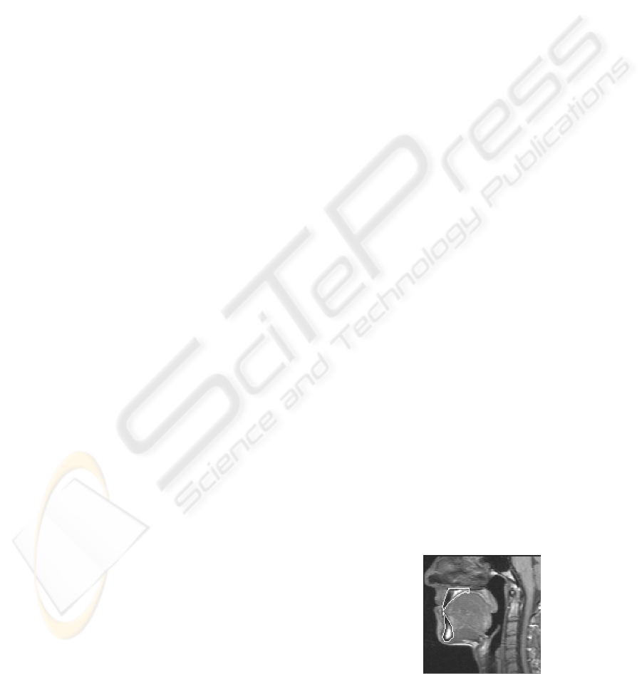
Initially, a single midsagittal T1-weighted image
was acquired with subjects instructed to rest with
mouth close and the tongue in full contact with the
teeth. This reference image was used for teeth space
identification and contour extraction (Figure 1).
Figure 1: Midsagittal reference image for teeth
identification and contours extraction.
IMAGAPP 2009 - International Conference on Imaging Theory and Applications
106
The used protocol set includes the parameters
shown in Table 1.
Table 1: MR protocol for vocal tract imaging.
Sagittal slices Coronal slices
TR = 443 ms TR = 470 ms
TE = 17 ms TE = 15 ms
ETL = 7 ETL = 7
SNR = 1 SNR = 1.03
3 mm thickness 6 mm thickness and 10 mm gap
Three slices Four slices
Matrix size 128 x 128
Resolution = 0.853 px/mm
Field of View (FOV) = 150 mm
2.2.2 Dynamic Study
The dynamic study was performed following the
same principle of MR cardiac analysis, with the
modification of a FLASH-2D sequence using the
patient’s heart beat as a trigger signal, a 300 mm
field of view and the acquisition parameters: TR =
60 ms and TE = 4.4 ms. The subjects tried to
synchronize the utterance of the syllables to their
own cardiac rhythm by means of the acoustic
monitoring of their own simple electrocardiogram
(ECG) through a synchronous sound emission
conveyed to the subject by an earphone.
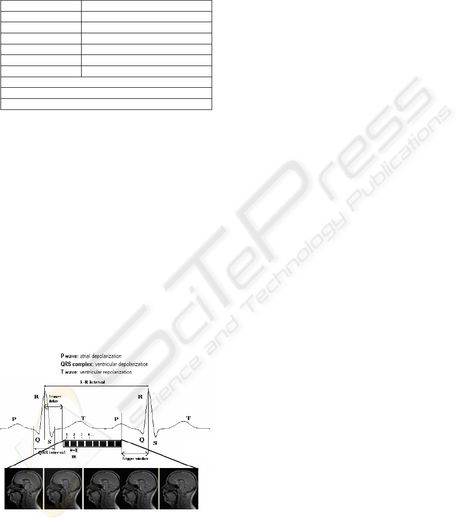
Each set of images from a single-slice
(midsagittal) of 6 mm thickness was collected during
12 to 22 seconds. For each sequence, a variable
number of images (4-6 images) were acquired with a
regularly increasing shift in synchrony from the start
of the cardiac cycle (Figure 2).
Figure 2: Diagram of the dynamic MR acquisition based
on ECG monitoring and synchronization.
The RR interval is the time duration between two
consecutive R waves (in ECG graph), and it is
usually the reference interval for programming the
slice acquisitions. It should be noted that depicted
the images were acquired in a single cardiac cycle
for the sake of representation, this is an under-
sampling method, assuming that the phenomenon is
stationary, which is quite good in cardiac analysis
but no so in repeated speech production. In fact, the
images were collected distributed along the time
with a period as short as possible, but always longer
than a cardiac cycle due to machine limitations.
2.2.3 Image Processing and Analysis
Few segmentation methods have been described in
speech studies for vocal tract contours extraction
from MR images. Briefly, those methods are based
on manual edition of curves, such as Bézier curves,
and threshold binarizations (Badin et al., 2000;
Engwall, 2004; Soquet et al., 2002). Soquet et al.
(1998) compared different approaches on the same
data in order to assess the accuracy of some manual
segmentation methods, and concluded that the
methods considered give comparable results and that
the threshold method is the one that presents lower
dispersion.
Here, image analysis and 3D model
reconstruction were accomplished in two stages:
Image segmentation using the Segmenting
Assistant, a 3D editing plug-in of Image J the
image processing software developed by the
National Institute of Health and subsequent
3D reconstruction;
Graphic representation and combination of
orthogonal stacks using the Blender software
for 3D graphics creation.
The histogram-derived threshold technique was
chosen for the segmentation of the airway from the
surrounding tissues. The extraction of the contours
of the vocal tract was then obtained by the following
sequence of procedures:
(a) Identification and closure of the vocal tract area
of interest, mandatory closure of the mouth, larynx,
vertebral column and velum, through the manual
superimposition of opaque objects;
(b) Manual overlapping of teeth image (done only
on the sagittal stacks), after extraction of the teeth
contours from the initially acquired sagittal
anatomic reference image;
(c) Extraction of the contours of the vocal tract, for
each image of 2D slices using the Image J semi-
automatic threshold technique.
The Figure 3 depicts the image segmentation
procedure used in sagittal and coronal slices during a
sustained nasal vowel. The closure of the vocal tract
MAGNETIC RESONANCE IMAGING OF THE VOCAL TRACT -
Techniques and Applications
107
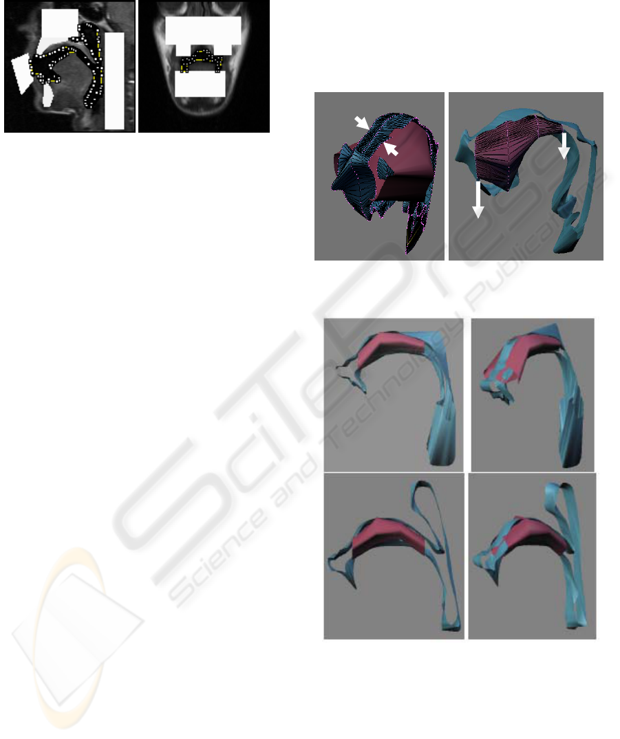
area was necessary to avoid contours to “escape”
and complicate the segmentation task.
Figure 3: Contours extraction from sagittal (right) and
coronal (left) slices after closure of the vocal tract area and
manual overlapping of teeth.
The contour extraction process resulted in a total
of 175 planar contours (maximally 7 contours for
each sound).
Outlines were subsequently used to generate a
3D surface, after importing the contours in .shapes
format, into the Blender software.
For each articulatory position, the next phase was
the combination of sagittal and coronal outlines (2D
curves). To make this possible, it was required that
the outlines be well aligned – this process is usually
known as image registration. In Computational
Vision, the term image registration means the
process of transforming the different sets of images
into one common coordinate system, what was here
necessary in order to be able to compare or integrate
the data obtained from different measurements.
3 RESULTS AND DISCUSSION
The static study was designed to obtain the
morphologic data of most of the range of the
articulators’ positions aiming the imaging
characterization of Portuguese sounds.
A variable
number of images were obtained by dynamic MR,
according to the cardiac cycle of each subject,
followed by the assembly of all images for sequence
visualization.
3.1.1 3D models
The following images (Figure 4) represent different
perspectives of the 3D model obtained for the vowel
[u]. In the presented images, the blue surface
represents the union of the three outlines extracted
from the sagittal stack. By other hand, the red
surface represents the union of the four outlines
extracted from the coronal stack.
The Figure 5 depicts two 3D models of the
vowels: corresponding to an oral sound (above) and
to a nasal sound (below). The different viewpoints
presented allow the identification of the velum
lowering, and especially the partial closure of the
oral cavity, comparatively to the oral sound. In
Portuguese there is a special interest in nasal sounds
due to their frequent use in common speech.
1
3
4
1
Figure 4: Surfaces representations for the 3D model of the
vowel [u].
Figure 5: Three-dimensional models of the oral (top) and
nasal (bottom) vowel [a] of European Portuguese.
In the 3D models obtained, some differences in
the vertical lengths between sagittal and coronal
stacks in some sounds were observed resulting in
some registration errors. This could reflect the
specific variability of the speaker in sound
production, due in this case, to the fact that
acquisitions of different orientations were separately
done (first sagittal images, and subsequently coronal
IMAGAPP 2009 - International Conference on Imaging Theory and Applications
108
images), to minimize the subjects’ effort needed for
an extra long utterance. On the other hand, the
segmentation process used had also some
implications for the determination of the vocal tract
area contour.
Furthermore, the coronal data is important for 3D
modelling of the vocal tract, because some
articulatory situations lead to occlusions in the
midsagittal plane, while lateral channels are
maintained open (e.g. lateral consonants, nasals
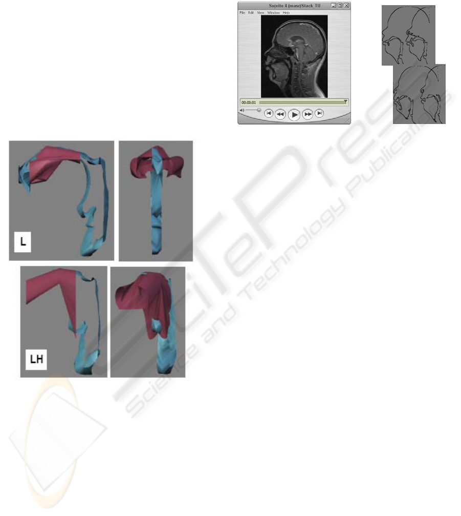
vowels). The models shown in Figure 6 intend to
demonstrate the relevance of coronal stacks for the
characterization of lateral consonants of Portuguese.
Although the 3D models built are not yet
completely closed, it can be observed several
essential features needed for the articulatory
description of speech.
Figure 6: Three-dimensional models of the laterals
consonants [l] and [lh] of European Portuguese.
3.1.2 Image Sequence during Speech
A variable number of images (sagittal slices) were
obtained in dynamic studies, according to the
cardiac cycle (heart frequency) of each subject,
followed by the assembly of all images for image
sequences visualization. The best image features
were obtained for the syllable /tu/, as illustrate in
Figure 7, because the articulatory positions are very
different between single sounds, compared with the
other syllables.
When considering the differences verified
amongst the subjects, by image comparison, the
dynamic studies demonstrated the actual variability
in sounds production between subjects, not only due
to anatomic differences, but also because each
subject uses different strategies for motion control
and articulation.
Figure 7: Contours extracted from midsagittal images
obtained in the dynamic study by the repetition of the
sequence /tu/.
4 CONCLUSIONS
AND FUTURE WORK
In our study, a considerable number and diversity of
images were acquired aiming at not only
morphological but also a dynamic characterization,
by exploring various MR techniques. We tried to
acquire a number of images with enough anatomic
resolution, maximum vocal tract extension of
representative speech gestures, minimizing speaker
effort (reducing hyperarticulation). The image data
was analysed and processed resulting in the
reconstruction of 3D models for the entire corpus
(3D geometrical database).
For almost all 3D models obtained for
Portuguese sounds the morphologic data showed
that both orientations slices (sagittal and coronal) are
useful for the knowledge of the vocal tract shape
during speech production. Articulators’ positions are
better demonstrated in sagittal images, and the
coronal images allow the observation of the lateral
dimension of oral cavity.
The completion of the construction of the
surfaces for the hybrid models made from sagittal
and coronal stacks is the next step in the way to
obtain a complete 3D anatomical model of the vocal
tract, prepared for the subsequent prediction of the
acoustic output.
The extension of the dynamic sequences
obtained to other sequences is also important in
terms of coverage of the study, and will be done in
the near future.
MAGNETIC RESONANCE IMAGING OF THE VOCAL TRACT -
Techniques and Applications
109

Other problems related with the image
registration and with acoustic recording of speech
are being investigated until now, aiming to be
solving in a future work.
ACKNOWLEDGEMENTS
The images considered were acquired at the
Radiology Department of Hospital S. João, Porto,
with the collaboration of Isabel Ramos (Professor
from Faculdade de Medicina da Universidade do
Porto and Department Director) and the technical
staff, which are gratefully acknowledged.
REFERENCES
Avila-García, M.S., Carter, J.N., Damper, R.I., 2004.
Extracting Tongue Shape Dynamics from Magnetic
Resonance Image Sequences. Transactions on
Engineering, Computing and Technology V2,
December, 288-291.
Badin P., Bailly G., Raybaudi M., Segebarth C., 1998. A
three-dimensional linear articulatory model based on
MRI data. 3rd ESCA / COCOSDA Int. Workshop on
Speech Synthesis, Australia, 249-254.
Badin, P., Borel, P., Bailly, G., Revéret, L., Baciu, M.,
Segebarth, C., 2000.Towards an audiovisual virtual
talking head: 3D articulatory modeling of tongue, lips
and face based on MRI and video images. 5th Speech
Production Seminar, Germany, 261-264.
Badin, P., Serrurier, A., 2006. Three-dimensional
Modeling of Speech Organs: Articulatory Data and
Models. IEICE Technical Committee on Speech,
Japan, 29-34.
Baer, T., Gore, J.C., Gracco, L.C., Nye, P.W., 1991.
Analysis of Vocal Tract Shape and Dimensions using
Magnetic Resonance Imaging: Vowels. J. Acoust. Soc.
Am., 90, 799-828.
Behrends J., Wismuller A., 2001. A Segmentation and
Analysis Method for MRI data of the Human Vocal
Tract, FIPKM-37, 179-189.
Demolin D., Metens T., Soquet A., 1996. Three-
dimensional Measurement of the Vocal Tract by MRI.
4th Int. Conf. on Spoken Language Processing (ICSLP
96), USA, 272-275.
Demolin, D., Metens, T., Soquet, A., 2000. Real time MRI
and articulatory coordinations in vowels. 5 th Speech
Production Seminar. Germany.
Engwall, O. , 2003. A revisit to the Application of MRI to
the Analysis of Speech Production - Testing our
assumptions. 6th Int. Seminar on Speech Production.
Sydney.
Engwall, O., 2000a. A 3D Tongue Model based on MRI
data. 6th Int. Conf. on Spoken Language Processing
(ICSLP), China, 901-904.
Engwall, O., 2000b. Are static MRI representative of
dynamic speech? Results from a comparative study
using MRI, EPG and EMA. 6th Int. Conf. on Spoken
Language Processing (ICSLP), China, 17-20.
Engwall, O., 2004. From real-time MRI to 3D tongue
movements. ICSLP 2004, vol. II, October, Korea,
1109-1112.
Kitamura T., Takemoto H., Honda K., Shimada Y.,
Fujimoto I., Syakudo Y., Masaki S., Kuroda K., Oku-
uchi N., Senda M., 2005. Difference in vocal tract
shape between upright and supine postures:
Observations by an open-type MRI scanner.
Acoustical Science and Technology, 26(5), 465-468.
Mády, K., Sader, R., Zimmermann, A., Hoole, P., Beer,
A., Zeilhofe, H., Hannig, C., 2001. Use of real-time
MRI in assessment of consonant articulation before
and after tongue surgery and tongue reconstruction.
4th Int. Speech Motor Conf. Netherlands, 142-145.
Martins, P., Carbone, I.C., Pinto, A., Silva, A., Teixeira,
A.J., 2008. European Portuguese MRI based speech
production studies. Speech Communication, 50, 925-952.
Narayanan, S., Nayak, K., Lee, S., Sethy, A., Byrd, D.,
2004. An Approach to Real-time Magnetic Resonance
Imaging for Speech Production. Journal Acoustical
Society of America, 115(4), 1771-1776.
Rua, S.M., Freitas, D.R., 2006. Morphological Dynamic
Imaging of Human Vocal Tract. Computational
Modelling of Objects Represented in Images:
Fundamentals, Methods and Applications
(CompIMAGE), Portugal, October 20-21, 381-386.
Serrurier, A. & Badin, P., 2005. A Three-dimensional
Linear Articulatory Model of Velum based on MRI
data. Interspeech 2005: Eurospeech, 9th Europ. Conf.
on Speech Communication and Technology, Portugal,
2161-2164.
Soquet, A., Lecuit, V., Metens, T., Demolin, D., 2002.
Mid-sagittal cut to area function transformations:
Direct measurements of mid-sagittal distance and area
with MRI. Speech Communication, 36, 169-180.
Soquet, A., Lecuit, V., Metens, T., Nazarian, B., Demolin,
D., 1998. Segmentation of the Airway from the
Surrounding Tissues on Magnetic Resonance Images:
A comparative study. ICSLP. Sydney, 3083-3086.
Takemoto, H., Honda, K., 2003. Measurement of
Temporal Changes in Vocal Tract Area Function
during a continuous vowel sequence using a 3D Cine-
MRI Technique. 6th Int. Seminar on Speech
Production, Australia, 284-289.
Teixeira, A. et al., 2002. SAPWindows – Towards a
Versatile Modular Articulatory Synthesizer.
Proceedings of 2002 IEEE Workshop on Speech
Synthesis. Portugal, 31-34.
Teixeira, A., Moutinho, L.C., Coimbra, R.L., 2003.
Production, Acoustic and Perceptual Studies on
European Portuguese Vowels Height. 15th Int.
Congress of Phonetic Sciences, Barcelona, 3033-3036.
Teixeira, A., Vaz, F., 2001. European Portuguese Nasal
Vowels: An EMMA Study. Eurospeech 2001. Aveiro,
Portugal, 1483-1486.
IMAGAPP 2009 - International Conference on Imaging Theory and Applications
110
