
OPTIMUM DCT COMPRESSION OF MEDICAL IMAGES
USING NEURAL NETWORKS
Adnan Khashman and Kamil Dimililer
Intelligent Systems Research Group (ISRG), Near East University, Lefkosa, Northern Cyprus, Turkey
Keywords: X-ray medical images, Optimum image compression, Neural Network, DCT Image Compression.
Abstract: Medical imaging requires storage of large quantities of digitized data Efficient storage and transmission of
medical images in telemedicine is of utmost importance however,. Due to the constrained bandwidth and
storage capacity, a medical image must be compressed before transmission or storage. An ideal image
compression system must yield high quality compressed images with high compression ratio; this can be
achieved using DCT-based image compression, however the contents of the image affects the choice of an
optimum compression ratio. In this paper, a neural network is trained to relate the x-ray image contents to
their optimum compression ratio. Once trained, the optimum DCT compression ratio of the x-ray image can
be chosen upon presenting the image to the network. Experimental results suggest that out proposed system,
can be efficiently used to compress x-rays while maintaining high image quality.
1 INTRODUCTION
X-rays or radiographs are images produced on a
radiosensitive surface, such as a photographic film,
by radiation other than visible light, especially by x-
rays passed through an object or by photographing a
fluoroscopic. These images, commonly referred to
as x-rays, are usually used in medical diagnosis,
particularly to investigate bones, dental structures,
and foreign objects within the body. X-rays are the
second most commonly used medical tests, after
laboratory tests.
Recently, teleradiology, which is one of the most
used clinical aspects of telemedicine, has received
much attention. Teleradiology is the transmission of
radiologic images from a site of image acquisition to
a remote location for interpretation in hospitals such
as computerized tomography (CT) scans, magnetic
imaging (MRI), ultrasonography (US), and x-rays.
These radiological images are needed to be
compressed before transmission to a distant location
or due to the bandwidth or storage limitations (Singh
et al., 2007).
There has been a rapid development in
compression methods to compress large data files
such as images where data compression in various
applications has become more vital (Nadenau et al.,
2003). Efficient methods of compression, to
compress and store or transfer image data files while
retaining high image quality and marginal reduction
in size are needed due to the improvements of
technology (Ratakonda and Ahuja, 2002).
The discrete cosine transform (DCT) is possibly
the most popular transform used in compression of
images in standards like Joint Photographic Experts
Group (JPEG). In DCT-based compression the
image is split into smaller blocks for computational
simplicity. The blocks are classified on the basis of
information content to maximize compression ratio
without sacrificing diagnostic information (Singh et
al., 2007). DCT-based medical image compression
has been investigated by several researchers. For
example, in (Chikouche et al., 2008) DCT-based
compression was applied to IRM type medical
images. In (Prudhvi Raj and Venkateswarlu, 2007)
a medical image compression application based on
3-dimensional DCT was proposed. In (Zukoski et
al., 2006) region based medical image compression
has been applied to choose the clinically relevant
regions as defined by radiologists. In (Shih and Wu,
2005) another region of interest based medical
image compression based on genetic algorithms was
also investigated.
The use of DCT and artificial neural networks
has also been investigated in search for optimum
compression methods. In (Dokur, 2008) MR and CT
medical images were compressed using DCT and
neural networks. In (Ashraf and Akbar, 2006)
90
Khashman A. and Dimililer K. (2009).
OPTIMUM DCT COMPRESSION OF MEDICAL IMAGES USING NEURAL NETWORKS .
In Proceedings of the 11th International Conference on Enterprise Information Systems - Artificial Intelligence and Decision Support Systems, pages
91-96
DOI: 10.5220/0001865600910096
Copyright
c
SciTePress
another application of neural networks in medical
image compression was also proposed. In (Meyer-
Base et al., 2005) topology-preserving neural
networks were applied for medical image
compression by a ‘‘neural-gas’’ network. In (Liying
and Khashayar, 2005) different image compression
techniques were combined with neural network
classifier for various applications. In (Soliman and
Omari, 2006) a neural network model called direct
classification was also suggested to compress image
data. In (Ciernak, 2004) periodic vector quantization
algorithm based image compression was suggested
and was based on competitive neural networks
quantizer and neural networks predictor.
More works using neural networks for image
compression applications emerged lately, such as
those in (Ashraf and Akbar, 2005), (Northan and
Dony, 2006), (Vilovic, 2006), and (Veisi and
Jamzad, 2007). Recently, a neural network based
DCT compression system that finds the optimum
compression ratios for a variety of images was also
suggested (Khashman and Dimililer, 2007), where
the evaluation method of the neural network-
obtained optimum compression results was based on
the comparison criteria; which was suggested in
(Khashman and Dimililer, 2005).
The aim of the work presented within this paper
is to develop a medical image compression system
using Discrete Cosine Transform and a neural
network. Our proposed method suggests that a
trained neural network can learn the non-linear
relationship between the intensity (pixel values) of
an x-ray image and its optimum compression ratio.
Once the highest compression ratio is obtained,
while maintaining good image quality, the result
reduction in x-ray image size, should make the
storage and transmission of x-rays more efficient,
thus providing compressed images with good quality
and satisfactory information for the medics.
The paper is organized as follows: Section 2
describes the x-ray image database which is used for
the implementation of our proposed system. Section
3 presents the x-rays compression system;
describing image pre-processing and the neural
network design and implementation. Section 4
introduces the method used to evaluate the results
and provides an analysis of the system
implementation. Finally, Section 5 concludes the
work that is presented within this paper and suggests
further work.
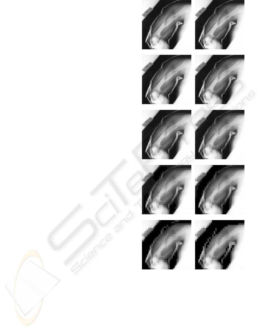
Original Image 10%
20 % 30%
40 % 50%
60 % 70%
80 % 90%
Figure 1: An original x-ray image and its DCT
compression at nine ratios.
2 X-RAY IMAGE DATABASE
The development and implementation of the
proposed medical x-rays compression system uses
60 x-ray images from our medical image database
which were obtained from the Radiology
Department at the Famagusta General Hospital
(Famagusta, Cyprus), which contains x-ray images
OPTIMUM DCT COMPRESSION OF MEDICAL IMAGES USING NEURAL NETWORKS
91
of fractured, dislocated, broken, and healthy bones
in different parts of the body. DCT compression has
been applied to 50 radiographs using nine
compression ratios (10%, 20%, ..., 90%) as shown in
an example in Figure 1.
The optimum DCT compression ratios for the 50
x-ray images were determined using the optimum
compression criteria based on visual inspection of
the compressed images as suggested in (Khashman
and Dimililer, 2005), thus providing 50 images with
known optimum compression ratios and the
remaining 10 images with unknown optimum
compression ratios. The image database is then
organized into three sets:
• Training Set: contains 25 images with known
optimum compression ratios which are used for
training the neural network within the radiograph
compression system. Examples of training images
are shown in Figure 2a.
• Testing Set 1: contains 25 images with known
optimum compression ratios which are used to
test and validate the efficiency of the trained neural
network. Examples of these testing images are
shown in Figure 2b.
• Testing Set 2: contains 10 images with unknown
optimum compression ratios which are used to
further test the trained neural network. Examples of
these testing images are shown in Figure 2c.
Examples of original x-ray images and their
compressed versions using their optimum
compression ratios while training the neural network
are shown in Figure 3.
3 X-RAY IMAGE COMPRESSION
SYSTEM
The optimum x-ray image compression system uses
a supervised neural network based on the back
propagation learning algorithm, due to its
implementation simplicity, and the availability of
sufficient “input/target” database for training this
supervised learner. The neural network relates the x-
ray image intensity (pixel values) to the image
optimum compression ratio having been trained
using images with predetermined optimum
compression ratios. The ratios vary according to the
variations in pixel values within the images. Once
trained, the neural network would choose the
optimum compression ratio of an x-ray image upon
presenting it to the neural network by using its
intensity values.
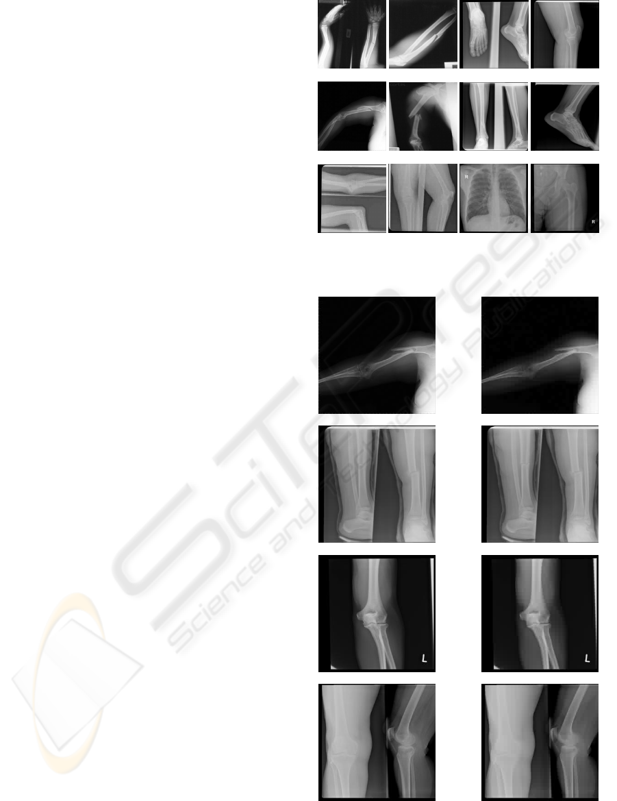
a
b
c
Figure 2: (a) Training Set examples (b) Testing Set 1
examples, (c) Testing Set 2 examples.
X-ray 1 40% Compression
X-ray 2 10% Compression
X-ray 3 30% Compression
X-ray 4 20% Compression
Figure 3: Examples of Training Set images and their
optimum compression ratios.
ICEIS 2009 - International Conference on Enterprise Information Systems
92
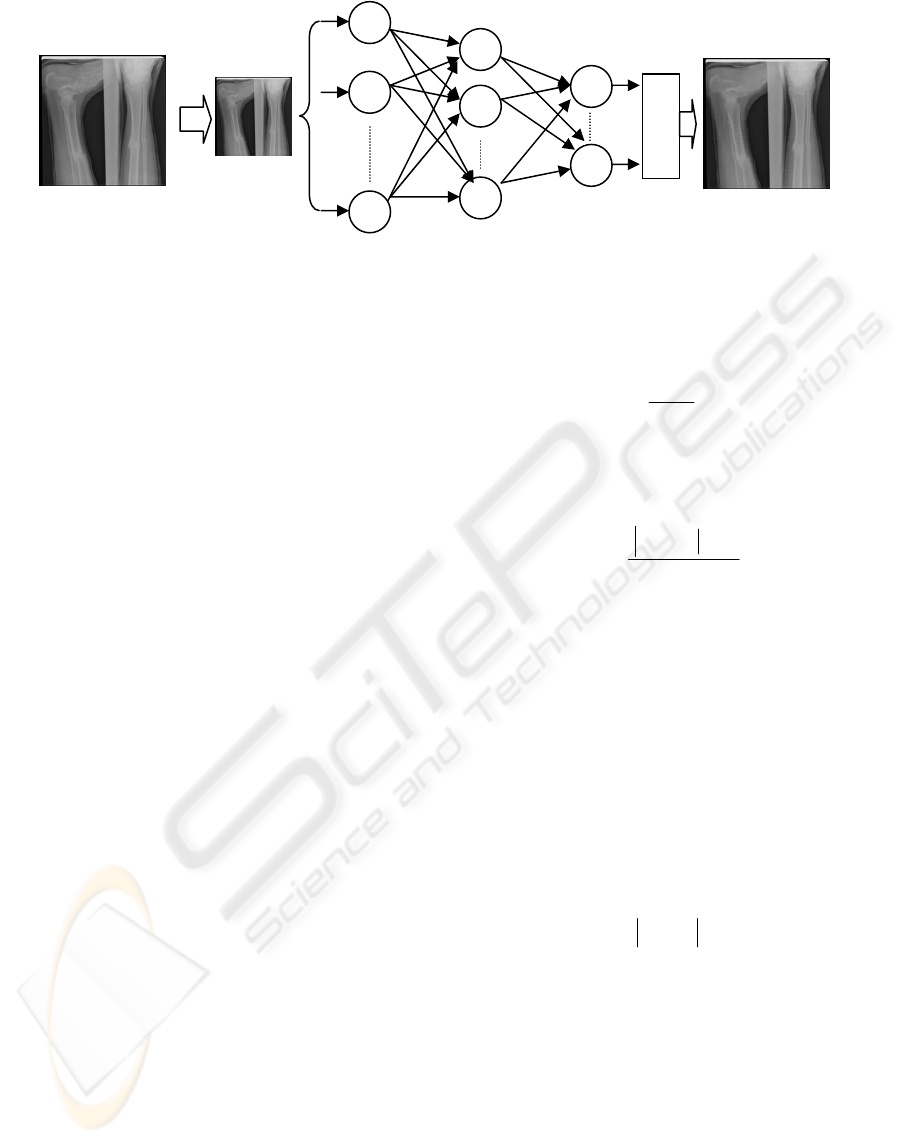
Adobe Photoshop was used to resize the original
images of size (256x256) pixels into (64x64) pixels.
Further reduction to the size of the images was
attempted in order to reduce the number of input
layer neurons and consequently the training time,
however, meaningful neural network training could
not be achieved thus, the use of whole images of the
reduced size of 64x64 pixels.
The size of the input x-ray images affects the
choice of the number of neurons in the neural
network’s input layer, which has three layers; input,
hidden and output layers. Using one-pixel-per-
neuron approach, the neural network’s input layer
has 4096 neurons, its hidden layer has 50 neurons,
which assures meaningful training while keeping the
time cost to a minimum, and its output layer has nine
neurons according to the number of the considered
compression ratios (10% - 90%).
During the learning phase, the learning
coefficient and the momentum rate were adjusted
during various experiments in order to achieve the
required minimum error value of 0.003; which was
considered as sufficient for this application. Figure 4
shows the topology of this neural network, within
the x-ray image compression system.
4 RESULTS AND DISCUSSIONS
The evaluation of the training and testing results was
performed using two measurements: the recognition
rate and the accuracy rate. The recognition rate is
defined as follows: where RR
OHC
is the recognition
rate for the neural network within the radiograph
compression system, I
OHC
is the number of optimally
compressed x-ray images, and I
T
is the total number
of x-ray images in the database set.
100∗
⎟
⎟
⎠
⎞
⎜
⎜
⎝
⎛
=
T
ODC
ODC
I
I
RR
(1)
The accuracy rate RA
OHC
for the neural network
output results is defined as follows:
(
)
100*
10
1
⎟
⎟
⎠
⎞
⎜
⎜
⎝
⎛
∗−
−=
T
ip
ODC
S
SS
RA
(2)
where S
P
represents the pre-determined (expected)
optimum compression ratio in percentage, S
i
represents the optimum compression ratio as
determined by the trained neural network in
percentage and S
T
represents the total number of
compression ratios.
The Optimum Compression Deviation (OCD) is
another term that is used in our evaluation. OCD is
the difference between the pre-determined or
expected optimum compression ratio S
P
and the
optimum compression ratio S
i
as determined by the
trained neural network, and is defined as follows:
(
)
10∗−=
ip
SSOCD
(3)
The OCD is used to indicate the accuracy of the
system, and depending on its value the recognition
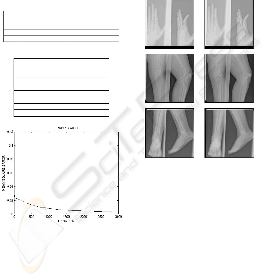
rates vary. Table 1 shows the three considered
values of OCD and their corresponding accuracy
rates and recognition rates. The evaluation of the
system implementation results uses (OCD = 1) as it
provides a minimum accuracy rate of 89% which is
considered sufficient for this application.
The neural network learnt and converged after
2960 iterations or epochs, and within 774 seconds,
whereas the running time for the generalized neural
networks after training and using one forward pass
Original Image
(256x256) pixels
Input Hidden Output
Layer Layer Layer
O
D
C
R
1
2
4096
1
2
50
1
9
Reduced Size
Image (64x64)
pixels
Figure 4: X-ray Image Compression System.
Compressed Image
OPTIMUM DCT COMPRESSION OF MEDICAL IMAGES USING NEURAL NETWORKS
93
was 0.015 seconds. These results were obtained
using a 2.0 GHz PC with 2 GB of RAM, Windows
XP OS and Matlab 2008b software. Table 2 lists the
final parameters of the successfully trained neural
network, whereas Figure 5 shows the error
minimization curve of the neural network during
learning.
Table 1: Accuracy and recognition rates for OCD.
OCD
Accuracy Rate
(RA
OD
C
)
Recognition Rate
(RR
OD
C
)
0 100 % 15/25 (60 %)
1 89 % 24/25 (96 %)
2 78 % 25/25 (100 %)
Table 2: Neural network final training parameters.
Input nodes 4096
Hidden nodes 50
Output nodes 9
Learning rate
0.003
Momentum rate
0.4
Error
0.003
Iterations 2960
Training time (seconds) 774
Run time (seconds)
0.015
Figure 5: Neural network learning curve.
The trained neural network recognized correctly
the optimum compression ratios for all 25 training
images as would be expected, thus yielding 100%
recognition of the training set. Testing the trained
neural network using the 25 images from Test Set 1
that were not presented to the network before
yielded 96% recognition rate, where 24 out of the 25
images with known optimum compression ratios
were assigned the correct ratio.
The trained neural network was also
implemented using the remaining 10 images with
unknown optimum compression ratios from the
testing set. The results of this application are
demonstrated Figure 6 which shows examples of the
optimally compressed x-ray images as determined
by the trained neural network.
X-ray 1 10% Compression
X-ray21 20% Compression
X-ray 3 30% Compression
Figure 6: Examples of Testing Set 2 image compression
using the trained neural network.
5 CONCLUSIONS
A novel method to medical x-ray image compression
using a neural network is proposed in this paper. The
method uses DCT-based compression with nine
compression ratios and a supervised neural network
that learns to associate the grey x-ray image
intensity (pixel values) with a single optimum
compression ratio.
The implementation of the proposed method uses
DCT image compression where the quality of the
compressed images degrades at higher compression
ratios due to the nature of the lossy compression.
The aim of an optimum ratio is to combine high
compression ratio with good quality compressed x-
ray images, thus making the storage and
transmission of images more efficient.
ICEIS 2009 - International Conference on Enterprise Information Systems
94

The proposed system was developed and
implemented using 60 x-ray images of fractured,
dislocated, broken, and healthy bones in different
parts of the body. The neural network within the x-
ray image compression system learnt to associate the
25 training images with their predetermined
optimum compression ratios within 774 seconds.
Once trained, the neural network could recognize the
optimum compression ratio of an x-ray image within
0.015 seconds
In this work, a minimum accuracy level of 89%
was considered as acceptable. Using this accuracy
level, the neural network yielded 96% correct
recognition rate of optimum compression ratios. The
successful implementation of our proposed method
using neural networks was shown throughout the
high recognition rates and the minimal time costs
when running the trained neural network.
Future work will include the implementation of
this method using wavelet transform compression
and comparing its performance with DCT-based x-
ray image compression using larger database.
REFERENCES
Ashraf, R., Akbar, M., 2005. Absolutely lossless
compression of medical images. In EMBS 2005,
Proceedings of IEEE 27th Conference on Engineering
in Medicine and Biology, 4006-4009.
Ashraf, R., Akbar, M., 2006. Adaptive Architecture
Neural Nets for Medical Image Compression. In
ICCIMA 2007, IEEE International Conference on
Engineering of Intelligent Systems, 1-4.
Ciernak, R., 2004. Image Compression algorithm based on
neural networks. In Lecture Notes in Artificial
Intelligence. Vol. 3070, Springer-Verlag, Berlin
Heidelberg, 706-711.
Chikouche, D., Benzid, R., Bentoumi M., 2008.
Application of the DCT and Arithmetic Coding to
Medical Image Compression. In ICTTA 2008, 3
rd
International Conference on Information and
Communication Technologies: From Theory to
Applications, 1-5.
Dokur, Z., 2008. A unified framework for image
compression and segmentation by using an
incremental neural network. In An International
Journal on Expert Systems with Applications, 34, 611-
619.
Khashman, A., Dimililer, K., 2005. Comparison Criteria
for Optimum Image Compression, In EUROCON’05,
Proceeding of IEEE International Conference, 935-
938.
Khashman, A., Dimililer, K., 2007. Neural Networks
Arbitration for Optimum DCT Image Compression. In
EUROCON’07, Proceeding of IEEE International
Conference, 151-156.
Liying, M., Khashayar, K., 2005. Adaptive Constructive
Neural Networks using Hermite Polynomials for
Image Compression. In Lecture Notes in Computer
Science, Vol. 3497, Springer-Verlag, Berlin
Heidelberg, 713-722.
Meyer-Base, A., Jancke, K., Wismuller, A., Foo, S.,
Martientz, T., 2005. Medical image compression using
topology-preserving neural networks. In Engineering
Applications of Artificial Intelligence, 18(4), 383-392.
Nadenau, M.J., Reichel, J., Kunt, M., 2003. Wavelet
Based Color Image Compression: Exploiting the
Contrast Sensitivity Function. In IEEE Trans. Image
Processing, 12(1), 58-70.
Northan, B., Dony, R.D., 2006. Image Compression with a
multiresolution neural network. In Canadian Journal
of Electrical and Computer Engineering. 31(1), 49-58.
Raj, N.P., Venkateswarlu, T., 2007. A Novel Approach to
Medical Image Compression using Sequential 3D
DCT. In ICCIMA 2007, International Conference on
Computational Intelligence and Multimedia
Applications, 3, 146-152.
Ratakonda, K., Ahuja, N., 2002. Lossless Image
Compression with Multiscale Segmentation. In IEEE
Trans. Image Processing, 11(11), 1228-1237.
Shih, F.Y., Wu, Y., 2005. Robust watermarking and
compression for medical images based on genetic
algorithms. In An International Journal on
Information Sciences, 175, 200-216.
Singh, S., Kumar, V., Verna, H.K., 2007. Adaptive
Threshold-based block classification in image
compression for teleradiology. In Computers in
Biology and Medicine, 37, 811-819.
Soliman, H.S., Omari, M., 2006. A neural networks
approach to image compression. In Journal of Applied
Soft Computing, 6(3), 258-271.
Veisi, S., Jamzad, M., 2007. Image Compression with
Neural Networks Using Complexity Level of Images.
In ISPA 2007, Proceedings of the 5th IEEE
International Symposium on image and Signal
Processing and Analysis, 282-287.
Vilovic, I., 2006. An Experience in Image Compression
Using Neural Networks. In ELMAR-2006,
Proceedings of IEEE 48th International Symposium
on Multimedia Signal Processing and
Communications, 95-98.
Zukoski, M.J., Bould, T., Iyriboz, T., 2006. A Novel
Approach to Medical Image Compression. In
International Journal on Bioinformatics Research and
Applications, 2(1), 89-103.
OPTIMUM DCT COMPRESSION OF MEDICAL IMAGES USING NEURAL NETWORKS
95
