
Shape and Semantics
for 3D Anatomical Structure Retrieval
Davide Moroni
1
, Mario Salvetti
2
and Ovidio Salvetti
1
1
Institute of Information Science and Technologies (ISTI),
Italian National Research Council (CNR), Pisa, Italy
2
Department of Mathematics, University of Pisa, Pisa, Italy
Abstract. In this paper, we propose a framework for the description of anatomi-
cal structures based on topological and geometrical features and on semantic an-
notation. The main goal is to enhance the description of an anatomical structure
–understood as a 3D model– with other non-geometrical pieces of information
relevant to the particular problem-context. Hybrid methods for similarity searches
are then introduced and shown to be able to support effective case-based reason-
ing procedures. The approach is illustrated with examples from several medical
application fields in order to discuss its potential impact.
1 Introduction
In the field of medical imaging, the analysis of the shape of anatomical structures may
shed new light in understanding their functional properties and may be used to support
the decision-making processes in diagnosis and treatment. For example, it has been
shown that irregular growth of the hippocampus is strongly correlated to epilepsy [1].
Thus, the development of computerized tools for hippocampus segmentation and re-
construction together with retrieval of similar cases in an annotated database may be
used as a clinical decision support in preoperative planning. Further, in the field of neu-
rology, modelling and simulation of intracranial phenomena –such as haemorrhages,
neoplasm and hematoma– may be used to analyze the influences they have on neuro-
functional structures in the brain [2]. In cardiology, the modelling of the heart may be
used at different levels to analyze local and global myocardial performance and define
new quantitative parameters for injury evaluation [3].
Imaging modalities are the fundamental source of raw data on which to perform
such kind of shape analysis. Nowadays, the more and more increasing number of tech-
niques (e.g. MRI, fMRI, CT, Ultrasound, PET, SPECT, PAM and fPAM, . . . ) is able to
provide reliable anatomical images together with various and rich functional informa-
tion, ranging from perfusion to metabolism, from absorption to diffusion. In addition,
some of these modalities allow to acquire video sequences of dynamic phenomena,
involving for example the deformation of anatomical structures, and, thus, high dimen-
sional data (2 or 3 spatial dimensions plus time).
Further, such diagnostic resources are becoming more and more accessible and
common. This results in the production, on a daily basis, of a bunch of heterogeneous
Moroni D., Salvetti M. and SSalvetti O. (2009).
Shape and Semantics for 3D Anatomical Str ucture Retrieval.
In Proceedings of the 2nd International Workshop on Image Mining Theory and Applications, pages 73-83
DOI: 10.5220/0001962500730083
Copyright
c
SciTePress

image data, which undoubtedly represent a great richness for case-based reasoning and
data-mining procedures.
However, the organization and exploitation of such data cannot be fully accom-
plished unless with the help of suitable tools for image analysis, shape representation
and annotation.
In the last decade, much effort has been spent in developing methods for the geo-
metric representation of 3D models, especially from a general-purpose perspective (see
e.g. [4]). Among other scopes (e.g. classification and categorization), such methods
are usually employed to accomplish the retrieval of similar shapes from an annotated
database. Given a query shape, the retrieved shapes can then be used for comparison
purposes; additional pieces of information available for the retrieved shapes (for exam-
ple as semantic tags) may also be used as “clues” for analyzing the case at hand.
The impact of such shape description and retrieval techniques has been clearly
strong; still, apart from several general-purpose approaches not suited for medical ap-
plications, the attempts have been confined to particular and restricted problems without
moving towards generic anatomical structures retrieval.
In addition, the association of keywords to 3D models is still a subjective process,
often not even based on editable catalogue and, so, exhibiting very limited reusability.
Actually, the formalization of the process of geometric and semantic annotation is seen
as a necessary condition for enabling cross-modal access to 3D model/image reposito-
ries.
This being the general setting, the main aim of this paper is to present a framework
for the description of anatomical structures, based both on topological and geometri-
cal features and on semantic annotation. We argue that a 3D model –representing an
anatomical structure– may be enhanced with other non-geometrical pieces of informa-
tion relevant to the particular problem-context. Hybrid methods for similarity searches
are then introduced and shown to be able to support effective case-based reasoning pro-
cedures. This point is illustrated with three examples from various medical application
fields, namely neurology, radiotherapy and cardiology.
2 Anatomical Structure Representation
Following the ideas of Grenander and Miller [5], an anatomical structure O embedded
in the background space Ω ⊂ R
d
(d = 2, 3) may be modeled as a collection:
O =
{
(V
α
, P
α
)
}
α=1,2,...,k
where each V
α
is a smooth manifold (possibly with boundary) embedded in Ω and
P
α
: V
α
→ R
d(α)
is a smooth property function assuming its values in a suitable prop-
erties space. Notice that the collection
{
V
α
}
α=1,2,...,k
represents the underlying purely
geometrical structure, while the property functions are a structured representation of the
local attributes to the geometrical structure.
The smoothness assumption is a quite common hypothesis in computational anatomy
and it is satisfied in practice to a large extent; it implies for example that differential
geometric properties can be computed everywhere. We use, moreover, collection of
manifolds -instead of a single one- to be able to describe structure subparts (possibly
74

of different dimensionality) by attaching them specific salient attributes via a dedicated
property function. For example, in heart left ventricle modeling, the structure of interest
is the myocardium, that can be modeled as a 3D manifold, whose boundaries are two
surfaces: the epicardium and the endocardium. It is convenient to attach to the boundary
surfaces a different (actually richer) set of attributes than those used for internal points.
Time-resolved structures O = (O
t
)
t=0,1,...
may be represented as a temporal sequence of
structures satisfying some smoothness constraints. Each O
t
=
{
(V
α
t
, P
α
t
)
}
1≤α≤k
should
be regarded as the snapshot of the deforming structure at time t.
We require that each manifold V
α
t
appearing in the snapshot at time t can be smoothly
deformed into V
α
t+1
in the subsequent snapshot. Such modelization is thus adaptable
and flexible, permitting to represent geometrically 2D, 3D and time-resolved anatomi-
cal structures and their properties in just one framework. Since methods for recovering
such representation from raw image data have been extensively described in [6, 7], we
focus in the section below on shape characterization for anatomical structures.
3 Shape Characterization
The aim of this section is to provide methods for the representation of a structure which
are suitable for similarity searches and data mining procedures. After having recon-
structed an anatomical structure as a collection
{
V
α
}
α=1,2,...,k
, a first step toward char-
acterization consists in assigning a significant property function P
α
: V
α
→ R
d(α)
to
each manifold V
α
. Three types of properties may be considered:
– intensity-based properties;
– local shape descriptors;
– local dynamic behavior descriptors (for time-resolved structures only).
Intensity-based properties include gray level value, gradients, textures and the like. Such
intensity information may be extracted from the anatomical image leading to structure
reconstruction and may correspond to densitometric properties. In addition, data col-
lected from other imaging modalities may be fused (after performing some context-
dependent registration procedure) so as to further annotate the structure; for example,
in the case of the heart, information regarding perfusion and metabolism, obtained e.g.
by means of PET imaging, can be referred to the reconstructed myocardium. Geomet-
ric based properties, belonging to the second type, may be extracted directly from the
collection of manifolds {V
α
}, and are essential to describe locally the shape of the struc-
ture. It is possible to distinguish between context independent features (automatically
computable for every manifold of a given dimensionality, such as Gaussian principal
curvatures, shape index and curvedness [8] for surfaces) and problem-specific proper-
ties.
Notice however that this first characterization (given by collection of manifolds de-
scribed by functions) is not suited for data mining or similarity searches. The reason
is twofold. First, the given description of the whole structures has a dimensionality too
high to make the problem computationally feasible. Even worse, a pointwise character-
ization does not permit, at least in a straightforward manner, the comparison of anatom-
ical structures belonging to different patients, because a point-to-point correspondence
75

should be first established.
These issues could be addressed in two ways. The first solution is by using a deformable
model (given for example by mass-spring models as presented in [2]) and normalizing
every instance of anatomical structure to that model: in this way anatomical structures
(belonging to the same family) are uniformly described and can be then compared
according to the feature-vector paradigm. The second solution, which can be used in
tandem with the first, is to use derived global features, not depending of any problem-
specific model of the anatomical structure. Generally, the first solution is able to perform
a fine discrimination among the dataset; the latter instead may be used to make a first
coarse discrimination in order to drastically reduce the search space. We review in the
sections below some recent methods for extracting such kind of more intrinsic features
using a geometrical approach (Section 3.1) and a topological one (Section 3.2).
3.1 Geometrical Approaches
One of the most simple but flexible geometric approach for the computation of global
features not depending on any problem-specific model is given by the so-called prop-
erty spectrum [7]. By definition, the property spectrum is the probability density func-
tion (PDF) of a given component of the property function P
α
(·). The property spec-
trum captures how the property is globally distributed; thus, comparison of different
property spectra is directly feasible; to reduce dimensionality, moreover, it could be ef-
fective to compute the momenta of the PDF (mean, variance,. . . ). However, property
spectrum does not convey any information at all about regional distribution of the prop-
erty. In clinical applications, this is a drawback which cannot be ignored: actually a
small highly anomalous region may not affect appreciably the property spectrum, but
its clinical relevance is, usually, not negligible. Hence, spatial distribution of proper-
ties has to be analyzed. One approach would be to estimate multidimensional property
spectrum. In this way, spatial relationship between different kinds of features could be
implicitly encoded. For example, considering the cords going from the center of mass
of a structure to its boundary, we may use the cord length and orientation as a property
function defined on the structure boundary. See Figure 1.
Then, the associated multidimensional PDF implicitly codifies the elongation axis
of the structure. A major issue in dealing with such sort of multidimensional shape
distributions is the accurate estimation of the PDF. Some methods, based on the fast
Gauss transform, have been reported [9]. Although, this approach may be conceivable
for general-purpose 3D structure indexing and retrieval, it has low relevance in medical
applications, due to the too implicit encoding and the poor characterization capabil-
ities of local anomalous regions. In the same vein, approaches which do not need a
refined model of the structure (e.g., Gaussian image, spherical harmonics, Gabor spher-
ical wavelets and other general purposes shape descriptors used for content-based image
retrieval) may be suitable.
However, the introduction of models (or templates) for the anatomical structures
under examination should be considered so as to gain more description capability in
problem-specific scenarios. Indeed, using matching techniques, the template could be
propagated to any particular instance of the anatomical structure to be studied. Then,
76

O
Orientation
Fig. 1. An example of multivariate property function, encoding important spatial relationship
among features. We consider for each point of the structure the cord joining it with the center
of mass O of the structure. The length and orientation of such cord define a function in a 3-
dimensional property space.
using suitable averaging schemes for the property function on a model primitive (con-
sisting for example in a tetrahedron or in a polygon), a highly discriminative feature
vector may be coded.
3.2 Topological Approaches
The most known and well-established topological methods for the description of 3D
models include the medial axis transform [10–12], skeletal structures and Reeb graphs
[13]. Very often the extraction process is greatly sensitive to small perturbations in the
object to be studied. For this reason, in classical approaches, ad hoc cleaning of the
datasets has been used to achieve stability in the computation of the derived features.
Since topological noise is not always easily distinguishable from non-spurious topo-
logical features, multi-scale analysis should be generally preferred to ad hoc cleaning.
Persistency theory is a quite recent theory (see e.g. [14]) which offers the possibility to
build multi-scale hierarchical representations for 3D models, and to analyze and track
their topological features. In particular, persistency may describe at which scale a topo-
logical feature (e.g. a hole) is created and when it is annihilated (e.g. when the hole
has been filled) in a multi-scale representation. Topological features having long lives
are more robust and, likely, more salient. The interest in persistency theory has brought
several research groups to extend these ideas from the native 3D objects to derived
spaces in which geometrical properties are coded in a more explicit way. For example
in [15] the original object is replaced by its tangent complex, which, informally, may
be considered as the original space extended with the tangent directions in everyone of
its points. A multi-scale representation of the tangent complex is then obtained by con-
sidering a filtration based on the curvature. Thus, a blending of geometric information
and topology is achievable by persistency analysis. Such synergy between the descrip-
tion power of geometry and the discriminative power of algebraic topology invariants
is clearly appealing for shape characterization and retrieval applications. For example,
77

in [16], the use of multidimensional Morse function and persistency is suggested for
building multiscale representation of anatomical structures, while in [17], persistency
of is applied to the skeletal graph of 3D models. The freedom in the choice of the func-
tion driving skeletal extraction coupled with the filtrations arising from a wide-ranging
collection of shape descriptors allows the construction of suitable similarity measures
for 3D models.
3.3 Extension to Time-resolved Anatomical Structure
In [6] an extension to 3D+1 anatomical structures was proposed, with particular refer-
ence to the study of periodically deforming structures (such as the heart and the lungs).
Indeed, assuming that the deformations among the different phases of the cycle fit into a
smooth family, modal analysis may be used to reduce the problem dimensionality, since
the anatomical structure has mainly low frequency excited deformation modes. See [3]
for an application to the study of the heart deformation pattern.
4 Anatomical Structure Semantic Annotation
The shape characterization features introduced so far may be understood as low level
features, which do not explain the case at hand, but just provide some –more or less
transparent– encoding of its appearance. Semantic annotation, on the other side, aims at
providing a meaningful, human understandable description of the case under examina-
tion. Semantic annotation generally consists in attaching (in a manual or assisted way)
textual labels (e.g. keywords) to objects. Such textual labels, however, should have fur-
ther structure in order to define a more powerful, clear and reusable language. Among
other models, ontologies appeared in artificial intelligence as computational artefacts
used for building conceptual models of a domain of discourse [18]. They can come in
different forms with increasing level of complexity, ranging from simple catalogues of
terms to thesauri till complex models with logical constraints that allow automated rea-
soning. Basically, an ontology contains a vocabulary of terms that states what exists in
the domain and, then, what can be predicated about.
In this sense, the Foundational Model of Anatomy (FMA, see [19]) defines concepts
and relations among anatomical structures; for instance relations such as has part are
included in FMA (e.g. Heart has part Heart Left Ventricle) and may be directly ex-
ploited in our framework for cross-modal access (for example a model of the heart
clearly represents also the heart left ventricle). Besides the use of a general medical on-
tology – like UMLS Semantic Network [20] – additional ontologies may be considered
to code reference terms for a specific domain, thus resulting in the use of a suite of
ontologies to gain modularity, scalability and extensibility.
5 Similarity Searches
The use of both shape and semantic tags for the annotation of anatomical structures
results into the possibility to gain cross-modal access to a repository, for example by
supporting the following two search strategies:
78

(a) (b) (c) (d)
Fig. 2. Sample neurofunctional systems: anterolateral (a), limbic (b), pyramidal (c) and auditorial
(d) systems.
– Searches based on similarity metrics for the feature vectors
– Concept-based searches
Well-established techniques are available for tuning similarity metrics for a given dataset
of feature vectors (see e.g. [4, 9]). Technologies are also available and mature to perform
concept-based searches using terms coded in an ontology and their semantic relations
(see e.g. the infrastructure presented in [21]). Our principal interest here is to use these
two methods for querying and accessing data and metadata in a hybrid way, for ex-
ample for supporting context-sensitive searches, as we describe next. Suppose indeed
that a query anatomical structure consists in a 3D model and that the user has already
stated that this model corresponds to a brain butterfly. In a basic way, the two searches
strategies above may be both triggered and, then, the two resulting sets of most similar
cases are compared to end up with just one set of most similar cases. An improvement
to this basic method consists in letting the semantic tag to influence the search based
on feature vector similarity. For example, the availability of the brain butterfly tag
may drive the system to the definition of a particular subset of features to be computed
and to the selection of a similarity metric ad hoc for the semantic class of brain but-
terflies. Other type of context-sensitive searches may take into account the scope of the
query (e.g. corresponding to a particular decisional task such as diagnosis or prognostic
stratification) or general information about the patient (e.g. anamnesis). In both cases,
similar influences of the semantic tags on the similarity metric may be envisaged.
6 Application Scenarios
The aim of the following sections is to briefly discuss sample scenarios for the applica-
tion of the ideas of this paper to neurology, radio-therapy and cardiology respectively.
6.1 Neurology
A still interesting problem in neurosurgery is represented by the morphological evalua-
tion of intracranial lesions, especially for what regards the identification of surroundings
neurofunctional systems and vascular structures. For these reasons, neurofunctional at-
lases have been introduced since long time (see e.g. [22]). With this respect, the pro-
posed framework may be used for supporting case-based reasoning in the following
79

way. After having prepared a repository containing 3D models of neurofunctional sys-
tems (some samples are shown in Figure 2) and associated metadata (in the form of
geometric & topological features and semantic tags), several statistical similarity met-
rics may be learned, each one specifically designed for some particular sub-populations
in the dataset. For example, apart from a general-purpose similarity metric suitable for
all the anatomical structures in the repository, a specific similarity metric for the limbic
system may be included. This specialized similarity metric should be designed so as to
have a better retrieval performance on the subset of anatomical structure corresponding
to the limbic system. After this preparation step, the repository may be interrogated at
different context level; for example for answering a query consisting in just a 3D model
with no further information, the general purpose similarity metric should be used; if
semantic tags are available, more specialized similarity metrics together with concept-
based searches should be triggered instead. Of course, some strategies for on-line up-
dating the learned metrics on the basis of new annotated cases may be conceived.
6.2 Image Guided Radiation Therapy
Image-Guided Radiation Therapy (IGRT; see e.g. [23] for some introductional tech-
nical aspects) is a recent technique for the treatment of cancer, involving a workflow
even more complex than conventional conformal or intensity-modulated radio-therapy.
Indeed, in IGRT, high-resolution imaging modalities (usually CT) are used first for ac-
quiring reliable anatomical images and reconstructing the boundary of the tumor and
of the sensible nearby structures (e.g. the spinal cord or genital organs). Then, a radio-
therapy plan is made, trying to maximize fraction delivery to tumoral cells and minimiz-
ing risk of exposing sensible structures. After the registration of radio-therapy plan to
physical world coordinates, the actual treatment delivery starts and is repeated for sev-
eral days or weeks (without repeating from scratch the radio-therapy planning). During
radio-therapy delivery, in addition, images (normally Cone-Beam CT images) of the pa-
tient anatomy are acquired on-line and may be used to make more effective and safe the
treatment, by checking that sensible structures are not irradiated (e.g. due to registra-
tion errors) and tuning the radio-therapy plan according to the actual growth/shrinkage
of the tumor during the treatment period. With respect to this scenario, the proposed
framework could be used to integrate the several available pieces of information, some
of which are depicted in Figure 3.
In particular, we may foreseen the following applications:
– Using as a query the reconstruction of a tumor, the framework may support retrieval
of already-available radio-therapy plans. Retrieved plans should be suitable as a
starting point for working out a new plan.
– The availability of annotated sensible structures may be used to setting up a deci-
sion support system to prevent their exposition to radiation. Such decision support
should run in the background and should present its suggestions in the proper con-
text during treatment planning and tuning. Since the latter is made on-line, preven-
tion of trivial mistakes is particularly important.
– Finally, retrieval using as query a temporal sequence of the tumor (for instance
depicting the tumor before treatment, after one week of treatment and after two
weeks) may be useful to evaluate the success of the therapy.
80
Shape Integration
Integrate the realm of shapes in the problem context
Gain support for:
More Powerful KDD
Case
reasoning
Effective Decision
Support
Demographics
/ History
LAB
Planning
Functional
Imaging
3D Model
Fig. 3. Integration of shape information in the general process of IGRT.
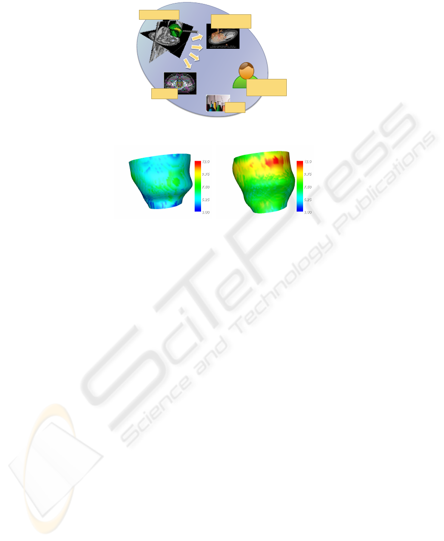
Fig. 4. Wall thickness at end diastole and systole, shown as a property function for the epicardial
surface.
Notice that the above applications demand for the management of scope-sensitive
queries, as defined in Section 5. Of course, if a sufficiently rich dataset is available, such
procedures may also take into account patient demographic/anamnesis information, to-
ward a more personalized treatment.
6.3 Cardiology
The analysis of the shape of the heart and of its deformation pattern may be used to sup-
port case-based reasoning procedures for diagnosis of several cardiological pathologies
and for preoperative planning. For example, we consider here ventricular dyssynchrony.
Dyssynchrony is a complex phenomenon whose origins are to be tracked back to elec-
trical conduction disturbances that affect both regional and global function of the heart
and which results into incoordinate ventricular wall motion due to activation delay. De-
spite its relevance, the only dyssynchrony marker that has received some consensus is
an ECG-derived parameter, which however is poorly correlated to the outcome of resyn-
chronization therapy, usually consisting in pacing device implantation [24]. Since there
is a lack of generally accepted evidence about dyssynchrony therapy, the retrieval of
similar cases from a repository may help the physician to make appropriate therapeutic
choices. Indeed, after having reconstructed a time-resolved model of the heart of the
patient and having computed meaningful features (like activation time, wall motion and
thickening; see e.g. Figure 4), the patient anatomy, enriched possibly with demographic
and drug assumption information, may be compared with other patients which already
underwent resynchronization therapy.
81

7 Conclusions and Further Work
In this paper, a framework for the description of anatomical structures has been pre-
sented. The framework takes into account 1) topological and geometrical features and
2) semantic annotation. In this way, a 3D model representing an anatomical structure
may be extended with non-geometrical pieces of information, ranging from metadata
inherent to the instance of the structure (such as textual description of a lesion) to other
patient-specific data (regarding age, sex, anamnesis,. . . ).
Possible hybrid methods for similarity searches are then envisaged and shown to be
useful for refined scope-sensitive retrieval. In this way, enhanced support for case-based
reasoning procedures is provided, as illustrated with three sample scenarios extracted
from various medical application fields.
In the future, we plan to integrate this work with other already developed infras-
tructures. Indeed, the presented framework may happily coexist with the system pre-
sented in [21], which introduces and uses an ontology for shape descriptors that –after
a suitable extension– may be profitable also for the scopes of this paper. Another im-
provement towards a more ontology-centered framework is seen in the exploitation of
the algorithm ontology presented in [25], so as to provide semantic insight also in the
feature extraction process.
Acknowledgements
This work was partially supported by European Project HEARTFAID “A knowledge
based platform of services for supporting medical-clinical management of the heart
failure within the elderly population”(IST-2005-027107).
References
1. Keim, D.: Efficient geometry-based silmilarity search of 3D spacial databases. In: Proceed-
ings of the 1999 ACM SIGMOID, New York, ACM Press (1999) 419–430
2. Di Bona, S., Lutzemberger, L., Salvetti, O.: A simulation model for analyzing brain struc-
tures deformations. Physics in Medicine and Biology 48 (2003) 4001–4022
3. Moroni, D., Colantonio, S., Salvetti, M., Salvetti, O.: Shape modeling for the analysis of
heart deformation patterns. In Gurevich, I., Niemann, H., Salvetti, O., eds.: First Interna-
tional Workshop on Image Mining: Theory and Applications (IMTA 2008). INSTICC press,
Funchal, Madeira (2008) 37–50
4. Bustos, B., Keim, D.A., Saupe, D., Schreck, T., Vranic, D.V.: Feature-based similarity search
in 3d object databases. ACM Comput. Surv. 37 (2005) 345–387
5. Grenander, U., Miller, M.I.: Computational anatomy: an emerging discipline. Q. Appl. Math.
LVI (1998) 617–694
6. Colantonio, S., Moroni, D., Salvetti, O.: Shape comparison and deformation analysis in
biomedical applications. In: Eurographics Italian Chapter Conference. (2006) 37–43
7. Moroni, D., Perner, P., Salvetti, O.: A general approach to shape characterisation for biomed-
ical problems. International Journal of Signal and Imaging Systems Engineering 1 (2008)
30–35
8. Koenderink, J.J., van Doorn, A.J.: Surface shape and curvature scales. Image Vision Comput.
10 (1992) 557–564
82

9. Akg
¨
ul, C.B., Sankur, B., Schmitt, F., Yemez, Y.: Multivariate density-based 3d shape de-
scriptors. In: Shape Modeling International. (2007) 3–12
10. Matheron, G.: Examples of topological properties of skeletons & On the negligibility of the
skeleton and the absolute continuity of erosions. In: Image Analysis and Math. Morphology:
Theoretical Advances. Volume 2. Academic Press (1988) 216 256.
11. Blum, H.: A transformation for extracting new descriptions of shape. In: Computer Methods
in Images Analysis. (1977) 153–171
12. Blum, H., Nagel, R.: Shape description using weighted symmetric axis features. Pattern
Recognition 10 (1978) 167–180
13. Biasotti, S., Giorgi, D., Spagnuolo, M., Falcidieno, B.: Reeb graphs for shape analysis and
applications. Theor. Comput. Sci. 392 (2008) 5–22
14. Edelsbrunner, H., Harer, J.: Persistent homology — a survey. In Twenty Years After, eds. J.
E. Goodman, J. Pach and R. Pollack, AMS. (2007)
15. Collins, A., Zomorodian, A., Carlsson, G., Guibas, L.: A barcode shape descriptor for curve
point cloud data. Computers and Graphics 28 (2004) 881–894
16. Moroni, D., Salvetti, M., Salvetti, O.: Multi-scale representation and persistency for shape
description. In Perner, P., Salvetti, O., eds.: MDA. Volume 5108 of Lecture Notes in Com-
puter Science., Springer (2008) 123–138
17. Biasotti, S., Giorgi, D., Spagnuolo, M., Falcidieno, B.: Size functions for comparing 3D
models. Pattern Recognition (2008)
18. Gruber, T.R.: A translation approach to portable ontology specifications. Knowledge Acqui-
sition 6 (1993) 199–221
19. Rosse, C., Jos
´
e L. V. Mejino, J.: A reference ontology for biomedical informatics: the foun-
dational model of anatomy. J. of Biomedical Informatics 36 (2003) 478–500
20. UMLS: http://www.nlm.nih.gov/research/umls/. (2008)
21. Colantonio, S., Salvetti, O., Tampucci, M.: An infrastructure for mining medical multimedia
data. In Perner, P., ed.: ICDM. Volume 5077 of Lecture Notes in Computer Science., Springer
(2008) 102–113
22. Kretschmann, H., Weinrich, W.: Neuroanatomy and Cranial Computed Tomography. Thieme
(1986)
23. Censor, Y., Jiang, M., Louis, A., eds.: Mathematical Methods in Biomedical Imaging and
Intensity-Modulated Radiation Therapy (IMRT). Edizioni della Normale (2008)
24. Boyer, K.L., Gotardo, P.F.U., Saltz, J.H., Raman, S.V.: On the detection of intra-ventricular
dyssynchrony in the left ventricle from routine cardiac MRI. In: ISBI. (2006) 169–172
25. Colantonio, S., Gurevich, I.B., Martinelli, M., Salvetti, O., Trusova, Y.: Thesaurus-based
ontology on image analysis. In Falcidieno, B., Spagnuolo, M., Avrithis, Y.S., Kompatsiaris,
I., Buitelaar, P., eds.: SAMT. Volume 4816 of Lecture Notes in Computer Science., Springer
(2007) 113–116
83
