
INTEGRITY AND AUTHENTICITY OF QUALITY ASSURANCE
AND CONTROL IN AN IMAGING EXAMINATION WORKFLOW
Chung-Yueh Lien
Institute of Biomedical Engineering, National YangMing University
No. 155, Sec. 2, Linong st., Beitou District, Taipei 112, Taiwan
Chia-Hung Hsiao
Department of Medical Informatics, TzuChi University, Hualien City, Taiwan
Tsung-Lung Yang
Department of Radiology, Kaohsiung Veterans General Hospital, Kaohsiung City, Taiwan
Tsair Kao
Department of Biomedical Engineering, Hungkuang University, Taichung, Taiwan
Keywords: Security, DICOM, Digital Signature, Key Object Selection, Modality Performed Procedure Steps, PACS.
Abstract: In this paper, we evaluated the implementation of a digital signature for medical imaging quality assurance
and control (QA/C) by a technician in accordance with the Digital Image and Communication in Medicine
(DICOM). After QA/C, a set of DICOM images were collected into a DICOM Key Object Selection (KOS)
document with digital signatures. The digital signature was implemented by RSA public-key cryptography
combined with a public-key health certificate and health professional card (HPC) to digitally sign a series of
DICOM images. Our method includes the DICOM Modality Performed Procedure Steps (MPPS)
mechanism that assures the image transmission completeness and accuracy in an image examination
workflow. The results show that the method is more efficient and requires less loading time to create the
technician’s signature in an imaging examination workflow.
1 INTRODUCTION
In the past decade, many institutions have
accommodated the increased variety of imaging
modalities and their communication protocols in
accordance with Digital Image and Communication
in Medicine (DICOM) to create a filmless
environment. Medical imaging quality assurance
and control (QA/C) is a key factor for a high-quality
filmless medical environment. Ensuring quality of
services for medical image transmission among
modality, QA/C station, and image archive has
become an important issue. The DICOM storage
commitment service allows the modality to verify
that images have been sent to an image archive
before deleting the images locally. Image loss can
happen when a technician does not check to see
whether the image has been sent to the QA/C site.
With the DICOM Modality Performed Procedure
Step (MPPS) service, the image transmission
completeness and accuracy in an imaging
examination workflow are assured (Moore 2003;
Noumeir 2005). Physicians will have more
confidence in using a picture-archiving and
communication system (PACS) with MPPS.
Security protection is a necessary requirement in
a filmless environment. DICOM has also adopted
public-key cryptography for protecting medical
images transmitted in PACS (ACM-NEMA 2009;
Schüze et al. 2004). Based on public-key
infrastructure (PKI), mechanisms needed to comply
with medical information security regulations could
155
Lien C., Hsiao C., Yang T. and Kao T. (2010).
INTEGRITY AND AUTHENTICITY OF QUALITY ASSURANCE AND CONTROL IN AN IMAGING EXAMINATION WORKFLOW.
In Proceedings of the Third International Conference on Health Informatics, pages 155-158
DOI: 10.5220/0002689501550158
Copyright
c
SciTePress
be implemented (Brandner et al. 2002; Cao et al.
2003). In PACS, digital-signature technology can be
implemented to assure the integrity of medical
images and to authenticate the operators in the
workflow of medical image examination (Brandner
et al. 2002). The report showed that the use of a
digital signature can reduce time needed for
reporting, thereby increasing efficiency (Lepanto et
al. 2003).
DICOM defined a specific instruction regarding
digital signatures in DICOM Part 15: Security and
System Management Profiles (ACM-NEMA 2009).
However, most digital signatures deal only with
single images and do not provide a satisfactory
solution for multiple images (Kobayashi et al. 2009).
The DICOM Supplement 86 offered a solution by
creating the digital signature in structured reports
(SR) with selected images. In this paper, we propose
a novel approach to assuring the integrity and
authenticity of QA/C in an imaging examination
workflow. Using the MPPS mechanism, the QA/C
site receives a complete set of DICOM images
created from a certain modality and creates a signed
key object selection (KOS) document with secure
references to all of the DICOM images that
comprise the examination.
2 METHODS
A general description of a modality acquisition
system consists of modality, QA/C site, MPPS
manager, and image archive (Figure 1). The
modality receives an imaging request from the
modality worklist server. After imaging, the
modality generates an MPPS list and then forwards
the list to the MPPS manager. The MPPS manager
uses the list to record the status of each modality.
The QA/C site receives the images transmitted from
modality and checks the MPPS status to assure that
the transformation of images has been completed. A
technician can manually read and adjust the
examination data such as the number of images, and
the window level at the QA/C site.
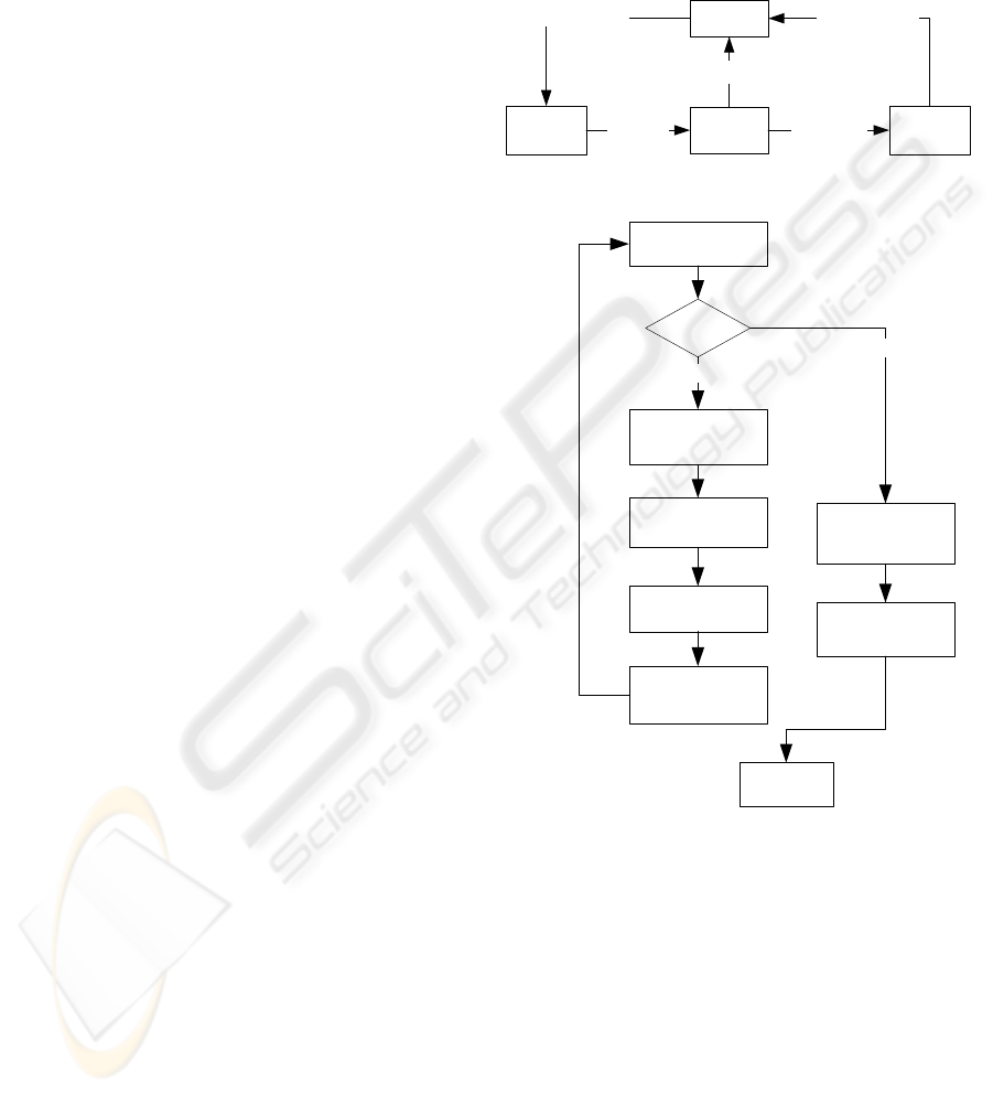
Figure 2 shows the flowchart of the digital
signature of the DICOM KOS document by
technician at the QA/C site. A DICOM KOS
document consists of the signed information of all
images, and the QA/C technician will digitally sign
the DICOM KOS document with the QA/C
signature. If images are transferred completely, the
image archive will update the MPPS status to the
MPPS manager as “COMPLETE,” and the MPPS
status of modality also will be updated; images are
deleted from the cache of modality consequently.
The mechanism of MPPS ensures the completeness
of image transmission. After transmission is
complete, the technician creates the QA/C digital
signature for the QA/C result. The images and
signatures are forwarded to the image archive.
Modality QA/C SiteSend images
MPPS
Manager
Update MPPPS Status
Image Archive
Send Images &
Signature
Update MPPS Status
Check MPPPS Status
Figure 1: System overview.
Check MPPS status
Receive images
Is finish ?
NO
Create DICOM
KOS signature
YES
Send signature to
image archive
Create secure
references
send images to image
archive
Operate QA/C
Finish
Figure 2: The block diagram of a DICOM KOS document
generation that is digitally signed by technician at the
QA/C site.
2.1 Digital Signature of DICOM KOS
Document
After the QA/C site receives all of the images from
the modality, the MAC (message authentication
code) references of all of the images are also
collected into a set of DICOM data objects in a
DICOM KOS document. In order to increase the
signing performance at the QA/C site, we did not
directly sign all images, but we indirectly signed a
DICOM KOS document, which also protects the
HEALTHINF 2010 - International Conference on Health Informatics
156
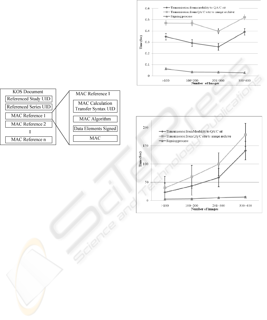
data integrity. The DICOM KOS document contains
multiple MAC references (Figure 3). The MAC
reference contains four attributes that represent
signed information for each image: 1) MAC
Calculation Transfer Syntax UID presents the
encode type of MAC; 2) MAC algorithm presents
the algorithm used in generating the MAC; 3) Data
Elements Signed presents a list of data element tags
in the order they appear at the top level of the
referenced image to identify the signed range; and 4)
the MAC presents the digest value of the referenced
image. The digest value is calculated by a hash
function that creates a “digital fingerprint” of an
image. The DICOM supports three hash algorithms:
RIPEMD-160, MD5, and SHA-1.
Figure 3: The architecture of a DICOM KOS document
containing n MAC references.
2.2 Data Elements Signed
The attribute “Data Elements Signed” is used to
select the range data elements to be included with
the image before signing. In accordance with
different departmental policies and roles, the data
elements signed attributes can be adjusted and well-
defined, creating a customized information base
designed by each institution using the DICOM
digital signature system. Only the selected data
elements are created or modified by the signer
according to his/her responsibility.
3 RESULTS
For this evaluation, all attributes of a DICOM-
formatted image were selected to be included in the
digital signature. The hash algorithm is SHA1 with a
160-bit output. The digital signature was
implemented by RSA public-key cryptography
combined with a public-key health certificate and
health professional card (HPC). The result of MAC
calculation was stored as a MAC Parameters
Sequence (4FFE, 0001), and the digital signature
was stored in as a Digital Signature Sequence
(FFFA, FFFA), as defined in DICOM part 15.
Figure 4: Time required per image for image transmission
among modality, QA/C site, image archive, and signing
process at the QA/C site.
Figure 5: Time required per exam for image transmission
among modality, QA/C site, image archive, and signing
process at the QA/C site.
Fifty-three CT examinations were recorded at the
QA/C site. The number of images for each
examination ranged from 30 to 394. Two CT
scanners (Toshiba Aquilion 64 and Siemens
Sensation 16) support MPPS and automatic DICOM
transfer. The image archive was installed on a PC in
the department of radiology. All stations were
connected via 100 MB-based Ethernet to the PACS.
Figure 4 illustrates the time required per image
for image examination. Figure 5 illustrates the time
dependence on the number of images in an
examination. We calculated the percentage of time
spent on each transition in an imaging examination
workflow. The average percentage of signing time
was 5.12±1.73%; transmission from modality to
QA/C site was 38.55±10.22%; and transmission
INTEGRITY AND AUTHENTICITY OF QUALITY ASSURANCE AND CONTROL IN AN IMAGING
EXAMINATION WORKFLOW
157

from QA/C site to image archive was
56.33±10.57%, respectively.
4 DISCUSSION
The delivery time of images is an important issue in
the department of radiology. Several steps are
involved: The images are created by a modality and
the image is transferred to the QA/C site. The
technician performs QA/C of the images, combines
the images with the study, then sends the images to
be stored to the image archive. The introduction of
digital signatures should avoid much extra loading
time in a normal workflow. Although the signing
time increases depending on the number of images,
the percentage of time spent loading is still less. The
impact of digital signatures in an imaging
examination workflow was significant in our
evaluation.
The implementation of digital signatures in
DICOM is not yet widespread. The main reason is
that the public-key infrastructures are not well
accepted in the domain of healthcare. Several
hospitals have followed the DICOM security profile
to sign medical images in their systems. However, it
is difficult to use the recommended DICOM
signature specification in the workflow of image
examination. It is not necessary to sign each image
in a study, which reduces the signing time.
Specifically, the technician can sign only one image
using DICOM KOS document while inserting secure
references into all of the DICOM images that
comprise one examination. The results of the present
study show that this method is more efficient and
requires less loading time to create the technician’s
signature.
5 CONCLUSIONS
In PACS, the security protection of medical images
is very important. Although the DICOM regulates
the digital signature for a single image, it can be
improved for implementation in an imaging
examination workflow. The implementation of
digital signatures for QA/C by a technician
following the DICOM Supplement 86 with MPPS
mechanism offers a satisfactory solution for multiple
images. These results show that this method is more
efficient and requires less extra load to create the
technician’s signature.
ACKNOWLEDGEMENTS
This work was supported by the National Science
Council of Taiwan under Grant NSC 97-2114-E-
010-002. The authors would like to acknowledge the
technical support provided by Mr. Wei-Chung Chen
of Department of Radiology, Kaohsiung Veterans
General Hospital.
REFERENCES
ACM-NEMA, 2009. Digital Imaging and
Communications in Medicine [online]. Available from:
http://medical.nema.org/dicom/ [Accessed 11 July
2009].
Brandner, R., M. Van der Haak, et al., 2002. Electronic
signature for medical documents - Integration and
evaluation of a public key infrastructure in hospitals.
Methods of Information in Medicine 41(4), 321-330.
Cao, F., H. K. Huang, et al., 2003. Medical image security
in a HIPAA mandated PACS environment.
Computerized Medical Imaging and Graphics 27(2-3):
185-196.
Kobayashi, L., Furuie S., et al., 2009. Providing Integrity
and Authenticity in DICOM Images: a Novel
Approach. IEEE Transactions on Information
Technology in Biomedicine 13(4), 582-589.
Lepanto, L., 2003. Impact of Electronic Signature on
Radiology Report Turnaround Time. Journal of
Digital Imaging 16(3), 306-309.
Moore, S. M., 2003. Using the IHE Scheduled Work Flow
Integration Profile to Drive Modality Efficiency.
Radiographics 23(2), 523-529.
Noumeir, R., 2005. Benefits of the DICOM Modality
Performed Procedure Step. Journal of Digital Imaging
18(4), 260-269.
Schüze, B., Kroll, M., et al., 2004. Patient data security in
the DICOM standard. European Journal of Radiology
51(3), 286-289.
HEALTHINF 2010 - International Conference on Health Informatics
158
