
FREQUENCY DOMAIN ANALYSIS AS RISK PREDICTOR
OF SUDDEN CARDIAC DEATH FROM LONG-TIME
ECG RECORDINGS
Diego Garc
´
ıa, C
´
esar S
´
anchez and Ra
´
ul Alcaraz
Innovation in Bioengineering Research Group, University of Castilla-La Mancha, Cuenca, Spain
Keywords:
Sudden cardiac death, Signal processing, Electrocardiogram, Frequency analysis.
Abstract:
Sudden Cardiac Death (SCD) is a disease that may not only affect patients with cardiovascular pathologies,
but also to apparently healthy patients. Thereby, identification of patients with a high potential of suffering
SCD is crucial for their treatment with adequate therapies. To this respect, in the present work, different
signal processing tools were applied to surface electrocardiographic (ECG) recordings to develop markers
which can clearly differentiate between subjects without cardiovascular pathologies and patients who died of
SCD. Precisely, the proposed indexes were the Spectral Concentration (SC) around the main frequency peak,
which reached a sensitivity of 100.00% and a specificity of 88.89%, and the Mean Frequency Distance (MFD)
between the first spectral peaks, which provided a sensitivity of 95.00% and a specificity of 100.00%.
1 INTRODUCTION
Sudden cardiac death (SCD) is nowadays a surprising
episode that causes death to 350,000 people per year
only in the USA (Al-Khatib et al., 2007). Accord-
ing to recent statistics, it is one of the main causes of
death for developed countries in apparently healthy
population (Chugh et al., 2000). Moreover, 50% of
deaths in patients with cardiovascular pathologies suf-
fered SCD (Al-Khatib et al., 2007). Only 5% of
patients who experienced SCD survived (Al-Khatib
et al., 2007), but many of them could have been saved
by cardioversion, especially those for which SCD was
the first symptom.
Hence, risk stratification is a determining factor to
reduce sudden cardiac mortality. To this respect, sev-
eral medical diagnosis techniques are currently used.
Thus, a low left ventricular ejection fraction (≤ 35 %)
is the gold standard, and subjects with NYHA class
II and III symptoms are at higher risk for SCD (Al-
Khatib et al., 2007). However, these methods have a
low predictive capacity (Arya et al., 2006). Thereby,
during the last years, many new lines of investigation
have been developed to find effective markers which
are able to predict SCD with anticipation. Most of the
recently proposed predictors are based on the time do-
main analysis, such as QRS duration, QT dispersion,
ST abnormalities, T-wave alternans, late potentials,
or heart rate variability complexity (Al-Khatib et al.,
2007; Engel et al., 2004). Nevertheless, these indica-
tors have also shown a low diagnostic accuracy (Durin
et al., 2008). Overall, in this work, two promising in-
dexes, developed from the frequency domain analy-
sis of ECG signals, are proposed to detect subjects at
high risk.
2 MATERIALS
For the study, two databases available from Phys-
ioBank (Goldberger et al., 2000) were used: the MIT-
BIH Normal Sinus Rhythm (NSRDB) and Sudden
Cardiac Death Holter (SCDHDB). The first set in-
cludes ECG recordings, with a length between 20 and
24 hours, belonging to five men aged 26 to 45 and
thirteen women aged 20 to 50. They are referred to
the Arrhythmia Laboratory at Beth Israel Deaconess
Medical Center, and were found not to have signifi-
cant arrhythmias. This group of 18 ECG recordings
of normal sinus rhythm was considered as reference.
The second database contains 23 Holter signals asso-
ciated to patients who suffered SCD during the 24-h
monitoring. They were mainly obtained in the 1980’s
in Boston area hospitals. In the analysis, 20 record-
ings were only considered, given that paced patients
were discarded.
424
García D., Sánchez C. and Alcaraz R. (2010).
FREQUENCY DOMAIN ANALYSIS AS RISK PREDICTOR OF SUDDEN CARDIAC DEATH FROM LONG-TIME ECG RECORDINGS.
In Proceedings of the Third International Conference on Bio-inspired Systems and Signal Processing, pages 424-427
DOI: 10.5220/0002742604240427
Copyright
c
SciTePress

3 METHODOLOGY
To predict SCD, the 60 minutes preceding ventricu-
lar fibrillation (VF) were taken from each SCDHDB
recording. For the NSRDB signals, 60 minutes start-
ing at an random instant were selected. In order to
unify the sampling rates of the considered segments,
the first lead was resampled at 1 kHz. Addition-
ally, to improve later analysis, interferences present
in the ECG were removed with four filters (S
¨
ornmo
and Laguna, 2005): an 8th-order Chebyshev low-pass
filter ( f
o
= 100Hz), a 3rd-order Butterworth high-
pass filter ( f
o
= 0.50Hz), a 2nd-order IIR notch fil-
ter ( f
c
= 60Hz), and an envelope detector for baseline
wander subtraction.
The power spectral density (PSD) of each chosen
segment was computed over 4096 samples-length in-
tervals, obtained with a Hamming window and over-
lapped 2048 samples, making use of an 8192-points
Fast Fourier Transform (FFT). Finally, the Spectral
Concentration (SC) around the main frequency peak
and the mean frequency distance (MFD) between the
N first were computed for each interval, thus obtain-
ing two numerical series.
3.1 SC Around the Main Frequency
Peak
The Spectral Concentration, SC[x(t)], around the
main frequency peak, f
p
, has been previously used
as a performance indicator of the atrial activity ex-
traction in atrial fibrillation and tachyarrhythmias
episodes (S
´
anchez et al., 2004; Castells et al., 2005).
It has been defined as:
SC[x(t)] =
1,17· f
p
∑
f =0,82·f
p
x
P
( f )
f
s
/2
∑
f =0
x
P
( f )
(1)
where f
s
is the sampling rate of the analyzed segment,
x(t) and x
P
( f ) is its PSD: x
P
( f ) = ℜ
2
DFT {x(t)}
.
This index relates present energy on the peak band to
the rest of the distribution of interest. In this sense,
an ideal sinus signal has a SC of 1, because all spec-
tral content is on its own oscillation frequency. Simi-
larly, ECG signal with a ventricular flutter presents a
SC value very high, because this arrhythmic episode
is highly regular (Castells et al., 2005). On the con-
trary, low SC values means that only an small part of
the PSD is concentrated around main frequency peak,
which suggests the existence of other notable peaks in
the rest of the spectrum.
3.2 MFD between the First Spectral
Peaks
The MFD[x(t),N] is defined as the mean frequency
distance between the N first peaks of the PSD. Its
computation requires a smooth spectrum to determine
with high accuracy the frequency peaks. Because of
the PSD provided by Fast Fourier Transform (FFT)
shows transitory peaks that may distort the index out-
come, a smoothing spectrum technique consisting in
a low-pass filter with a 40 samples-length Hamming
window was applied (Proakis and Manolakis, 2007).
This parameter can be used to estimate the PSD dis-
persion. Thus, a low value implies close frequency
peaks and, therefore, an concentrated spectrum. On
the contrary, a high value indicates a high dispersion
and a PSD with considerable empty gaps between fre-
quency peaks.
4 RESULTS
Table 1 shows the obtained SC and MFD values for
all the recordings. To associate every ECG with an
only value, the mean for the time series provided by
each parameter was computed. The differences be-
tween the values obtained for the two groups, see
Fig. 1, were statistically significant, given that the sig-
nificance level, obtained by a t Student test, was p <
0.0001 for both markers. Receiver Operating Charac-
teristics (ROC) curve was used to obtain the discrim-
ination threshold between healthy patients and those
who died by SCD (Lasko et al., 2005). In addition,
the predictive ability of both indicators was also ob-
tained with this curve. Thus, sensitivity was consid-
ered as the number of healthy patients correctly clas-
sified, whereas specificity represented the percentage
of patients who died by SCD correctly discerned. Fig
2 shows the ROC curves for the two makers, and Ta-
ble 2 presents their values obtained of sensitivity and
specificity.
As can be appreciated, the SC revealed only two
falses positives (signals #19140 and #19093) with an
area under ROC curve (AUC) of 0.9694 and an ac-
curacy of 94.74% (36 out of 38). WIth regard to the
MFD, only one false negative (signal #39), an AUC
of 0.9944 and an accuracy of 97.37% (37 out of 38)
were provided. Since the failures obtained with both
tests were associated to different signals, a combin-
ing discriminator considering a positive result with
SC > 16.25% and MFD > 3.75Hz, would have an ac-
curacy of 100% (38 out of 38).
FREQUENCY DOMAIN ANALYSIS AS RISK PREDICTOR OF SUDDEN CARDIAC DEATH FROM LONG-TIME
ECG RECORDINGS
425

Table 1: SC and MFD results for all the analyzed record-
ings.
NSRDB SCDHDB
Signal SC MFD Signal SC MFD
# (%) (Hz) # (%) (Hz)
16265 11.9 2.704 30 26.1 5.848
16272 10.1 2.289 31 27.0 5.070
16273 12.2 3.404 32 49.4 5.747
16420 14.0 1.719 33 20.5 4.798
16483 11.5 2.423 34 16.3 5.960
16539 10.5 1.988 35 27.0 4.327
16773 12.7 3.070 36 19.1 5.514
16786 12.9 3.705 37 25.1 4.453
16795 14.9 2.917 38 67.7 4.523
17052 11.3 1.656 39 25.8 3.788
17453 16.1 3.340 41 34.5 5.154
18177 12.0 2.428 43 22.3 3.777
18184 12.1 1.729 44 16.3 5.022
19088 12.7 2.722 45 16.9 4.619
19090 10.9 2.228 46 16.4 3.952
19093 18.4 2.068 47 37.8 4.294
19140 22.2 3.661 48 24.0 4.428
19830 16.2 1.901 50 24.5 4.967
51 33.8 4.314
52 20.9 4.214
Mean 13.5 2.553 Mean 27.5 4.724
Std 3.1 0.674 Std 8.2 0.685
(a) SC (b) MFD
Figure 1: SC and MFD for NSRDB and SCDHDB (‘box-
and-whiskers’ plot).
4.1 Numerical Evolution of Markers
Given that results showed that SCD could be suc-
cessfully predicted with the SC and MFD, both pa-
rameters were computed for a longer time interval.
Thus, the 300 minutes preceding VF from 13 SCD-
HDB were selected. In addition, for all the NSRDB
signals, 300 minutes starting at a random instant were
also chosen. For this case, the time course provided
Table 2: Area Under ROC Curve (AUC), threshold, sensi-
tivity and specificity for SC and MFD tests.
AUC Threshold Sensitivity Specificity
SC 0.9694 16.25 % 100.00% 88.89%
MFD 0.9944 3.750 Hz 95.00% 100.00%
Figure 2: ROC Curves.
Figure 3: SC numerical evolution.
Figure 4: MFD numerical evolution.
BIOSIGNALS 2010 - International Conference on Bio-inspired Systems and Signal Processing
426
by both markers is displayed in Figs. 3 and 4.
5 DISCUSSION AND
CONCLUSIONS
In the present work spectral processing has been used
to obtain two indexes that reveal statistically signifi-
cant differences (p < 0.0001) between patients who
suffered SCD and a sample of completely healthy
subjects.
Both parameters showed values substantially
higher for patients who suffered SCD than for healthy
subjects. This fact suggests that the ECG of healthy
subjects is characterized by a higher harmonic con-
tent. Consequently, it could be considered that fre-
quency peaks of relative high amplitude in the ECG
of healthy patients are disappeared in subjects who
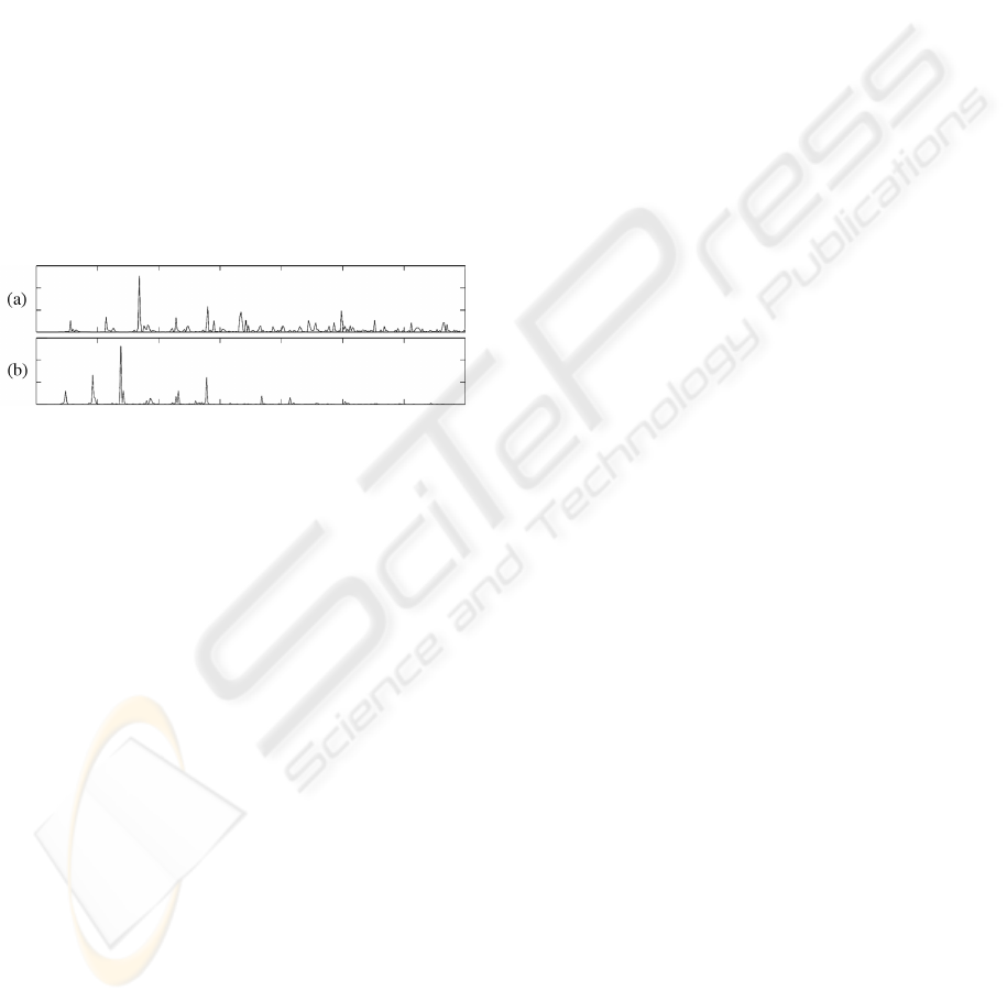
suffered SCD, such as Fig. 5 shows.
Figure 5: Typical PSD of an ECG for the (a) NSRDB and
(b) SCDHDB.
On the other hand, the time course of the markers
showed very constant values during the last 5 previous
hours preceding the death of the patient. Therefore,
SCD could be predicted with an anticipation above 5
hours, which suggests the problem that causes SCD,
it can be congenital or acquired during subject’s life.
Finally, given that the analyzed database are lim-
ited, the results should be considered with caution.
Nevertheless, the work suggests that SC and MFD can
initiate new lines of research as non-invasive predic-
tors of SCD. In this sense, a wider data set allowing
a more rigorous statistical analysis should be required
in order to provide confidence in the robustness of the
proposed parameters.
ACKNOWLEDGEMENTS
This work was supported by the projects PII1C09-
0036-3237 and PII2C09-0224-5983 from Junta de
Comunidades de Castilla-La Mancha.
The authors would like to acknowledge the invalu-
able helpful support received from Dr. J.L. Bardaj
´
ı,
Dr. M.L. L
´
opez, Dr. F. Madero, and Eng. M.E.
Garc
´
ıa.
REFERENCES
Al-Khatib, S. M., Sanders, G. D., Bigger, J. T., Buxton,
A. E., and et al (2007). Preventing tomorrow’s sud-
den cardiac death today: part i: Current data on risk
stratification for sudden cardiac death. Am Heart J,
153(6):941–950.
Arya, A., Haghjoo, M., and Sadr-Ameli, M. A. (2006). Risk
stratification for arrhythmic death after myocardial in-
farction: current perspective and future direction. Int
J Cardiol, 108(2):155–164.
Castells, F., Rieta, J. J., Millet, J., Zarzoso, V., and Asso-
ciate (2005). Spatiotemporal blind source separation
approach to atrial activity estimation in atrial tach-
yarrhythmias. IEEE Trans Biomed Eng, 52(2):258–
267.
Chugh, S. S., Kelly, K. L., and Titus, J. L. (2000). Sudden
cardiac death with apparently normal heart. Circula-
tion, 102(6):649–654.
Durin, O., Pedrinazzi, C., Donato, G., Pizzi, R., and Inama,
G. (2008). Usefulness of nonlinear analysis of ecg
signals for prediction of inducibility of sustained ven-
tricular tachycardia by programmed ventricular stim-
ulation in patients with complex spontaneous ventric-
ular arrhythmias. Ann Noninvasive Electrocardiol,
13(3):219–227.
Engel, G., Beckerman, J. G., Froelicher, V. F., Yamazaki,
T., and et al (2004). Electrocardiographic arrhythmia
risk testing. Curr Probl Cardiol, 29(7):365–432.
Goldberger, A. L., Amaral, L. A., Glass, L., Hausdorff,
J. M., Ivanov, P. C., and et al (2000). Physiobank,
physiotoolkit, and physionet: Components of a new
research resource for complex physiologic signals.
Circulation, 101(23):E215–E220.
Lasko, T. A., Bhagwat, J. G., Zou, K. H., and Ohno-
Machado, L. (2005). The use of receiver operat-
ing characteristic curves in biomedical informatics. J
Biomed Inform, 38(5):404–415.
Proakis, J. G. and Manolakis, D. K. (2007). Digital Signal
Processing. Principles, Algorithms and Applications.
Pretence Hall.
S
´
anchez, C., Rieta, J. J., Castells, F., Alcaraz, R., and Mil-
let, J. (2004). Wavelet domain blind signal separation
to analyze supraventricular arrhythmias from Holter
registers. Conf Proc Independent Component Analy-
sis and Blind Signal Separation, 5:1111–1117.
S
¨
ornmo, L. and Laguna, P. (2005). Bioelectrical Signal Pro-
cessing in Cardiac and Neurological Applications. El-
sevier Academic Press.
FREQUENCY DOMAIN ANALYSIS AS RISK PREDICTOR OF SUDDEN CARDIAC DEATH FROM LONG-TIME
ECG RECORDINGS
427
