
CEREBRAL CORRELATES OF THE CONTINOUS
PERFORMANCE TEST-IDENTICAL PAIRS VERSION
An fMRI Study
J. M. Serra-Grabulosa
Departament de Psiquiatria i Psicobiologia Clínica, Universitat de Barcelona, Barcelona, Spain
Institut d’Investigacions Biomèdiques August Pi i Sunyer (IDIBAPS), Barcelona, Spain
A. Adan
Departament de Psiquiatria i Psicobiologia Clínica, Universitat de Barcelona, Barcelona, Spain
C. Falcón
Institut d’Investigacions Biomèdiques August Pi i Sunyer (IDIBAPS), Barcelona, Spain
CIBER-BBN, Barcelona, Spain
N. Bargalló
Secció de Neuroradiologia, Servei de Radiologia, Centre de Diagnòstic per la Imatge (CDI)
Hospital Clínic de Barcelona, Barcelona, Spain
J. Solé-Casals
Digital Technologies Group, University of Vic, Vic, Spain
Keywords: CPT, CPT-IP, Sustained attention, fMRI, Prefrontal cortex, Parietal cortex.
Abstract: One of the most used paradigms in the study of the attention is the Continuous Performance Test (CPT).
The Identical Pairs version of the CPT (CPT-IP) has been used to evaluate attention deficits in
developmental, neurological and psychiatric disorders. Since both dyscalculia and ADHD (attention deficit
hyperactivity disorder) show attentional and numerical processing deficits, it would be interesting to
evaluate functional brain patterns related to the CPT-IP in a task which uses numerical stimuli. In this sense,
the aim of our study was to design a task to evaluate sustained attention using functional magnetic
resonance imaging. This task has to be sensitive to evaluate later dyscalculic and ADHD subjects. Forty
right-handed, healthy subjects (20 women; age range 18–25) were recruited to participate in the study. A
CPT-IP implemented as a block design was used to assess sustained attention in the fMRI session. Results
showed the CPT-IP task used activates a network of frontal, parietal and occipital areas and could be related
to executive, attentional and numerical processing functions.
1 INTRODUCTION
Sustained attention is the ability to maintain an
adequate status monitoring certain events or stimuli
prolonged in time. Among the various tests used to
evaluate sustained attention highlights the
Continuous Performance Test, Identical Pairs
Version (CPT-IP) (Cornblat, 1989), a serial visual
detection task where stimuli mainly require
sustained attention and working memory effort for
its realization. This task was designed initially to
detect attentional deficits in patients diagnosed with
schizophrenia or depression. Subsequently it has
491
M. Serra-Grabulosa J., Adan A., Falcón C., Bargalló N. and Solé-Casals J. (2010).
CEREBRAL CORRELATES OF THE CONTINOUS PERFORMANCE TEST-IDENTICAL PAIRS VERSION - An fMRI Study.
In Proceedings of the Third International Conference on Bio-inspired Systems and Signal Processing, pages 491-496
DOI: 10.5220/0002758604910496
Copyright
c
SciTePress

been used to study cognitive deficits in disorders
such as Alzheimer's (White and Levin, 1999),
Parkinson (Kelton et al., 2000), dyscalculia (Lindsay
et al., 2001) and specially with Attention Deficit
Hyperactivity Disorder (Hill et al., 2003).
Dyscalculia and ADHD have a high comorbidity
(25%) (Shalev et al., 1995; Barkley, 2003). Previous
studies indicate that dyscalculic and ADHD subjects
show attentional deficits. Specifically, dyscalculic
subjects show attentional deficits when number
stimuli are used (Lindsay et al., 2001; Passolunghi
and Siegel, 2001). However, until today there are no
neuroimaging studies addressed to evaluate
similarities in functional brain correlates of
attentional deficits in pure dyscalculic, ADHD-
dyscalculic, and ADHS subjects. For this purpose, it
would be necessary to design a CPT-IP fmri task
which uses numerical stimuli.
In this sense, the aim of our study is to design a task
to evaluate sustained attention using functional
magnetic resonance imaging. This task has to be
sensitive to evaluate dyscalculic and ADHD
subjects, evaluating attentional deficits related to
numerical stimuli. Moreover, it has to activate the
attentional network found previously (Posner and
DiGirolamo, 2000; Lawrence et al., 2003; Ogg et al.,
2008). Specifically, studies which used a CPT-IP
task found a pattern of activation that includes
prefrontal and parietal superior areas (Pardo et al.,
1991; Fan et al., 2005).
2 MATERIALS AND METHODS
2.1 Participants
Forty right-handed healthy undergraduate students
(20 women; age range 18–25, mean (±S.D.) 19.6
(±1.7)) were recruited from the University of
Barcelona. Subjects with chronic disorders, nervous
system disorders or history of mental illness were
excluded, as well as habitual drinkers and those on
medication. The study was approved by the ethics
committee of Hospital Clínic de Barcelona. Written
consent was obtained from all participants, who
were financially rewarded for taking part.
2.2 fMRI Procedure
The fMRI session started between 9 am and 9:30
am. Participants had to perform a series of
alternating CPT-IP and control tasks in a block
design. After an initial accommodation block of 35
seconds, 9 CPT-IP blocks were alternated with 9
control blocks. The CPT-IP task was a modification
of the Cornblatt task (Cornblatt et al., 1989), similar
to that described in Strakowski et al., (2004).
Specifically, in the CPT-IP task, subjects were
presented with a series of 27 four-digit numbers
(from 1 to 9 without repetition in the same number)
and were asked to respond by pressing a button as
faster as possible when the same number occurred
twice sequentially. In each CPT-IP block, only 4
numbers were repeated in relation to the previous
number. The control task consisted of the number ‘1
2 3 4’ presented at the same rate and intervals as the
CPT-IP to the subjects. The CPT-IP and control
tasks were given in alternating blocks of 20 s each
with numbers being presented for 450 ms at 750 ms
intervals. Thus, the duration of the acquisition
protocol was 8 min and 6 s and yielded 243 whole-
brain volumes. Instructions were displayed on the
screen for a period of 5 seconds before each CPT-IP
and control block. Stimulus presentation was
triggered by the MRI-scanner. The Presentation
program, version 0.76, (Neurobehavioral System,
USA) was used to develop the stimuli task. Prior to
the fMRI scanning, subjects were given instructions
and undertook a trial version of the task to ensure
they had understood.
2.3 MRI Acquisition
The study was performed in a 3 T MRI scanner
(Magnetom Trio Tim, Siemens Medical Systems,
Germany) at the Centre for Image Diagnosis of the
Hospital Clínic (CDIC) using the blood-oxygen
level-dependent (BOLD) fMRI signal. The MRI
protocol included an fMRI dataset of 243 volumes
of 36 axis slices each (using a gradient-echo echo-
planar imaging – EPI sequence) and a high-
resolution 3D structural dataset (T1-weighted
Magnetization Prepared Rapid Gradient Echo – MP-
RAGE image) for coregistering with the fMRI
images. The acquisition parameters for the fMRI
were: repetition time (TR) = 2000 msec; echo time
(TE) = 29 msec; percentage phase field of view =
100; matrix size = 128 × 128; slice thickness = 3.75
mm; interslice gap = 0.75 mm; flip angle = 90°. The
parameters for the structural images were: TR =
2300 msec, TE = 2.98 msec, inversion time (TI) =
900 msec; FOV = 25.6 × 25.6 cm; matrix size = 256
× 256; flip angle = 9°; slice thickness = 1 mm.
BIOSIGNALS 2010 - International Conference on Bio-inspired Systems and Signal Processing
492
2.4 Behavioural Data Analysis
Three different measures were obtained from the
CPT-IP task: accuracy (number of correctly
identified items, hits), false positives (the number of
incorrect “yes” responses, commissions) and the
number of omissions. Reaction time was also
measured by calculating the mean reaction time (in
milliseconds) for target stimuli.
2.5 fMRI Data Analysis
For image processing, Statistical Parametric
Mapping (SPM5, Wellcome Department of
Cognitive Neurology, London) was used. The
images of each subject were corrected for motion
and realigned to remove any minor motion-related
signal change. All volumes for each subject were
normalized into an EPI template supplied with
SPM5. During spatial normalization, all scans were
resampled to 2-mm
3
isotropic voxels. Low-
frequency noise was removed with a high-pass filter
(128 s) applied to the fMRI time series at each
voxel. Lastly, the images were smoothed with an 8
mm full-width half maximum (FWHM) Gaussian
filter.
Statistical analyses were first performed at a single-
subject level. A linear contrast was performed
comparing the activation during the CPT-IP blocks
and control blocks for each subject and fMRI
session. We then performed a “CPT-IP block >
control block” contrast to obtain the pattern of brain
activity reflecting sustained attention processes.
Analyses were performed considering all voxels
constituting the brain and results were interpreted at
a voxel level of P < 0.001 (uncorrected) considering
only clusters of ≥ 15 contiguous voxels at a
corrected P-value < 0.05 cluster level. The
anatomical location of the cerebral activated areas
was determined by the Montreal Neurological
Institute (MNI) coordinates.
3 RESULTS
3.1 Behavioural Measures
Behavioural results in the CPT-IP task showed a
good performance in all subjects. Specifically, in
number of hits (mean 28,47; s.d. = 5,45) (79% of
hits), comissions (mean = 10,03; s.d. = 4,00) and
omissions (mean = 1,21; s.d. = 0,74). Mean of
reaction time was 501,52 ms (s.d. = 57,74). No
gender differences were observed.
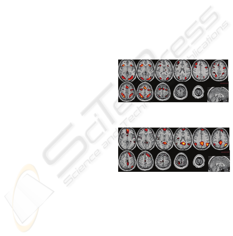
3.2 fMRI Results
The contrast ‘CPT-IP blocks > control blocks’ was
computed in each group to investigate task-related
effects. Activations were located (Fig. 1) bilaterally
in frontal (including ventral -BA 11-; dorsolateral -
BA 10, BA 46-; ventrolateral -BAs 44, 45-;
premotor -BA6- and the anterior cingulate cortex -
BA 32-); in parietal (BA 7) and occipital cortex (left
BA 18 and bilateral BA 19).
On the other hand, the contrast ‘control blocks >
CPT-IP blocks’ showed a pattern of bilateral
activation (Fig.2) in angular gyrus (BA 39); in
posterior cingulate gyrus (BA 23); in left frontal
gyrus (BA 10) and in inferior and medial temporal
gyrus (BA 21 and 20).
Figure 1: Areas of significantly greater brain activity on
the contrast “CPT-IP task vs. control task” (group fMRI
data).
Figure 2: Areas of significantly greater brain activity on
the contrast “control task vs. CPT-IP task” (group fMRI
data).
4 DISCUSSION
The aim of our study was to design a task to evaluate
sustained attention using functional magnetic
resonance imaging. This task would have to be
sensitive to evaluate dyscalculic and ADHD subjects
and could help to better understand common and
individual deficits. It is important, as discalculia can
be expressed as a pure and very specific
developmental disorder. But most of times,
CEREBRAL CORRELATES OF THE CONTINOUS PERFORMANCE TEST-IDENTICAL PAIRS VERSION - An fMRI
Study
493

dyscalculic people also show other cognitive
deficits, as working memory deficits, attention
deficits, spatial processing deficits or grapheme-
phoneme association deficits. In these cases, and
particularly in ADHD comorbidity cases, it is
necessary to investigate which cerebral regions are
related to the attention and working memory deficit,
searching for common and uncommon brain areas.
Until today, it is not well understood if dyscalculia
cognitive deficits are the result of a unique or
multiple pathophysiology (Rubinsten and Henik,
2009). Evaluation of these deficits by using an fMRI
approach could contribute to clarify it. Specifically,
CPT-IP could help to better understand neural
substrate of attention and working memory deficits
in dyscalculic and ADHD subjects.
In relation to our study, behavioural results showed a
good performance in all subjects. Since performance
was around 80%, it indicates that can be used to
discriminate between different levels of
performance.
Analysis of task-related effects indicated that the
CPT-IP task used activates a network of frontal,
parietal and occipital areas. This activation pattern is
similar to the activation pattern observed previously
in the evaluation of sustained attention (Pardo et al.,
1991; Coull et al., 1996; Casey et al., 2001;
Lawrence et al., 2003; Fan et al., 2005), and it has
been related to executive, attentional and numerical
processing functions.
The frontal activation obtained was bilateral,
including ventral (BA 11), dorsolateral (BA 10, BA
46), ventrolateral (BAs 44, 45), premotor (BA6) and
the anterior cingulate cortex (BA 32). Ventral and
lateral prefrontal areas would be related to executive
and working memory functions (Raz and Buhle,
2006; D’Esposito, 2008). In addition, activation of
Broca’s area (BA 44) could be related to the verbal
working memory processes involved in the CPT-IP
task, as it has been observed that this region is
activated by tasks which tax verbal working memory
incrementally (for a review see Grodzinsky and
Santi, 2008). Furthermore, the anterior cingulate
cortex (ACC) has been related to cognitive conflict
monitoring tasks, being important to facilitate
detection of the appropriate stimulus, while ignoring
others. Moreover, it has been related to error
detection and immediate-response re-adjustment
(Ridderinkhof et al., 2004; Raz and Buhle, 2006). In
cooperation with dorsolateral and ventral prefrontal
areas, the ACC has also been related to the
maintenance of numbers in working memory, thus
facilitating mental operations (Zago et al., 2008). In
our CPT-IP task, maintenance of numbers in
working memory was necessary, as the subjects
were asked to compare each number and decide if it
was the same as the previous one.
As in previous studies, bilateral parietal activation
was also observed in the CPT-IP task. Parietal
regions are related to both alerting and reorienting of
attention (Konrad et al., 2005; Raz and Buhle,
2006). They are also related to executive functions
such as allocation of attention and verbal working
memory processes, mediating the short-term storage
and retrieval of phonologically coded verbal
material (Jonides et al., 1998). Thus, parietal regions
could contribute to holding digits online during
verbal working memory tasks (Coull et al., 1996),
which was necessary in the CPT-IP task in our
study.
Another cluster of activation related to the CPT-IP
task was found in occipital areas, both in primary
and associative areas. As has been previously
reported (Ogg et al., 2008), this occipital activation
may reflect processes of analysis and identification
of the visually presented stimuli. Specifically in our
case, it may be reflecting the number processing
related to the CPT-IP task, as it has been observed
that the associative visual cortex contributes to
number identification (Dehaene et al., 2004;
Schmithorst and Brown, 2004) in addition to visual
letter (Vinckier et al., 2007), object (Grill-Spector et
al., 2001) and face recognition (Carlson et al., 2006).
In the control task, the activation of these visual
regions was minor, possibly due to the fact that the
recognition was easier as the stimulus was always
the same (‘1 2 3 4’).
On the other hand, the contrast ‘control blocks >
CPT-IP blocks’ showed activation in different brain
areas: angular gyrus, posterior cingulate gyrus, left
frontal gyrus and inferior and medial temporal
gyrus. These areas seems to have less metabolic
requirements in rest state (Lawrence et al., 2003;
Ogg et al., 2008), and have been found to be
deactivated when attentional effort to external
stimuli is needed (Raichle et al., 2001).
The significance of this deactivation is not
completely understood. However, it could reflect an
inhibition of processes that could interfere with the
correct execution of the task, as external and internal
monitoring. (Gusnard and Raichle, 2001). In this
sense, deactivation could optimize performance in
high attentional demanding tasks (McKiernan et al.,
2003).
BIOSIGNALS 2010 - International Conference on Bio-inspired Systems and Signal Processing
494

Finally, it is important to emphasize that there are
some limitations to our study. Firstly, introduction of
different degrees of difficulty in the CPT-IP task
might have given more sensitivity to our study.
Secondly, the use of multivariate analysis could
contribute to better delineate cerebral correlates of
sustained attention.
5 CONCLUSIONS
The CPT-IP task was associated with an attention
network, where activation corresponds with the
activations found in previous studies. This suggests
that the CPT-IP task was suitable for studying
sustained attention in dyscalculic and ADHD
subjects.
REFERENCES
Barkley, R. A., 2003. Issues in the diagnosis of attention-
deficit/hyperactivity disorder in children. Brain and
Development, 25, 7-83.
Carlson, T., Grol, M.J., Verstraten, F.A., 2006. Dynamics
of visual recognition revealed by fMRI. Neuroimage,
32 (2), 892-905.
Casey, B.J., Formans, S.D., Franzen, P., Berkowitz, A.,
Braver, T.S., Nystrom, L.E. et al., 2001. Sensitivity of
prefrontal cortex to changes in target probability: a
functional MRI study. Human Brain Mapping, 13 (1),
26-33.
Cornblatt, B.A., Lezenweger, M.F., Erlenmeyer-Kimling,
L., 1989. The Continuous Performance Test, Identical
Pairs Version: II. Contrasting attentional profiles in
schizophrenic and depressed patients. Psychiatry
Research, 29, 65–85.
Coull, J.T., Frith, C.D., Frackowiak, R.S.J., Grasby, P.M.,
1996. A fronto-parietal network for rapid visual
information processing: a PET study of sustained
attention and working memory. Neuropsychologia, 34,
1085-95.
D’Esposito, M. Working memory. (2008). En Handbook
of Clinical Neurology, Neuropsychology and
behavioral neurolgoy. G. Goldenberg i B. Miller
(Eds.).
Dehaene, S., Molko, N., Cohen, L., Wilson, A.J., 2004.
Arithmetic and the brain. Current Opinion in
Neurobiology, 14 (2), 218-24.
Fan, J., McCandliss, B. D., Fossella, J., Flombaum, J. I.,
Posner, M. I., 2005. The activation of attentional
networks. Neuroimage, 26, 471-9.
Grill-Spector, K., Kourtzi, Z., Kanwisher, N., 2001. The
lateral occipital complex and its role in object
recognition.Vision Research, 41(10-11), 1409-22.
Grodzinsky, Y., Santi, A., 2008. The battle for Broca's
region. Trends in Cognitive Sciences 12 (12), 474-80.
Gusnard, D.A., Raichle, M.E., 2001. Searching for a
baseline: Functional imaging and the resting human
brain. Nature Neuroscience Reviews, 2, 685-94.
Hill, D.E., Yeo, R.A., Campbell, R.A., Hart, B., Vigil, J,
Brooks, W., 2003. Magnetic ressonance imaging
correlates of Attention-Deficit/Hyperactivity Disorder
in children. Neuropsychology, 17 (3), 496-506.
Jonides, J.,Schumacher, E.H., Smith, E.E., Koeppe, R.A.,
Awh, E., Reuter-Lorentz, P.A., Marshuetz, C., Willis,
C.R., 1998. The role of parietal cortex in verbal
working memory. Journal of Neuroscience, 18, 5026-
34.
Kelton, M.C., Kahn, H.J., Conrath, C.L., Newhouse, P.,
2000. The effects of nicotine on Parkinson’s disease.
Brain and Cognition, 43, 274-82.
Konrad, K., Neufang, S., Thiel, C.M., Specht, K., Hanisch,
C., Fan, J., Herpertz-Dahlmann, B., Fink, G.R., 2005.
Development of attentional networks: an fMRI study
with children and adults. Neuroimage 28 (2), 429-39.
Lawrence, N.S., Ross, T.J., Hoffman, R., Garavan, H.,
Stein, E.A., 2003. Multiple neuronal networks mediate
sustained attention. Journal of Cognitive
Neuroscience, 15 (7), 1028-38.
Lindsay, R.L., Tomazic, T., Levine, M.D., Accardo, P.J.,
2001. Attentional function as measured by a continuos
performance task in children with dyscalculia. Journal
of Developmental and and Behavioral Pediatrics, 42
(8), 1049-56.
McKiernan, K.A., Kaufman, J.N., Kucera-Thompson, J.,
Binder, J.R., 2003. A parametric manipulation of
factors affecting task-induced deactivation in
functional neuroimaging. Journal of Cognitive
Neuroscience, 15, 394-408.
Ogg, R.J., Zou, P., Allen, D.N., Hutchins, S.B.,
Dutkiewicz, R.M., Mulhern, R.K., 2008. Neural
correlates of a clinical continuous performance test.
Magnetic Resonance Imaging, 26, 504-12.
Pardo, J.V., Fox, P.T., Raichle, M.E., 1991. Localization
of a human system for sustained attention by positron
emission tomography. Nature, 349, 61-4.
Passolunghi, M.C., Siegel, L.S., 2001. Short-term
memory, working memory, and inhibitory control in
children with difficulties in arithmetic problem
solving. Journal of Experimental Child Psychology,
80, 44-57.
Posner, M. I., DiGirolamo, G. J., 2000. Attention in
cognitive neuroscience: An overview. A Gazzaniga,
M. S. (Comp). The new cognitive neurosciences (pp
623-31). Cambridge: MIT Press.
Raichle, M.E., MacLeod, A.M., Snyder, A.Z., Powers,
W.J., Gusnard, D.A., Shulman, G.L., 2001. A default
mode of brain function. Proceedings of the National
Academy of Sciences, 98, 676-82.
Raz, A., Buhle, J., 2006. Typologies of attentional
networks. Nature Reviews Neuroscience, 7 (5), 367-
79.
Ridderinkhof, K.R., Ullsperger, M., Crone, E.A.
Nieuwenhuis, S., 2004. The role of the medial frontal
cortex in cognitive control. Science, 306, 443–7.
CEREBRAL CORRELATES OF THE CONTINOUS PERFORMANCE TEST-IDENTICAL PAIRS VERSION - An fMRI
Study
495

Rubinsten, O., Henik, A., 2009. Developmental
dyscalculia: heterogeneity might not mean different
mechanisms. Trends in Cognitive Sciences, 13 (2), 99-
9.
Schmithorst, V.J., Brown, R.D., 2004. Empirical
validation of the triple-code model of numerical
processing for complex math operations using
functional MRI and group Independent Component
Analysis of the mental addition and subtraction of
fractions. Neuroimage, 22 (3), 1414-20.
Shalev, R.S., Auerbach, J., Gross-Tsur, V., 1995.
Developmental dyscalculia behavioral and attentional
aspects: A research note. Journal of Child Psychology
and Psychiatry, 36, 1261-68.
Strakowski, S.M., Adler, C.M., Holland, S.K., Mills, N.,
DelBello, M.P., 2004. A preliminary FMRI study of
sustained attention in euthymic, unmedicated bipolar
disorder. Neuropsychopharmacology, 29 (9), 1734-40.
Vinckier, F., Dehaene, S., Jobert, A., Dubus, J.P., Sigman,
M., Cohen, L., 2007. Hierarchical coding of letter
strings in the ventral stream: dissecting the inner
organization of the visual word-form system. Neuron
55 (1), 143-56.
White, H.K., Levin, E.D., 1999. Four-week nicotine skin
patch treatment effects on cognitive performance in
Alzheimer’s disease. Psyichopharmacology, 143, 58-
165.
Zago, L., Petit, L., Turbelin, M.R., Andersson, F.,
Vigneau, M., Tzourio-Mazoyer, N., 2008. How verbal
and spatial manipulation networks contribute to
calculation: an fMRI study. Neuropsychologia, 46 (9),
2403-14.
BIOSIGNALS 2010 - International Conference on Bio-inspired Systems and Signal Processing
496
