
VERTEBRAL METRICS
Application of a Mechanical Instrument to Evaluate the Spinal Column in
Pregnant Women
Cláudia Quaresma, Mário Forjaz Secca
CEFITEC, Dep. of Physics, Faculdade de Ciências e Tecnologia, UNL
Quinta da Torre, P-2829-516, Caparica, Portugal
João Goyri O’Neill
Faculdade de Ciências Médicas, UNL, Quinta da Torre, P-2829-516, Lisboa, Portugal
Jorge Branco
Faculdade de Ciências Médicas, UNL / Maternidade Dr. Alfredo da Costa, Lisboa, Portugal
Keywords: Biomechanics, Spinal Column, Non-invasive instrument, Evaluation, Standing position, Pregnant women.
Abstract: The incidence of problems related to rachialgiae is so frequent and usual that it must be studied as if it were
an epidemic and social disease (Knoplich, 2003). It was built a completely mechanically and non-invasive
system, designed as Vertebral Metrics, which is able to identify the position x, y and z of each spine
apophysis, from the first cervical vertebra to the first sacral vertebra in standing position. The measuring
part is the “body” of the instrument, and the “support”. This devise was applied in a pregnant woman in four
moments of the pregnancy: 12; 20, 32 and 37 weeks. In the second moment of the evaluation of the spine
the curvatures decrease when compared with the other moments of the evaluation, where an increase of
these curvatures, related to rachialgie, is evident.
1 INTRODUCTION
Rachialgiae constitute a relevant problem in modern
society (Alexandre & Moraes, 2001). In many
women, this problem. appears for the first time
during pregnancy. 80% of pregnant women have
rachialgiae . where 50% of those remains affected
for the rest of their lives. This situation causes
serious troubles of absenteeism and consequently a
great loss for the labour market, under an economic
perspective.
In view of the number of women affected, and of
the economic implications of the problem, the
development an instrument that evaluates, in a
global way, the spinal column in standing position,
as become important in order to better understand
the behaviour of the spine during the gestational
period.
For that purpose a completely mechanical and
non-invasive instrument registered as Vertebral
Metrics, was built, which is able to identify the x, y
and z positions of each vertebra, from the first
cervical vertebra to the first sacral vertebra
(Quaresma et al 2009). After entering these data into
a mathematical model of the spine, the curvatures
and lateral deviations in the standing position can be
calculated (
Forjaz Secca et al, 2008).
The credibility of the instrument has been
recognized by means of the register of the patent as
well as the acceptance of the study with pregnant
women by the Ethics Committees of the
Maternidade Dr Alfredo da Costa and Regional
Health Administration of Lisboa and Vale do Tejo
(Quaresma et al 2009).
The validation process was successfully
.performed at Biomechanics Laboratory, of the
Faculty of Human Kinetics, at the
Technical
University of Lisbon, Portugal (Quaresma, 2009).
Vertebral Metrics provided evidence to be a
reliable, consistent and valid instrument in
comparison with an optoelectronic system, when
performed by one examiner (Quaresma, 2009).
143
Quaresma C., Forjaz Secca M., O’Neill J. and Branco J. (2010).
VERTEBRAL METRICS - Application of a Mechanical Instrument to Evaluate the Spinal Column in Pregnant Women.
In Proceedings of the Third International Conference on Biomedical Electronics and Devices, pages 143-146
DOI: 10.5220/0002759301430146
Copyright
c
SciTePress
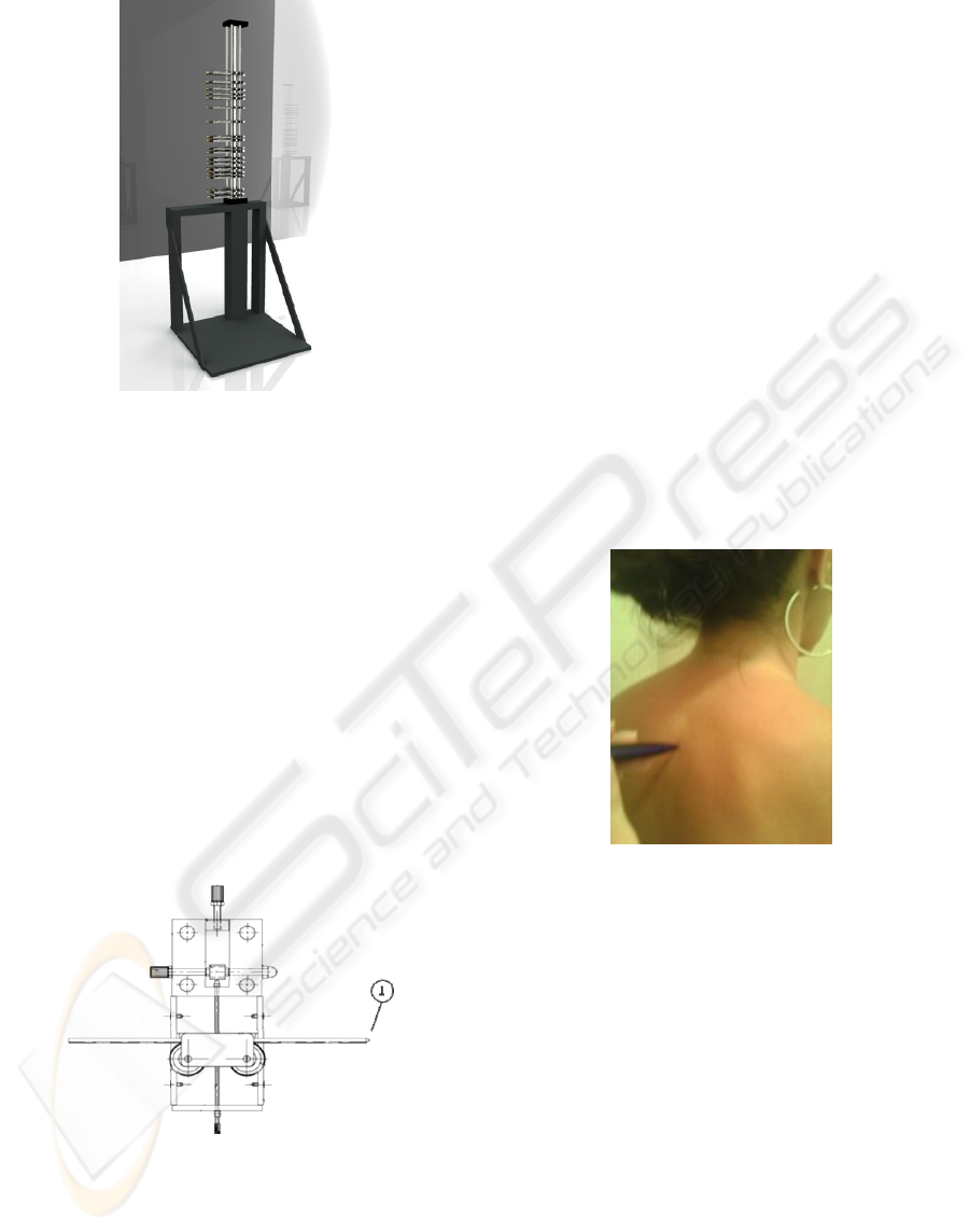
Figure 1: Image of Vertebral Metrics.
The main components of the apparatus (Figure 1)
are the “body” and the “support” (Forjaz Secca, et
al, 2008)
The “body” has a vertical piece, mounted in the
support, with 18 horizontal pieces called “2D
Positioner”. Each “2D Positioner” slides along the
scaled ruler attached on the vertical piece.
The “support” has two pieces, one vertical,
where the body of the instrument fits, and one
horizontal piece where the person to be evaluated
stands up.
All the 18 2D Positioners are identical and
adjustable, allowing the identification of the x, y,
and z positions of each vertebra (Quaresma, 2009)
The component 1 of the 2D Positioners is a
square sectioned rod, cone shaped, which is the
contact point for each vertebra (Figure 2)..
Figure 2: The “2D Positioner”.
Three of the “2D Positioner” are used in four
different ways (Quaresma et al 2009)..
The first is placed in the occipital region and is
used as a reference point during the data collection;
The second piece collects the data of the cervical
vertebrae:
The third, which is piece number fifteen,
collects the data from the first, second and third
lumbar vertebra;
Finally, the remaining horizontal pieces will
identify the position of all other vertebra of the
spinal column.
The x, y and z positions of each “2D P” are then
obtained and the 3 coordinates can be read using
different rulers on the device.
2 VERTEBRAL METRICS:
APLICATION
In order initiate the evaluation process the horizontal
pieces of the instrument are roughly adjusted,
according to the height of each woman
The examiner, starts by marking on the skin the
vertebral apophyses, from the first cervical vertebra
to the first sacral vertebra, using a washable pen
(Figure 3).
Figure 3: Marking the vertebral apophyses with a
washable pen.
Each pregnant woman stands up then on the
“support” of the apparatus with the posterior face of
the trunk facing the “body” of the Vertebral Metrics,
followed by adjustment of each “2D Positioner” to
each mark on the various points along the spinal
column(Quaresma et al 2009).
The evaluation starts by placing the first
horizontal piece in the occipital region.
Immediately afterwards the second, fourteenth
and the last “2D Positioner” are adjusted to the
seven cervical vertebra, twelve dorsal vertebra and
the first sacral vertebra. This points are used as
reference points to stabilize to position of the spine.
Subsequently the remaining “2D Positioner” are
adjusted
The x, y and z positions of each “2D Positioner”
are then obtained (Quaresma et al 2009).
BIODEVICES 2010 - International Conference on Biomedical Electronics and Devices
144
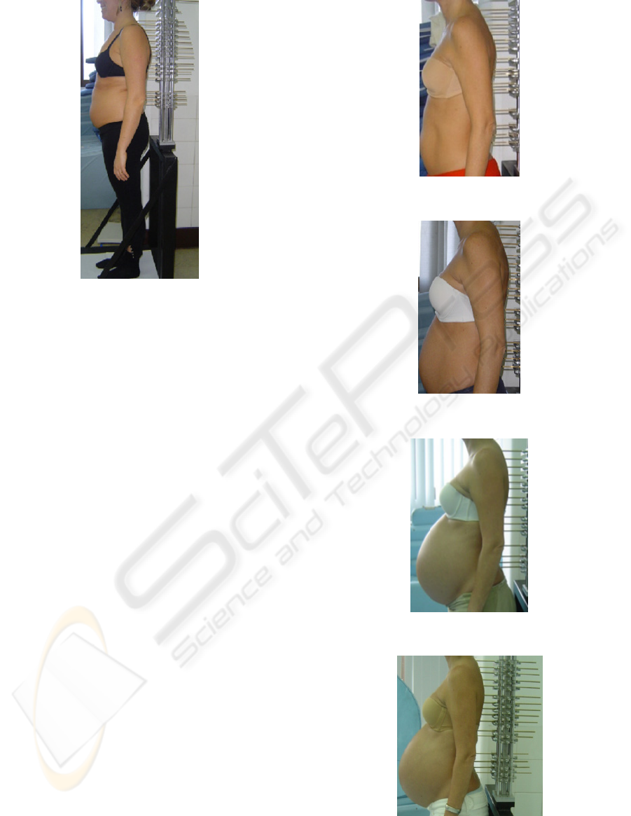
Figure 4: Example of application of Vertebral Metrics.
Despite the apparent complexity of the device and of
the respective measurements, each data collection
only lasts seven minutes (Figure 4).
The collected data are then recorded and
transferred to a specific data basis which includes
correction factors associated with the instrument
3 PREMILINARY RESULTS
This study is part of a broader analysis where we
intend to identify and describe the biomechanical
changes of the spinal column that occur throughout
pregnancy.
This paper presents the application of Vertebral
Metrics to one pregnant woman. Data collection was
made in three periods of the pregnancy:12, 20, 32
and 37 weeks of the gestation.
As can be observed in figures 5 to 8.in the
corresponding diagrams 1 to 4 .the curvatures of the
spine change during pregnancy. In the second
moment of the evaluation of the spine the curvatures
decrease when compared with the other moments of
the evaluation, where an increase of these
curvatures, related to rachialgie, is evident.
4 CONCLUSIONS
Vertebral Metrics is a non-invasive mechanical
instrument, which is able to identify the position x, y
and z of each vertebra in standing position
(Quaresma et al 2009).
Figure 5: Pregnant woman- 12 weeks of gestations.
Figure 6: Pregnant woman - 20 weeks of gestations.
Figure 7: Pregnant woman - 32 weeks of gestations.
Figure 8: Pregnant woman - 37 weeks of gestations.
VERTEBRAL METRICS - Application of a Mechanical Instrument to Evaluate the Spinal Column in Pregnant Women
145
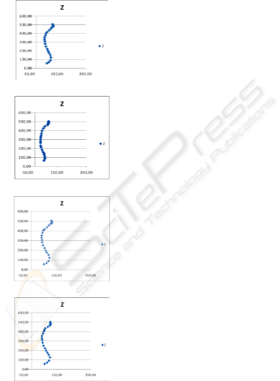
Diagram 1: Spine - 12 weeks of gestations.
Diagram 2: Spine - 20 weeks of gestations.
Diagram 3: Spine -32 weeks of gestations.
Diagram 4: Spine - 37 weeks of gestations.
This instrument allows a global simultaneous
assessment of the spine. Thus, identification of
dysfunctions and / or diseases of the spinal column
in pregnant women, will be shown on a full
diagnosis. Intervention programs, directly connected
to specific problems of each pregnant woman, may
then be elaborated and implemented (Quaresma et al
2009).
Vertebral Metrics was originally planned and
built to be applied to pregnant women, althout it can
be easily applied to the general population
(Quaresma et al 2009). This device can also be
applied in other situation, including the orthopaedic,
neurosurgical and rehabilitation area.
A automatic system to speed up the process of the
data collection is currently being developed.
Besides the mentioned importance of Vertebral
Metrics, it should be provided out that the present
instrument is easily carried making feasible its use in
any health institution.
REFERENCES
Alexandre, N.; Moraes, M. 2001. Modelo de avaliação
fisio-funcional da coluna vertebral. Rev. Latino-am
Enfermagem Março, 9 (2); 67-75.
Knoplich, J. 2003. Enfermidades da coluna vertebral..,
Robe Editorial. São Paulo, 3ªed
Forjaz Secca, M; Quaresma, C.; Santos, F.. 2008. A
Mechanical Instrument to Evaluate Posture of the
Spinal Column in Pregnant Women. Proc. IFMBE 4th
European Congress for Med. and Biol. Eng., Antwerp,
Belgium; 1781-1784
Quaresma, C.; Forjaz Secca, M.; O’Neill, J.; Branco, J.,
2009. Development a mechanical instrument to
evaluate biomechanically the spinal columnin
pregnant women. Proc. Internacional Conference
Biodevice, 310-113.
Quaresma, C.; Forjaz Secca, M.; O’Neill, J.; Branco.
2009. Métrica Vertebral: Intrumento de Avaliação
Biomecânica da Coluna Vertebral. III Congresso
Nacional de Biomecânica
Quaresma, C.; João, F. Fonseca, M. Forjaz Secca, M.
Veloso, A.; O’Neill, J.; Branco, J., 2009. Validação do
Métrica Vertebral Através de um Sistema
Optoeléctrico de Esterogrametria. Actas do
III.Congresso Nacional de Biomecânica
Quaresma, C.; João, F. Fonseca, M. Forjaz Secca, M.
Veloso, A.; O’Neill, J.; Branco, J., 2009. Validations
of Vertebral Metrics: a mechanical instrument to
evaluate posture osf the spinal column. O. Dössel and
W.C. Schlegel (Eds.): WC IFMBE Proceedings
25/VII, 711–713
BIODEVICES 2010 - International Conference on Biomedical Electronics and Devices
146
