
MRI
IMAGE ENHANCEMENT
A PDE-based Approach Integrating a Double-well Potential Function for Thin
Structure Preservation
A. Histace
ETIS UMR CNRS 8051, ENSEA-UCP, 6 avenue du Ponceau, 95014 Cergy, France
M. M
´
enard
L3i, University of La Rochelle, Pole Sciences et Technologie, 17000 La Rochelle, France
Keywords:
Image diffusion, Double well potential, Directional diffusion, Selectivity.
Abstract:
Non-linear or anisotropic regularization PDE’s (Partial Differential Equation) raised a strong interest in the
field of medical image processing. The benefit of PDE-based regularization methods lies in the ability to
smooth data in a nonlinear way, allowing the preservation of important image features (contours, corners or
other discontinuities). In this article, we propose a PDE-based method restoration approach integrating a
double-well potential as diffusive function. It is shown that this particular potential leads to a particular regu-
larization PDE which makes the integration of prior knowledge about the gradient intensity level to enhance
possible. The corresponding method shows interesting properties regarding stability and preservation of fine
structures. As a proof a feasibility, results of restoration are presented on natural images to show potentialities
of the proposed method. We also address a particular medical application: enhancement of tagged cardiac
MRI.
1 INTRODUCTION
In the particular field of image restoration, non-
linear or anisotropic regularization PDE’s are of pri-
mary interest. The benefit of PDE-based regular-
ization methods lies in their ability to smooth data
in a nonlinear way, allowing the preservation of im-
portant image features (contours, corners or other
discontinuities). In the particular domain of scalar
image restoration, a lot of studies have been pre-
sented in the literature so far: (Perona and Malik,
1990), (Alvarez et al., 1992), (Catt
´
e et al., 1992),
(Geman and Reynolds, 1992), (Nitzberg and Sh-
iota, 1992), (Whitaker and Pizer, 1993), (Weick-
ert, 1995), (Deriche and Faugeras, 1996), (Weickert,
1998), (Terebes et al., 2002), (Tshumperl
´
e and De-
riche, 2002), (Tschumperle and Deriche, 2005) for the
main references.
In the particular field of medical image process-
ing, PDE based approach for denoising are very
promising tools, but generally needs to be adapted to
the imaging context (PET, CT, Cone-beam CT, MRI,
etc.) since noise can be of very different types (Gaus-
sian, Poisson, Rayleigh). In (Histace et al., 2009), we
showed that, considering a particular general param-
eterizable PDE, it was possible to integrate selectiv-
ity regarding the gradient directions to diffuse or not
within the considered image. Qualitative and quanti-
tative results were also presented on a particular med-
ical application: enhancement of tagged cardiac MRI.
In this article, we propose a complementary PDE
to the one presented in (Histace et al., 2009) which en-
ables integration of selectivity regarding the intensity
of the gradient to restore and which makes the preser-
vation of thin structures from the diffusive effect pos-
sible. More precisely, we propose to make this selec-
tivity possible thanks to the integration of a double-
well potential diffusion function within the classical
Perona-Malik’s PDE (Perona and Malik, 1990). That
kind of approaches can be of interesting benefits for
medical image restoration and particularly for MRI
enhancement, since even thin structures can be of pri-
mary importance to establish the most appropriate di-
agnosis.
Our aims and motivation for such a study are
mainly to show that, firstly, such a choice can lead to
501
Histace A. and Ménard M. (2010).
MRI IMAGE ENHANCEMENT - A PDE-based Approach Integrating a Double-well Potential Function for Thin Structure Preservation.
In Proceedings of the International Conference on Computer Vision Theory and Applications, pages 501-508
DOI: 10.5220/0002887605010508
Copyright
c
SciTePress

a stable PDE-based approach for scalar image denois-
ing that can overpass classical approach of Perona-
Malik’s from which it is derived and which presents
instability problems as formerly shown in (Catt
´
e et al.,
1992). Secondly, we also want to show that this inte-
gration leads to a selective PDE-based approach that
overcomes classical mean curvature or tensor driven
diffusion problems, which in the particular case of di-
rectional diffusion are not suitable (see (Histace et al.,
2005) and (Terebes et al., 2002)) because they tend to
smooth transitions between patterns.
Regarding the medical application we want to ad-
dress in this article, we propose to tackle a known
problem of us: enhancement of tagged cardiac MRI.
Such a choice is guided by the fact that we have
already worked on that particular MR imaging se-
quence and that qualitative results have already been
computed for comparison. Moreover, as said in sec-
tion 3, this particular sequence of acquisition can be
of primary importance for the follow-up of cardiovas-
cular pathologies and totally fit the problem we want
to address: preservation of thin structures within the
enhanced data.
This article is organized as follows: In a section
two, we propose some recalls about PDE-based reg-
ularization approaches. Section three deals with the
general presentation of the tagged cardiac MR im-
ages problematic. Fourth section is dedicated to the
study of the double well function and of its mathe-
matical properties. Prospective results on “lena” are
also shown in this section. Section five deals with
the enhancement of tagged MRI: Comparative results
are shown and commented. Finally, the proposed ap-
proach and the obtained results are discussed in last
section.
2 PDE BASED
REGULARIZATION
APPROACH: A GENERAL
SCHEME
In (Deriche and Faugeras, 1996), authors propose a
global scheme for PDE-based restoration approaches.
More precisely, if we denote ψ(r,t) : R
2
× R
+
→
R the time intensity function of a corrupted image
ψ
0
= ψ(r,0), the corresponding regularization prob-
lem of ψ
0
is equivalent to the minimization problem
described by the following PDE:
∂ψ
∂t
= c
ξ
(k∇ψk)
∂
2
ψ
∂ξ
2
+ c
η
(k∇ψk)
∂
2
ψ
∂η
2
, (1)
where η = ∇ψ/k∇ψk, ξ⊥η and c
ξ
and c
η
are two
weighting functions (also called diffusive functions).
This PDE can be interpreted as the superposition
of two monodimensional heat equations, respectively
oriented in the orthogonal direction of the gradient
and in the tangential direction: It is characterized by
an anisotropic diffusive effect in the privileged direc-
tions ξ and η allowing a non-linear denoising of scalar
image.
Eq. (1) is of primary importance, for all classical
methods can be expressed in that global scheme: For
instance, if we consider the former anisotropic diffu-
sive equation of Perona-Malik’s (Perona and Malik,
1990) given by
∂ψ
∂t
= div(c(k∇ψk)∇ψ) , (2)
with ψ(r,0) = ψ
0
and c(.) a monotonic decreasing
function, it is possible to express it in the global
scheme of Eq. (1) with
(
c
ξ
= c(k∇ψk)
c
η
= c
0
(k∇ψk).|∇ψ| + c(k∇ψk)
(3)
Formulation of Eq. (1) is also interesting, for it makes
stability study of classical proposed methods possible.
More precisely, a stable PDE-based method for de-
noising will be characterized by a weighting function
c
η
positive for all values of k∇ψk as formerly shown
in (Catt
´
e et al., 1992). In practice, c
η
(.) function has
also to be of small values in order to diffuse the data in
the tangential direction of the image boundaries only.
This last property is of primary importance for the
preservation of thin structures since only a “small”
diffusive effect in the orthogonal direction of the cor-
responding gradient can lead to a low alteration of
them.
What we proposed in this article is a study for the
integration of a double well potential as a diffusive
function c(.) in Eq. (2).
3 TAGGED CARDIAC MRI
Mainly, to help cardiologists to establish a pre surgery
scheme for reperfusion of myocardial tissue after an
infarction, a study of the myocardial local viability is
necessary: Whereas classical cardiac MRI does not
make the study of the local contraction of the my-
ocardium possible, tagged cardiac MRI allows this lo-
cal estimation. More precisely, the classical SPAMM
(Space Modulation of Magnetization) acquisition pro-
tocol (Zerhouni et al., 1988) used for the tagging of
MRI data, displays a deformable 45-degrees oriented
VISAPP 2010 - International Conference on Computer Vision Theory and Applications
502
dark grid which describes the contraction of my-
ocardium (Fig. 1) on the images of temporal Short-
Axis (SA) sequences. This is the temporal tracking
of this grid that can enable radiologists to evaluate the
local intramyocardial displacements.
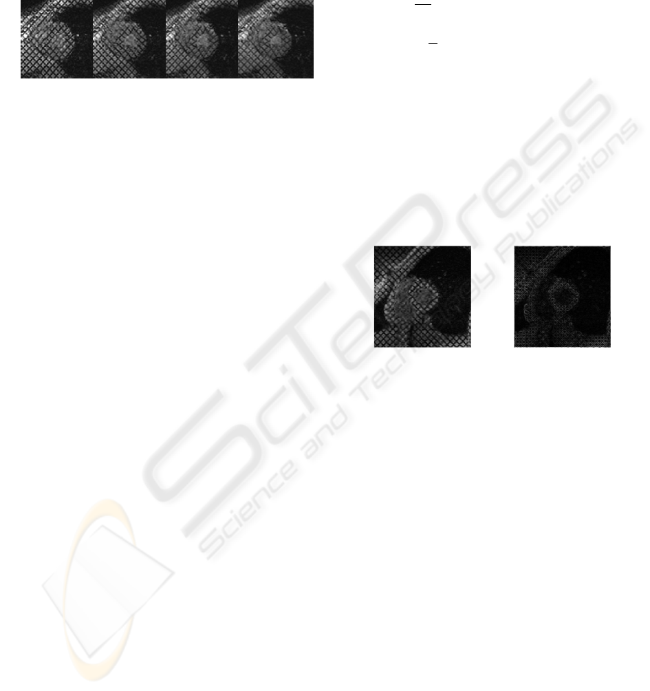
Figure 1: SA tagged MRI of the Left Ventricle (LV) ex-
tracted from a sequence acquired between end-diastole and
end-systole.
Tagged cardiac images present peculiar character-
istics which make the analysis difficult. More pre-
cisely, images are of low contrast compared with clas-
sical MRI, the level of corrupting noise is more im-
portant than with classical acquisition and their reso-
lution is only of approximately one centimeter. Nu-
merous studies were carried out concerning the anal-
ysis of the deformations of the grid of tag on SA se-
quences (see (Petitjean et al., 2005; Axel et al., 2007)
for a complete overview) but all have in common
the necessary enhancement of tagged cardiac images
which can be considered as thin oriented structures
since they are only three or four pixels wide.
Classically, in such a framework (oriented pat-
tern enhancement), the classical Edge Enhancing Dif-
fusion (EED) (Weickert, 1995) normally leads to
very satisfying results of regularization. However, as
shown in Fig. 10.(c), the poor quality of tagged car-
diac images makes the computation of the local struc-
ture tensor difficult and, as said in the introduction of
this article, that kind of approaches tends to always
smooth in the orthogonal direction of the image con-
tours: thin stucture are then altered.
As a consequence, diffusive restoration ap-
proaches like the Perona-Malik’s former one (Perona
and Malik, 1990) are more adapted to our purpose: A
non-linear smoothing of the data is performed by tak-
ing into consideration the local value of the gradient
intensity. If this value is small then the corresponding
data are diffused along the tangential direction of the
contour. On the contrary, if this value is important the
diffusive effect is stopped. That kind of approaches
makes the enhancement of the boundaries of the im-
age possible. Nevertheless, as one can notice on Fig.
2, due to the fact that norms of the gradient levels of
tagged MRI are very noisy, it is necessary to develop a
method that integrates within diffusion process more
than only this classical parameter: for instance, cal-
culation and integration of the direction of local gra-
dients of the grid could be of primary interest.
This can be achieved by considering some varia-
tions of the classical restoration approaches. We pro-
pose for example to consider a variant of the Perona-
Malik’s process (Perona and Malik, 1990) given by
∂ψ
∂t
= div(c(||A.∇ψ||)∇ψ) . (4)
with c(u) = e
−
u
2
k
2
(as proposed by Perona and Malik)
and A is a vector field defining the particular direc-
tion(s) to preserve from the diffusion process (in this
particular medical application, the gradient direction
of the grid). k represents here a soft threshold driv-
ing the decrease of c(.). In both cases, the directional
weighting of the diffusion process is driven by the
scalar product between the norm of the local gradient
and A. As a consequence when local gradient and A
are parallel, there is no diffusion, for c(||A.∇ψ||) = 0,
whereas all other directions are diffused: the grid of
tags is normally enhanced.
(a) (b)
Figure 2: (a) Original Image, (b) Norm of the corresponding
gradients.
Nevertheless, because of instability problems (see
section 4 for more details) of PM’s approach, it ap-
pears that process of Eq. (4) does not lead to inter-
esting results (see Fig. 10.(b)). Such a problem can
be overcome by a Gaussian filtering of the gradient
data as proposed in (Catt
´
e et al., 1992). But, such
a Gaussian filtering will also have for consequences
an increase of the values corresponding to the diffu-
sive effect in the orthogonal direction of the contours.
As explained before, the corresponding approach will
then not make preservation of thin structures possible.
Moreover, the classical c(.) function does not
allow to integrate within the iterative restoration
scheme selectivity regarding the preservation of par-
ticular gradient levels. However, such a selectivity
would be of significant benefits since value of the
tags’ gradient can be easily identified (Denney, 1999).
To overpass the drawbacks of Eq. (4) , we propose
to dethin c(.) as a double well potential function. This
particular function will make integration of gradient
level selectivity possible as well as the obtaining of a
stable PDE and the preservation of thin structures.
MRI IMAGE ENHANCEMENT - A PDE-based Approach Integrating a Double-well Potential Function for Thin Structure
Preservation
503
4 DOUBLE WELL POTENTIAL
AND CORRESPONDING PDE
4.1 Diffusive Function
The double well potential considered in this article is
dethind by the following function:
φ(u) =
Z
u
0
v(α − v)(v − 1)dv . (5)
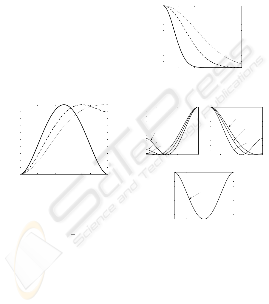
Some graphical representations of Eq. (5) for differ-
ent values of α are proposed Fig. 3.(a). The roots
of the corresponding non linear force (i.e. f (u) =
u(α − u)(u − 1)) 0, and 1 corresponds to the local
positions of the minima of the potential, whereas the
root α represents the position of the potential maxi-
mum. The non linearity threshold α dethins the po-
tential barrier between the potential minimum with
the highest energy and the potential maximum.
0 0.2 0.4 0.6 0.8 1
0
0.1
0.2
0.3
0.4
0.5
0.6
0.7
0.8
0.9
1
u
φ(u)
Figure 3: Plots of double well potential φ(.) of Eq. (5) for
different values of α ∈ [0.5,1]. Solid lines stand for α = 0.5,
dash-dotted lines for α = 0.7 and dotted lines for α = 1.
This function has to be compared with the classi-
cal Perona-Malik’s function c
PM
(.) given by:
c
PM
(u) = e
−
u
2
k
2
, (6)
with k a soft threshold defining selectivity of c
PM
(.)
regarding values of image gradients. Fig. 4 shows
graphical representations of c
PM
(.) dethind by Eq. (6)
for different values of k.
As one can notice on Fig. 4.(a), for k∇ψk →
0, c
PM
(k∇ψk) → 1, whereas for k∇ψk → 1,
c
PM
(k∇ψk) → 0. As a consequence, boundaries
within images which are on a threshold, function of
k, are preserved from the smoothing effect of Eq. (2).
Regarding Fig. 3, in order to preserve this major prop-
erty with integration of Eq. (5) as a diffusive function
in Eq. (2), it is necessary to dethin this diffusive func-
tion as follows:
c
DW
(u) = 1 − φ(u) . (7)
Graphical representations of c
DW
are proposed in Fig.
5.
0 0.2 0.4 0.6 0.8 1
0
0.1
0.2
0.3
0.4
0.5
0.6
0.7
0.8
0.9
1
u
c
PM
(u)
Figure 4: Plots of function c
PM
(.) of Eq. (6) for different
values of k. Solid lines stand for k = 0.2, dash-dotted lines
for k = 0.4, and dotted lines for k = 0.6.
0 0.2 0.4 0.6 0.8 1
0
0.1
0.2
0.3
0.4
0.5
0.6
0.7
0.8
0.9
1
u
c
DW
(u)
α=0.4
α=0.2
α=0.1
(a)
0 0.2 0.4 0.6 0.8 1
0
0.1
0.2
0.3
0.4
0.5
0.6
0.7
0.8
0.9
1
u
c
DW
(u)
α=1
α=0.8
α=0.6
(b)
0 0.2 0.4 0.6 0.8 1
0
0.1
0.2
0.3
0.4
0.5
0.6
0.7
0.8
0.9
1
u
c
DW
(u)
α=0.5
(c)
Figure 5: Plots of function c
DW
(.) of Eq. (5) for different
values of α: (a) 0 < α < 0.5, (b) 0.5 < α < 1, and (c) α =
0.5.
One can notice on Fig. 3 that φ(.) has been nor-
malized. As a consequence, we are able to ensure that
0 ≤ c
DW
(u) ≤ 1 for all values of u like classical PM’s
function of Eq. (2). For 0 ≤ α < 1, since c
DW
is issued
from a double well potential, selectivity of Eq. (2) is
more important and centered on a particular gradient
value function of α. For instance, for α = 0.5, only
gradients of value 0.5 are totally preserved from the
diffusive effect in the tangential direction. This can
be interpreted as an integration of gradient level se-
lectivity within the restoration process.
VISAPP 2010 - International Conference on Computer Vision Theory and Applications
504
Moreover, we are now going to show, that inte-
gration of c
DW
as diffusive function leads to interest-
ing stability property of corresponding PDE regarding
properties of the corresponding c
η
function.
4.2 Study of Stability
It is recognized that classical Perona-Malik’s PDE
presents instability problems. More precisely, as
shown in (Catt
´
e et al., 1992), sometimes noise can
be enhanced instead of being removed. This can be
explained considering Eq. (3). If we consider c
PM
(.)
function of Eq. (6), it appears that corresponding c
η
PM
function of Eq. (3), in the global scheme of Eq. (1),
can sometimes takes negative values (see Fig. 6 for
illustrations). This leads to local instabilities of the
Perona-Malik’s PDE which degrades the processed
image instead of denoising it.
0 0.2 0.4 0.6 0.8 1
−0.5
0
0.5
1
u
c
η
PM
Figure 6: Plots of function c
η
PM
for different values of k .
Solid lines stand for k = 0.2, dash-dotted lines for k = 0.4
and dotted lines for k = 0.6.
Now, if we calculate mathematical expression of
c
η
with c(.) = c
DW
(.) of Eq. (7), one can obtain that:
c
η
DW
(k∇ψk) = c
0
DW
(k∇ψk).|∇ψ| + c
DW
(k∇ψk) ,
(8)
Taking into account that |∇ψ| ∈ [0..1] and that
c
0
DW
(k∇ψk) is a one-order-less polynomial function
than c
DW
(k∇ψk), it happens that:
c
η
DW
(k∇ψk) ≈ c
DW
(k∇ψk) = c
ξ
(k∇ψk) . (9)
Considering Eq. (9), one can notice that cor-
responding c
η
function never takes negative values
(see Fig. 5 for illustrations): Diffusive process re-
mains stable for all gradient values of processed im-
age which is of primary importance.
Moreover, we can also notice that c
η
DW
is exactly
equal to 0 when c
ξ
DW
= 0. As a consequence, by a ju-
dicious choice of α, it becomes possible to completely
stop the diffusion process in the tangential and orthog-
onal directions of the contours at the same time. thin
structures characterized by an identified gradient level
can be preserved from any alterations.
4.3 Experimental Results
We propose in this section to make a visual and quan-
titative comparison between classical Perona-Malik’s
PDE of Eq. (2) with diffusive function c(.) = c
PM
(.)
of Eq. (6), and the following PDE given by:
∂ψ
∂t
= div(c
DW
(k∇ψk)∇ψ) . (10)
For practical numerical implementations, the pro-
cess of Eqs. (2) and (10) are sampled with a time step
τ. The restored images ψ(t
n
) are calculated at discrete
instant t
n
= nτ with n the number of iterations.
We propose to compare our proposed method with
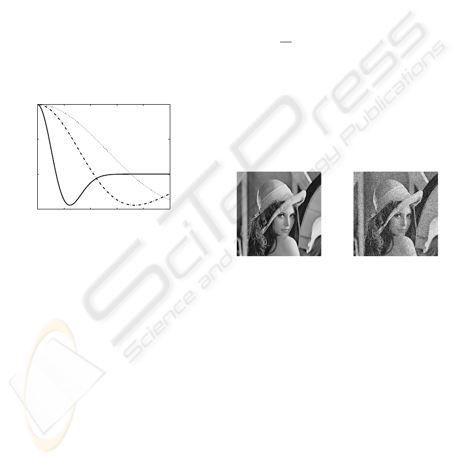
PM’s approach on the classical “lena” image. For
our purpose, this latter has been corrupted by a white
Gaussian noise of mean zero and standard deviation
σ (see Fig. 7).
(a) (b)
Figure 7: (a) Original image “lena” and (b) its corrupted
version ψ
0
. Corrupting noise is a white Gaussian one of
mean zero and standard deviation σ = 0.1.
Considering nature of non corrupted image (Fig.
7.(a)), quantification of the denoising effect of Eq.
(2) with c(.) = c
PM
(.) and c(.) = c
DW
(.), will be esti-
mated with a classical PSNR measurement.
Once again, because aim of this article is to show
potentiality of the described restoration method, only
optimal results for both compared approaches are pre-
sented Figs. 8 and 9.
One can notice on Figs. 8 and 9 that both visually
and quantitatively, it is possible to find a value of α
that can outperform results of optimal classical PM’s
approach. Although the number of iterations corre-
sponding to the optimal restoration results is more im-
portant with the proposed approach of Eq. (10) than
with PM’s approach, quantitatively speaking PSNR is
around 2dB higher, and visually speaking, boundaries
on Fig. 8.(b) are preserved in a better way from the
MRI IMAGE ENHANCEMENT - A PDE-based Approach Integrating a Double-well Potential Function for Thin Structure
Preservation
505
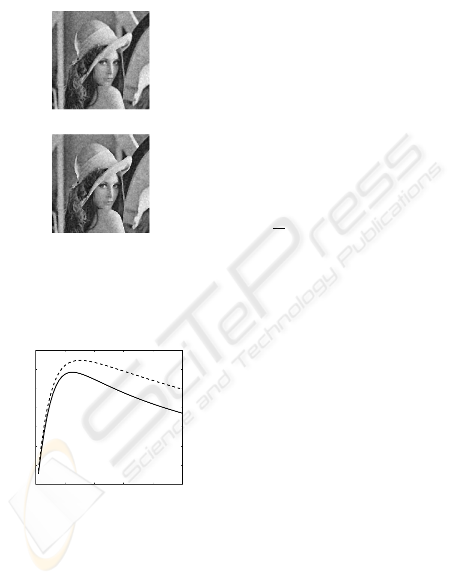
(a)
(b)
Figure 8: (a) Restored image with c(.) = c
PM
(.) (clas-
sical Perona-Malik’s approach), (b) Restored image with
c(.) = c
DW
(.) (proposed approach). The red circles high-
light some regions of interest where the preservation of
edges are better than with Perona-Malik’s approach. k is
equal to 1 for PM’s restoration approach, α is equal to 0.2
for the proposed approach (Eq. (10) (these values have been
empirically tuned). Time step τ = 0.05
0 10 20 30 40 50
138
140
142
144
146
148
150
152
iteration number (n)
PSNR (dB)
Figure 9: PSNR function of iteration number n, solid lines
stands for classical Perona-Malik’s approach, dotted line
stands for the proposed method (Eq. (10). k is equal to 1, α
is equal to 0.2 (these values have been empirically tuned
to obtained the best denoising effects). Time parameter
τ = 0.05 These two curves have been computed by calcula-
tion of the mean results obtained for one hundred different
realizations of the Gaussian corrupting noise.
diffusion effect (see red circles on Fig. 8 for particular
regions of interest).
Nevertheless, this example do not permit to di-
rectly appreciate the possible gradient level selectivity
of the proposed approach. To show it, we are now go-
ing to present some results dedicated to the targeted
medical application : enhancement of tagged cardiac
MRI.
5 TAGGED MRI ENHANCEMENT
We now focus this study on tagged cardiac MRI en-
hancement.
What we propose here is to compare enhancement
results obtained with: (a) the classical PM’s approach,
(b) the classical Weickert’s approach (Weickert, 1995)
(Edge Enhancing Diffusion-EED), (c) with PM’s ap-
proach integrating c
DW
(.) function (Eq. (10), and (d)
with the following PDE:
∂ψ
∂t
= div(c
DW
(||A.∇ψ||)∇ψ) . (11)
c
DW
function is set in order to entirely preserve the
gradient level of the tag from diffusion. To achieve
this, Fig. 2.(b) is processed so that gradient of the
tags are set to 0.5 and the parameter α is set to the
same value. This choice made for α is based on the
fact that for this particular value, we have shown that
the diffusion process is totally stop in both tangential
and orthogonal direction of the contours, and is the
most selective.
To obtain restoration results with Eq. (11) only
one direction of the grid has been taken into account
thanks a judicious computation of A. More precisely,
each local a priori direction of the corresponding gra-
dient has been estimated thanks to a frequential anal-
ysis of processed image (see (Histace et al., 2009) for
full detailed of the method). In order to compute a
precise estimation of A from the frequential analysis,
we propose to directly use the method of Rao (Rao
and Jain, 1992) and Terebes (Terebes et al., 2002).
As a consequence, each a priori gradient direction
computed from the frequential analysis is preserved
from diffusion effect thanks to A, and c
DW
(.) func-
tion makes the enhancement of the tag possible by
preserving the gradient level of tags from diffusion.
As one can notice, the grid enhancement per-
formed thanks to the classical PM’s approach (Fig.
10.(b)) presents strong instabilities. As a conse-
quence, the resulting enhanced grid is corrupted and
presents no real interest for the tracking of the grid.
Considering now the classical Weickert’s EED (Fig.
10.(c), one can clearly notice that the method fails
in enhancing the tag pattern for the reasons we men-
tioned earlier in the paper. Fig. 10.(d) shows re-
sults obtained with classical PM’s approach but with
VISAPP 2010 - International Conference on Computer Vision Theory and Applications
506
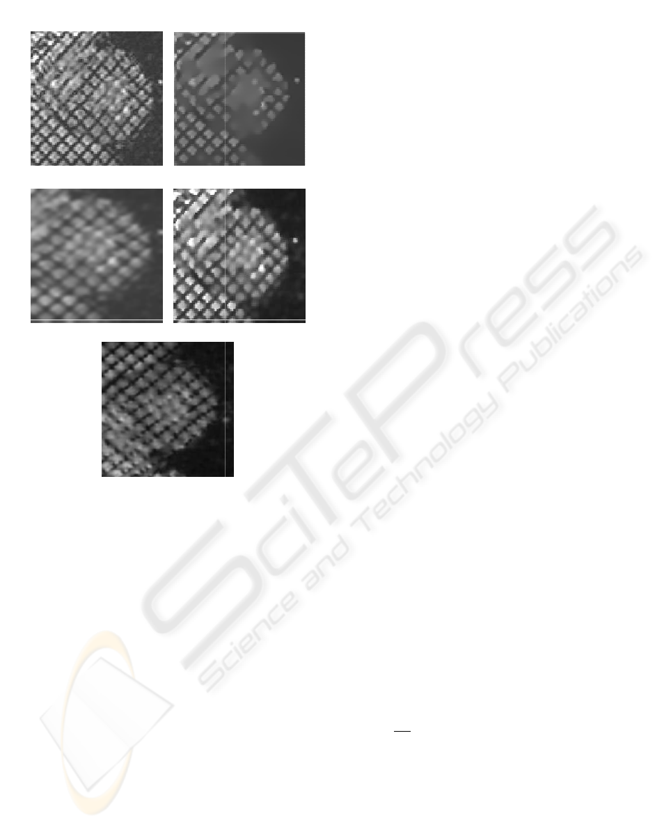
(a)
(c)
(e)
(b)
(d)
Figure 10: Tagged MRI restoration: (a) Original image, (b)
PM’s approach, (c) Weickert’s approach, (d) PM’s approach
with c(.) = c
DW
(.), (e) Result obtained with Eq. (11). “Op-
timal” visual results for each methods are shown.
c(.) = c
DW
(.). The first consequence of such a choice
for c(.) function is the absence of stability problems
within the iterative enhancing resulting process. As
one can see, visually speaking the grid is enhanced
and the corresponding boundaries are preserved from
the diffusion effect. Moreover, compare to Weickert’s
EED approach, thin structures (tags mainly) are bet-
ter preserved. If such a result is of real interest, en-
hancement effect can be outperformed by considering
Eq. (11). This time, result shown Fig. 10.(e) clearly
demonstrates the possibility of enhancing the tag pat-
terns by selecting (i) a particular direction, locally
computed thanks to a frequential analysis, and (ii)
a particular gradient-level characterizing the bound-
aries of the tags.
6 DISCUSSION AND
CONCLUSIONS
In this article, we have proposed an alternative diffu-
sive function for restoration of scalar images within
the framework of PDE-based restoration approaches.
The proposed diffusive function allows integrating
prior knowledge on the gradient level to restore thanks
parameter α of Eq. (7) and remains always stable on
the contrary of classical PM’s approach. Proposed
method also remains fast and easy to compute. Visu-
ally and quantitatively speaking, better restoration re-
sults have been obtained, but this point must be now
discussed. Since α parameter finally corresponds to
integration of prior information about gradient level
to preserve from the diffusion process, it would be
interesting to make an adaptive local use of the pro-
posed approach more than a global use.
If interesting visual and quantitative results have
been obtained on “lena” image thanks to a global
use of the proposed PDE (Eq. (10)), we have also
shown that a judicious tuning of this parameter in
terms of particular localization within the processed
image (Eq. (11) could lead to more interesting results
than classical approaches on a particular medical ap-
plication: enhancement of tagged cardiac MR images.
More precisely, thin structures are less altered by the
proposed diffusive scheme. Strategy for a local tun-
ing of α still to be now completely automatized. For
instance, in the framework of tagged cardiac MRI,
it could be of primary interest for the method to be
able to adapt the value of α to the fading of the tags
due to the non persistency of the magnetization corre-
sponding to the grid (see Fig. 1). More precisely, the
fact that this fading phenomenon can be analytically
studied would permit such an adaptive setting of α.
Moreover, if in this example we choose to select the
gradient-level, one could also think about integrating
a selectivity upon the grey-level to diffuse or not. This
can be achieved by considering a variant of Eq. (11)
given by
∂ψ
∂t
= c
DW
1
div(c
DW
2
(||A.∇ψ||)∇ψ) . (12)
In this equation, c
DW
2
, as shown in this article, permits
a selectivity regarding gradient-level, and c
DW
1
could
permit a selectivity in terms of grey-level intensity.
Considering the fact that the grey-level intensity of the
myocardium is different from the grey-level intensity
of the tags, this approach could be a good alternative
for enhancement of tagged cardiac MRI, but also for
MR images in general.
MRI IMAGE ENHANCEMENT - A PDE-based Approach Integrating a Double-well Potential Function for Thin Structure
Preservation
507

REFERENCES
Alvarez, L., Guichard, F., Lions, P., and Morel, J. (1992).
Image selective smoothing and edge detection by non-
linear diffusion (ii). Arch. Rationnal Mech. Anal.,
29(3):845–866.
Axel, L., Chung, S., and Chen, T. (2007). Tagged mri anal-
ysis using gabor filters. In Biomedical Imaging: From
Nano to Macro, 2007. ISBI 2007. 4th IEEE Interna-
tional Symposium on, pages 684–687.
Catt
´
e, F., Coll, T., Lions, P., and Morel, J. (1992). Im-
age selective smoothing and edge detection by nonlin-
ear diffusion. SIAM Journal of Applied Mathematics,
29(1):182–193.
Denney, T. (1999). Estimation and detection of myocar-
dial tags in MR images without user-defined myocar-
dial contours. IEEE Transactions on Medical Imag-
ing, 18(4):330–344.
Deriche, R. and Faugeras, O. (1996). Les edp en traitements
des images et visions par ordinateur. Traitement du
Signal, 13(6):551–578.
Geman, S. and Reynolds, G. (1992). Constrained restora-
tion and the recovery of discontinuities. IEEE Trans-
actions on Pattern Analysis and Machine Intelligence,
14(3):367–383.
Histace, A., Cavaro-M
´
enard, C., Courboulay, V., and
M
´
enard, M. (2005). Analysis of tagged cardiac MRI
sequences. Lecture Notes on Computer Science (Pro-
ceedings of the 3rd Functional Imaging and Mod-
elling of the Heart (FIMH) Workshop), 3504:404–
413.
Histace, A., M
´
enard, M., and Cavaro-M
´
enard, C. (2009).
Selective diffusion for oriented pattern extraction: Ap-
plication to tagged cardiac mri enhancement. Pattern
Recognition Letters, 30(15):1356–1365.
Nitzberg, M. and Shiota, T. (1992). Nonlinear image filter-
ing with edge and corner enhancement. IEEE Trans-
actions on Pattern Analysis and Machine Intelligence,
14(8):826–833.
Perona, P. and Malik, J. (1990). Scale-space and edge
detection using anistropic diffusion. IEEE Transca-
tions on Pattern Analysis and Machine Intelligence,
12(7):629–639.
Petitjean, C., Rougon, N., and Cluzel, P. (2005). Assess-
ment of myocardial function: A review of quantifica-
tion methods and results using tagged MRI. Journal of
Cardiovascular Magnetic Resonance, 7(2):501–516.
Rao, A. and Jain, R. (1992). Computerized flow field anal-
ysis: Oriented texture fields. Transactions on pattern
analysis and machine intelligence, 14(7).
Terebes, R., Lavialle, O., Baylou, P., and Borda, M. (2002).
Mixed anisotropic diffusion. In Proceedings of the
16th International Conference on Pattern Recogni-
tion, volume 3, pages 1051–4651.
Tschumperle, D. and Deriche, R. (2005). Vector-valued im-
age regularization with pde’s: A common framework
for different applications. IEEE Transactions on Pat-
tern Analysis and Machine Intelligence, 27:506–517.
Tshumperl
´
e, D. and Deriche, R. (2002). Diffusion PDEs on
vector-valued images. Signal Processing Magazine,
IEEE, 19(5):16–25.
Weickert, J. (1995). Multiscale texture enhancement. In
Computer Analysis of Images and Patterns, pages
230–237.
Weickert, J. (1998). Anisotropic Diffusion in image process-
ing. Teubner-Verlag, Stuttgart.
Whitaker, R. and Pizer, S. (1993). A multi-scale approach to
nonuniform diffusion. CVGIP:Image Understanding,
57(1):99–110.
Zerhouni, E., Parish, D., Rogers, W., Yang, A., and Shapiro,
E. (1988). Human heart : tagging with MR imaging
- a method for noninvasive assessment of myocardial
motion. Radiology, 169(1):59–63.
VISAPP 2010 - International Conference on Computer Vision Theory and Applications
508
