
EXPERIMENTAL RESEARCH VALIDATION FOR THE USE
OF 3D IN TEACHING HUMAN ANATOMY
Nady Hoyek, Christian Collet, Aymeric Guillot
Centre de Recherche et d’Innovation sur le Sport, Université Lyon 1, Lyon, France
Patrice Thiriet, Emmanuel Sylvestre
Innovation, Conception et Accompagnement à la Pédagogie, Université Lyon 1, Lyon, France
Keywords: Anatomy, 3D, Spatial ability, Learning, Teaching.
Abstract: ICAP department of Lyon 1 university developed instructional tools based on 3 dimensional (3D)
technologies to assist human anatomy teachers. Three experimental researches aimed to validate the use of
these tools. In study 1 we searched for correlations between spatial ability tests and anatomy examination
scores. In study 2 we evaluated the effects of specific spatial ability training on anatomy examination
results. Study 3 investigated the beneficial effects of using 3D tools during a short term learning session. We
found that spatial ability is a predictor of success in learning human anatomy, however the benefits from
using 3D tools is not effective during a 2-hours learning session.
1 INTRODUCTION
The effects of Mental Rotation (MR) and spatial
abilities on the medical field have been considered
in the literature. A large body of research has
provided evidence that the spatial ability was related
to the success in anatomy learning and procedures in
laparoscopic surgery, hence highlighting the crucial
role of individual spatial ability in human anatomy
learning (Garg et al. 2001; Hegarty et al. 2007;
Keehner et al. 2004; Risucci 2002; Wanzel et al.
2003; Rochford 1985). In 2005 instructional design
tools based on three dimensional (3D) technologies
was developed in Lyon 1 university for human
anatomy teaching. Since then, 3D videos as well as
other instructional tools (3D images, interactive
PDF, course book) are used during anatomy classes.
This instructional design is scientifically tested in
several didactic studies we conducted in order to
validate the use of 3D during human anatomy
courses. Our experimental researches intended
answering three main questions:
Is there any correlation between spatial
abilities tests scores and anatomy learning
scores?
What are the effects of MR training on
learning anatomy?
Is there any positive learning effect of using
3D tools in a 2 hours class as compared to the
use of classical 2D images?
We hypothesized that these instructional tools help
the students in forming a clearer mental
representation as well as in memorizing the
anatomical structures.
2 METHOD
Study 1 (Guillot et al. 2007)
A total of 184 undergraduate students took part in
the experiment. At the beginning of the functional
anatomy learning module, participants completed
spatial ability tests in a quiet room. The Group
Embedded Figures Test (GEFT) was used to
evaluate the degree of field dependence–
independence and the Vandenberg and Kuse Mental
Rotation Test (VMRT) evaluated MR ability. At the
end of the semester all the students completed the
anatomy examination consisting of a multiple choice
225
Hoyek N., Collet C., Guillot A., Thiriet P. and Sylvestre E..
EXPERIMENTAL RESEARCH VALIDATION FOR THE USE OF 3D IN TEACHING HUMAN ANATOMY.
DOI: 10.5220/0003337002250227
In Proceedings of the 3rd International Conference on Computer Supported Education (CSEDU-2011), pages 225-227
ISBN: 978-989-8425-50-8
Copyright
c
2011 SCITEPRESS (Science and Technology Publications, Lda.)
test made up of 220 propositions within a 60-min
period.
Study 2 (Hoyek et al. 2009)
32 undergraduate students attending functional
anatomy course took part in the experiment. They
were assigned in two groups. In the “MR training
group”, 16 students attended 12 MR training
sessions of 20 min each, three times per week. In the
“Anatomy Control group”, during equivalent time,
16 other students were enrolled in physical activities
that did not have any link with MR ability (e.g.,
gymnastics was proscribed). Before the first
functional anatomy learning session, all participants
completed the VMRT (pre-test). After the training
period all participants completed the VMRT
(posttest). The anatomy examination was finally
scheduled at the end of the learning module. It was
composed of questions that were considered as
requiring either MR or specific knowledge. To
evaluate the effect of the training sessions on MR
ability, the scores on the VMRT was compared in
both groups. Finally, the anatomy scores were taken
into account to investigate the effect of MR training
sessions on anatomy test.
Study 3
180 students enrolled in human anatomy module
were randomly assigned into 3 groups. The 2D
group learned the femur osteology using 2D black
and white images as learning tools. The 3D Video
group watched a 3D animated video of the femur
during learning. Finally the PDF group had an
interactive PDF of a 3D image of the femur as a
learning tool. The hall experience was administered
during a 2 hours practical class. All groups had the
same written support but different visual learning
tools. No explanation was given by the teachers, the
students were asked to learn the femur by
themselves using the common written support and
the defined visual tool of their respective group. At
the beginning of the experience, all participants
completed the VMRT as well as a general anatomy
test in order to make sure that they have the same
MR and anatomy level (pre-test). At the end of the
experience, all participants completed an
examination on the femur (post-test). To evaluate
the effects of each visual tool the femur examination
results were compared among the groups.
3 RESULTS
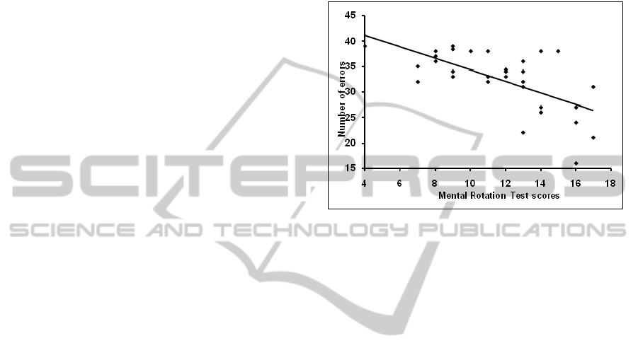
Study 1 (Guillot et al. 2007)
A significant correlation was shown between visuo-
spatial abilities and anatomy examination results for
both the GEFT and the VMRT (fig 1).
Figure 1: Correlation between anatomy examination errors
number and VMRT score. Students having good VMRT
scores made fewer errors on the anatomy examination.
Study 2 (Hoyek et al. 2009)
No significant difference was found between the
three groups, (F2, 45 = .12, p > .05, ns) on the
VMRT pre-test scores hence attesting for their
homogeneity. However the performance
enhancement was greater in the MR training group
compared to the anatomy control group (t = 4.14,
p <.001) suggesting a positive effect of MR training
sessions on VMRT performance. In MR questions,
the MR training group tended to score slightly better
than the anatomy group (F1, 29 = 3.52, p = .07). By
contrast, we did not found any statistical difference
regarding the specific knowledge questions (F1,
29 = .02, p = .8). Anatomy scores results are shown
in figure 2.
CSEDU 2011 - 3rd International Conference on Computer Supported Education
226

Figure 2: Anatomy examination scores. MR training group
(in red) had better results on the anatomy questions
requiring MR ability (right histogram).
Study 3
Unexpectedly no significant results were found
between the 3 groups on the post-test scores.
Consequently, using 3D visual tools was not
beneficial in this particular experimental design.
4 DISCUSSION
The correlations found in study 1 underscore the
advantage of students with high spatial abilities.
Such abilities could therefore be considered reliable
forecasters of success in acquiring human anatomy
knowledge. Furthermore, such predictive tests could
affect technical skills learning and training in
various scientific (e.g. architecture and design) and
medical disciplines, and help to identify students
who might need supplementary teaching modules.
Study 2 extended these results. Participants became
more accurate in solving the VMRT after practice
explaining a positive transfer of spatial reasoning.
Furthermore, after MR training, participants may
improve their ability to learn anatomical knowledge
by increasing their ability to make the anatomical
structures rotating. These results emphasize the
argument that spatial ability training as well as using
3D technologies may help student in various
scientific and medical disciplines. However the
unexpected results of study 3 are probably due to the
method. We argue that 2 hours are insufficient to
master 3D tools and then to acquire new anatomical
knowledge. In a future study we will evaluate the
effects of using 3D instructional tools over a hall
teaching semester.
REFERENCE
Garg, A. X., Norman, G., Sperotable, L. 2001. How
medical students learn spatial anatomy. Lancet, 357,
pp.363-364.
Guillot, A., Champely, S., Batier, C., Thiriet, P., Collet, C.
2007. Relationship Between Spatial Abilities, Mental
Rotation and Functional Anatomy Learning. Advances
in Health Sciences Education, 12, pp.491-507.
Hegarty, M., Keehner, M. & Cohen, C. 2007. The Role of
Spatial Cognition in Medicine: Applications for
Selecting and Training Professionals. In G. Allen (Ed)
Applied Spatial Cognition. Mahwah, NJ: Erlbaum,
285-316.
Hoyek, N., Collet, C., Rastello, O., Fargier, P., Thiriet, P.,
Guillot, A. 2009. Enhancement of Mental Rotation
Abilities and its Effect on Anatomy Learning.
Teaching and Learning in Medicine, 21 (3), pp.201-
206.
Keehner, M., Tendick, F., Meng, M. W., et al. 2004.
Spatial ability, experience, and skill in laparoscopic
surgery. American Journal of Surgery, 188, pp.71-75.
Risucci, D. A. 2002. Visual spatial perception and surgical
competence. The American Journal of Surgery, 184,
pp.291–295.
Rochford, K. 1985. Spatial learning disabilities and
underachievement among university anatomy students.
Medical Education, 19 (1), pp.13-26.
Wanzel, K. R., Hamstra, S. J., Caminiti, M. F., et al. 2003.
Visual-spatial ability correlates with efficiency of hand
motion and successful surgical performance. Surgery,
134 (5), pp.750-757.
EXPERIMENTAL RESEARCH VALIDATION FOR THE USE OF 3D IN TEACHING HUMAN ANATOMY
227
