
DIGITAL LARYNGOSCOPE
A New Force Measuring Laryngoscope
António Silva
1
, Pedro Amorim
2
, Manuel Quintas
1
, Luis Mourão
3
and Joaquim Gabriel
1
1
Labiomep, IDMEC – Pólo FEUP, Faculdade de Engenharia da Universidade do Porto, Porto, Portugal
2
Hospital Santo António, Centro Hospitalar do Porto, Porto, Portugal
3
Department of Biomedical Engineering, Escola Superior de Engenharia e Gestão Industrial,
Instituto Politécnico do Porto, Vila do Conde, Portugal
Keywords: Laryngoscopy, Anaesthesia, Force measurement, Medical devices.
Abstract: A laryngoscope is a medical device commonly used in most hospitals worldwide and used to conduct an
oral or endotracheal intubation which leads to changes in the patient parameters (heart rate, blood pressure,
etc.) due to the force applied on the tongue and other soft-tissues. However, these parameters are being
monitored continuously, and provide guidance for anaesthetists to control the drugs which may lead to an
inadequate dosage. This work aims to develop a laryngoscope capable of measuring the force applied during
a laryngoscopy. To measure the applied force, several solutions, based on different sensors, were analysed
and tested. The traditional laryngoscope xenon lighting lamp was replaced by a high bright LED which
result in a clear illumination and lower batteries consumption. A Bluetooth® communication module was
also include to allow a real-time force acquisition and display.
1 INTRODUCTION
1.1 Context
A laryngoscopy, as analyzed in this work, is a
medical procedure performed by anaesthetists in
order to achieve a good intubation and mechanical
ventilation, when the patients are subjected to
general anesthesia (GA). The anesthesia has three
goals: 1) disable the muscular activity to prevent
inadequate movement of the patient, 2) created an
unconsciousness (achieved by an hypnotic drug)
state and 3) avoid the sense of pain (analgesia).
1.2 Main Goals
This research work intends to achieve the following
objectives:
Develop a new laryngoscope capable of
measuring and record force in real-time;
Create wireless communication ability via
standard communication protocols;
Real-time force warnings based on trigger
values previously defined;
Keep the common laryngoscopy procedure,
weight and size;
Analysis software to view the recorded data;
Replace the usual Xenon light by the LED to
improves visibility.
2 ENDOTRACHEAL
INTUBATION
2.1 Laryngoscope
The laryngoscope is basically composed by two
parts: the handle and the blade (Figure 1). The
handle includes the lightbulb, the automatic switch
(to turn on/off the light), the batteries and the axis to
fit the blade. The blade possesses the correspondent
socket to connect to the handle and a metal protected
optical fiber to guide the light to the tip of the blade
when the laryngoscope is in working position (B).
Laryngoscope failure is extremely rare, but over
the years some have been registered, like
(Desmeules H. 1998). However, when it occurs, it is
in a very critical moment of the anaesthesia process,
since the patient is in apnoea without autonomous
breathing capability.
With the recent improvement in the overall
technologies, new laryngoscopes started to appear
368
Silva A., Amorim P., Quintas M., Mourão L. and Gabriel J..
DIGITAL LARYNGOSCOPE - A New Force Measuring Laryngoscope.
DOI: 10.5220/0003794303680371
In Proceedings of the International Conference on Biomedical Electronics and Devices (BIODEVICES-2012), pages 368-371
ISBN: 978-989-8425-91-1
Copyright
c
2012 SCITEPRESS (Science and Technology Publications, Lda.)
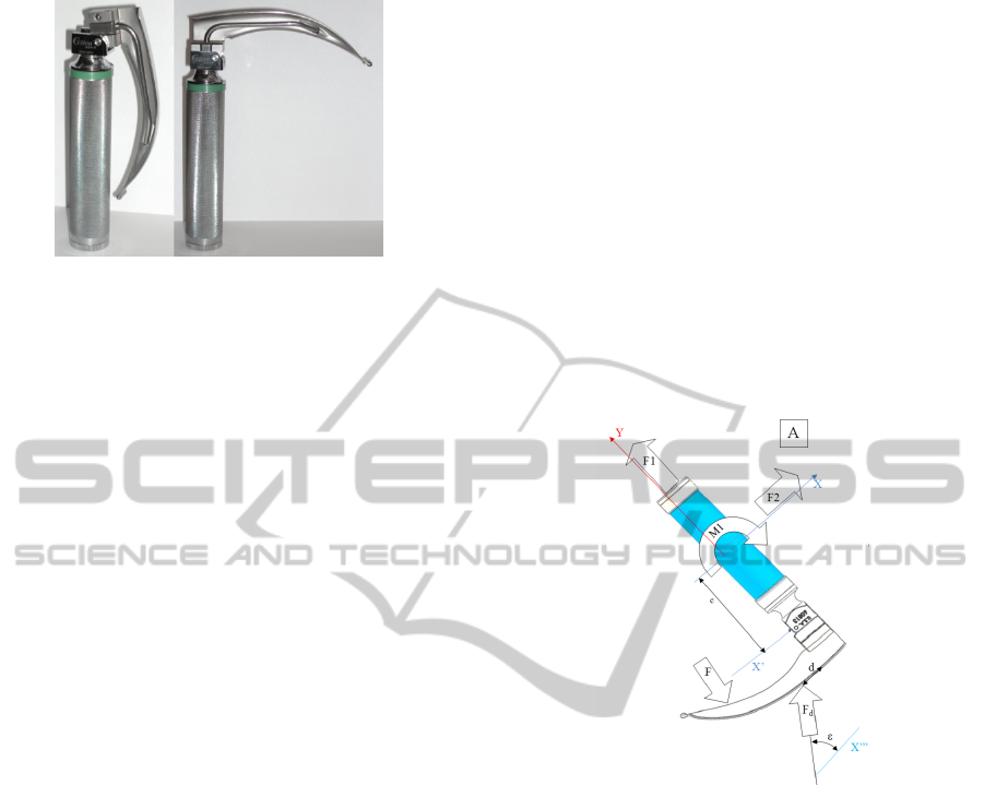
Figure 1: Laryngoscope handle and Macintosh blade size 3
A) closed position, B) opened position.
Whether may be improvements on the materials,
which may lead to disposable laryngoscopes, or
improvements in electronics that includes the video-
laryngoscopes (indirect laryngoscopies).
The disposable solution appears mostly because
of the wars, since it those situations it would be
difficult to have the sterilization equipment at a
hand.
2.2 Other Works
One of the first works on this field was (Grogono,
1983). He used strain gauges to measure the forces
in the patient mouth, Butt he did not explain how the
data was processed to get the force.
Another notorious work was done by (Hastings,
et al, 1996). He use springs and various sensors to
measure the force and torque aplied to the
laryngoscope. But, as previous work he did not tell
which was the precision or sensitivity of the
instrument.
On the other hand, there exist several studies
related to laryngoscopy force and pressure
measurements. But the target of this work was
studying the diference between experienced vs
novice users (Martin. J. L et al, 1994), nothing was
said about the device it self.
In the last years, some new devices arrived to the
market, called video laryngoscopes. These devices
use a small video camera that transmits the image to
a small screen outside of the patient mouth. Some
studies like (Ray, et al, 2009) tried to compare the
video-laryngoscopes with the standard
laryngoscopes, but without a way to measure the
damage or pressure applied to the patient, the
importance or utility of these studies is greatly
diminished.
2.3 Laryngoscopy
The laryngoscopy is a quick procedure which
requires very precise movement and force control
abilities, only achived by well trained professionals.
To perform a correct laryngoscopy the patient
has to lay down on his back and put his neck in a
hyperextension position, this causes the airway to be
in the straightest position. With the Laryngoscope in
the patients mouth, the endotracheal tube is
positioned at the entrance of the mouth and the
laryngoscope handle placed in a 45° (Figure 2 Y
axis) with the patient torso. The endotracheal tube is
then inserted between the vocal cords. This is when
most damage occurs, whether due to an excessive
force (F1) or by pulling the device in a wrong
direction (wrong amount of F2 or/and M1). This
leads to a larger force F that will hurt the patient or
produce an incorrect positioning of the blade.
Figure 2: Forces during laryngoscopy.
By analysing the laryngoscope internal forces, it
is easy to conclude that the force F and F
d
generate a
compression force in the contact point that occurs
when the laryngoscope is in the open position.
Based on previous studies, the position of the
force F along the blade can be estimated for normal
situations (Silva 2010). Measuring it in real time
may be possible but it requires the design of anew
laryngoscope blade.
To set the trigger value for the maximum force, a
wireless module was designed, in order to record
real data from laryngoscopies (Gabriel 2010). With
these results, a maximum force of 50 N in the tip of
the laryngoscope blade Macintosh type, size 3 was
set.
3 SENSOR POSSIBILITIES
The selection of the sensor that would lead to the
DIGITAL LARYNGOSCOPE - A New Force Measuring Laryngoscope
369

most precise and sensitive situation, three solutions
using the following sensors were evaluated:
1. Piezoresistive sensor
2. Hall effect sensor
3. Strain gauge sensor
In order to test these three sensors, it was build a
testing handle that permit the evaluation of all three
solutions, each one using a different sensor and a
different pin.
3.1 Overall Comparison
Like in the most projects, the sensor should not be
evaluated individually but together with the signal
conditioning circuit and overall characteristics. That
way, table 1 presents a general evaluation of the
idealized solution for each sensor.
Table 1: Overall sensors evaluation.
Sensor
Piezo-resistive Hall effect Strain gauge
Electric noise Medium Low Low
Space saving Medium Medium High
Reliability Low Medium High
Hysteresis High
Low (near
zero)
Low (near
zero)
Equivalent
noise (kgf)
0.2 0.36 0.02 – 0.03
It is notorious that the strain gauges present a far
better solution than any other sensor for this
particular application.
This is natural, since most of the commercial
load cells use also strain gauges. The downfall is
that due to the available size, a commercial load cell
did not fit inside the laryngoscope handle.
Despite the other solutions, it was chosen to
design a custom made load cell to fit in a new and
redesigned laryngoscope handle.
4 FINAL PROTOTYPE
4.1 General Features
The device could be divided in to three different
groups, mechanical parts, electronic circuits and data
acquisition and analyse software.
4.2 Mechanical Parts
The mechanical parts were in a first-hand simulated
using Solidworks®, this enabled the assembly
simulation and mechanical tests using the FEA
method.
The custom made load cell was also analysed
using a FEA that enabled the optimization of the
sensitivity vs mechanical strength to support the
forces applied to it (using a safety factor of 2.2). The
result was verified experimentally to prevent any
future failure.
4.3 Software
The goal of the software was to receive the data
from the digital laryngoscope interpret it and convert
the raw data so it could be displayed as force and
angles.
The final version could be installed in a
computer whether use Microsoft windows, Mac
OSX or Linux operating systems. This provides a
big flexibility whether it may be install in an
operation room or to analyse data in any other place.
The software is divided into four different tabs:
Setup, Reader, DL Analyser and Quick Help.
5 RESULTS
One of the most notorious differences is the
visibility increase provided by the illumination LED,
Figure 3.
Figure 3: Light visibility comparison.
The appearance of the Digital Laryngoscope,
Figure 4, is similar to the common one, being a little
longer (12 mm) and thicker, the diameter was
increased by 1.7 mm.
The device was tested in two different situations,
with an intubation simulator and real life situations.
In the simulator, trained doctors reached a
maximum force of 4.5 kgf (without warnings) and
non-experienced user were able to complete the
intubation with a maximum force around 2.7 kgf
(with the Digital Laryngoscope warnings).
The real life tests (Figure 5) served to validate
the device and the software. The global opinion was
that the Digital Laryngoscope was very similar to
Standard Laryngoscope Digital Laryngoscope
BIODEVICES 2012 - International Conference on Biomedical Electronics and Devices
370
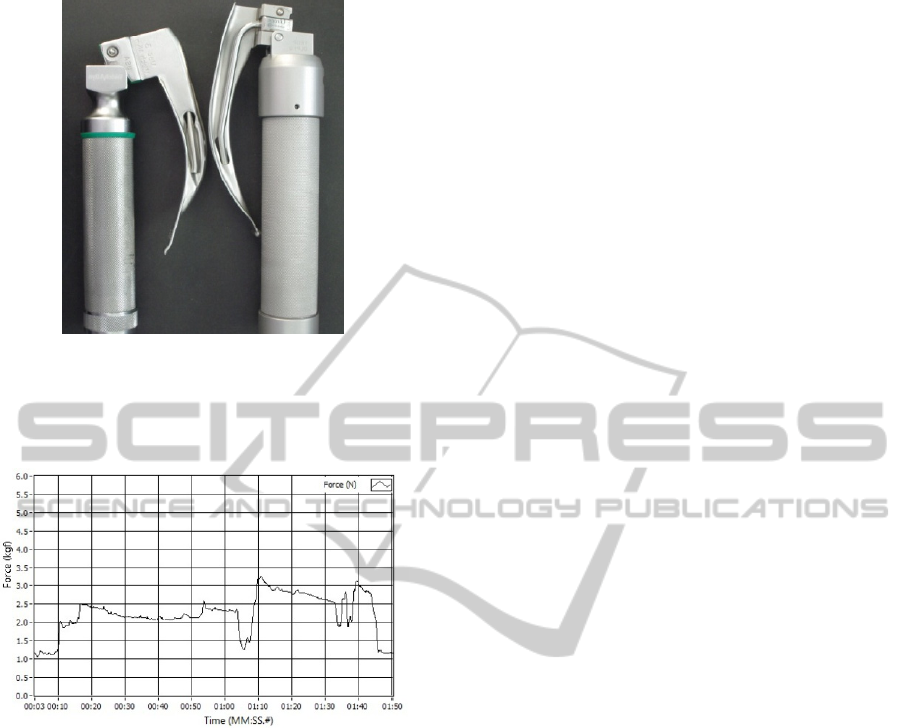
Figure 4: Standard Laryngoscope (left) and Digital
Laryngoscope (right).
the other not requiring any special training or
readjust of the global anaesthesia procedures.
Figure 5: Force example.
6 CONCLUSIONS
The Digital Laryngoscope was accepted by the
medical doctors with great enthusiasm. The overall
opinion was that the visibility improvement was
very good, greatly facilitating the intubation
procedure. The force warnings were very easy to
interpret and did not distract the doctor from the
main goal (look at the vocal cords).
Another positive aspect is that the this device is
completely compatible with the standards
laryngoscope blades, meaning it is not necessary to
buy a complete set of laryngoscope blades.
One other application for this device is in the
training and teaching of the intubation procedure.
The laryngoscope can be used at medical schools, to
train future doctors and to help them to avoid
possible damage in real patients. So, in relaxed and
control environment, it is possible to train the
intubation and have some numerical parameters that
can give a helpful feedback to their training.
ACKNOWLEDGEMENTS
This research was sponsored by FCT-Fundação para
a Ciência e a Tecnologia, under the project
PTDC/EEA-ACR/75454/2006.
REFERENCES
R. T. C. F. B. A. J. G. A. W. Grogono, "A measuring
laryngoscope handle: a device for measuring the
forces applied during laryngoscopy" Med. & Biol.
Eng. & Comput., 1983.
R. H. Hastings, et al., "Force, torque, and stress relaxation
with direct laryngoscopy," Anesthesia & Analgesia,
vol. 82, pp. 456-461, March 1, 1996 1996.
Martin. J. L. B. MD, et al., "Does experience influence
the forces exerted on maxillary incisors during laryn-
goscopy? A manikin study using the Macintosh
laryngoscope " pp. 1,2,3, 16, october, 1994 1994.
D. C. Ray, et al., "A comparison of McGrath and
Macintosh laryngoscopes in novice users: a manikin
study," Anaesthesia, vol. 64, pp. 1207-1210, 2009.
Desmeules H., T. P.-R. (1998). Laryngoscope blade
breakage during intubation.
Gabriel, J., Carlos Teixeira, António Silva, O. Postolache,
G. Postolache, Pedro Amorim (2010). Measuring
Force in a Laryngoscope. The Seventh IASTED
International Conference on Biomedical Engineering,
Innsbruck, Austria.
Silva, A. T., J.; Gabriel J.; Teixeira, C.; Amorim, P.;
Quintas, M.; Natal, R. (2010). Measuring the Pressure
in a Laryngoscope Blade. TMSi 2010 - 6th
International Conference on Technology and Medical
Sciences, Faculty of Engineering, University of Porto,
Portugal.
DIGITAL LARYNGOSCOPE - A New Force Measuring Laryngoscope
371
