
OPTICAL SPECTROSCOPY
AND OBSTACLES BY NON-INVASIVE DETECTION OF
GLUCOSE CONCENTRATION BY HOME MONITORING
O. Abdallah, Q. Qananwah, A. Bolz
Institute of Biomedical Engineering IBT, Karlsruhe Institute of Technology KIT, Karlsruhe, Germany
J. Hansmann, H. Walles, T. Hirth
Fraunhofer Institute for Interfacial Engineering and Biotechnology IGB, University of Stuttgart, Stuttgart, Germany
Keywords: Glucose Management, Raman Scattering IR Spectroscopy, Fluorescence Spectroscopy, Blood Components
Concentration Monitoring, High Signal to Noise Ratio, Parameters Affecting In-vivo Glucose
Measurement.
Abstract: Tight glycemic monitoring and control is the main goal in successful diabetes management to avoid its
complications. Frequent blood glucose measurements with a combination of regimented diet, exercise and
insulin administration can accomplish this task. Different methods are applied for non-invasive
measurement of blood glucose concentration. Despite the great interest and the intensive research in this
field since 1980s, there is no convenient device at the market that can measure the glucose concentration
non-invasively in an easy manner. This paper discusses the different methods for detecting the glucose
concentration. Elastic and inelastic (Raman) scattering as well as fluorescence and IR Spectroscopy
measurements well be shown and discussed for the development of a compact non-invasive device for home
monitoring. In conclusions, an optical multi-sensor measuring the fluorescence and light scattering in the
tissue optical window in and around visible range (360 nm – 1200 nm) taking the perturbation factors into
account is promising and under development.
1 INTRODUCTION
Diabetes risk lies in its complications like heart
diseases and infarcts, stroke, blindness, kidney
disease, nerve disease, diabetic foot and amputation.
The current applied invasive methods are
intermittent, inconvenient and painful, having
infection risk, blood loss and time delay, need
consumables materials, needles and strips. The
invasive method cannot be applied continuously, and
hence hypo- or hyperglycemia may be not detected.
An easy accessible and low-cost method for
continuous glucose concentration monitoring and
diabetes management will be a great help for more
than 250 millions of diabetic patients worldwide to
avoid the risks and the complications caused by
hyper- or hypoglycaemia. Standard treatment
includes lifestyle changes, medication and frequent
monitoring of blood glucose levels.
Insulin and other
diabetes medications are designed to lower the blood
sugar level when diet and exercise alone aren't
sufficient for managing diabetes. By the
development of a compact system, different LASER
diodes (LD`s) or light emitting diodes (LED`s) in
the range of UV and NIR will be used. Light
scattering and fluorescence spectroscopy can be
applied for non-invasive measurement of blood
components like glucose concentration or the early
detection of pathological variations like melanoma.
The complications of diabetes are largely
avoidable and may be reversed by strict control of
blood sugars through medication and diet. Patients
with type 2 diabetes mellitus are at increased risk for
macrovascular disease complications (Gaster, 1998;
Mezzetti, 2000; Pambianco, 2006). Detection of
blood contents like Glucose non-invasively in an
easy manner can reduce morbidity and mortality by
diabetics.
291
Abdallah O., Qananwah Q., Bolz A., Hansmann J., Walles H. and Hirth T..
OPTICAL SPECTROSCOPY AND OBSTACLES BY NON-INVASIVE DETECTION OF GLUCOSE CONCENTRATION BY HOME MONITORING.
DOI: 10.5220/0003796902910296
In Proceedings of the International Conference on Bio-inspired Systems and Signal Processing (BIOSIGNALS-2012), pages 291-296
ISBN: 978-989-8425-89-8
Copyright
c
2012 SCITEPRESS (Science and Technology Publications, Lda.)

1.1 Non-invasive Methods for
Detecting Glucose Concentration
In-vitro Glucose concentration measurements
depend on chemical principles. The non invasive
monitoring methods are much more difficult to
apply with the required accuracy. Glucose
concentrations in blood are very low compared for
example with that of hemoglobin. The effect of
glucose on the measured signals is too low and
hence a high amplification is needed, which means
that other background and surrounding noises will
also be amplified and hence the measurement will be
very sensible for tiny perturbations.
The different methods for non-invasive glucose
concentration montoring are discussed in diverse
literature (Khalil, 2004; Yamakoshi, 2006; Maruo,
2003; Tura, 2010). Raman and fluorescence
spectroscopy as examples of promising optical
methods will be briefly discussed in the next section.
1.2 Optical Spectroscopy
The optical methods rely on the interaction between
light and tissue. They are widely used by different
research groups and companies.
1.2.1 Raman Spectroscopy
Raman spectroscopy measures scattered light that
has been influenced by the oscillation and rotation of
the scattered molecules. Various Raman techniques
have been attempted in blood, water, serum, plasma
solutions and the eye, but multiple problems remain
before human studies can be accomplished.
Analytical problems include instability in the laser
wavelength and intensity, errors due to other
chemicals in the tissue sample and long spectral
acquisition times. The applying of special types of
Raman spectroscopy can greatly enhance the signal
noise ratio, the resolution and sensitivity. Resonance
Raman (RR) scattering, surface enhanced Raman
spectroscopy (SERS)
12
, coherent anti-Stokes Raman
scattering spectroscopy (CARS), and Stimulated
Raman scattering (SRS) are examples of the Raman
enhancement methods.
1.2.2 Fluorescence Spectroscopy
Fluorescence spectroscopy and time resolved
fluorescence are dominant methodologies and used
extensively not only in biochemistry and biophysics,
but also in biotechnology, medical dioagnostics and
genetic analysis (Moschou, 2004, Pickup, 2005,
Lakovics, 2006). The technique is extremely
sensitive. There are increasing examples of even
single-molecule detection using fluorescence
methods. Many studies indicate that fluorescent
technology has real sensitivity especially in low
glucose ranges. In addition, since near-infrared light
passes through several centimeters of tissue, with the
appropriate choice of fluorophore, molecules can in
theory be excited and the emission interrogated from
outside the body providing the potential for
completely non-invasive sensing. A convenient way
of classifying fluorescence-based glucose sensors
that involve measurements of fluorescence is either
according to the type of molecular receptor for
glucose, or whether cells or tissues are used to signal
glucose concentrations and/or glucose metabolism.
A review of the principles of operation and current
status of the various approaches to fluorescence-
based glucose sensing are described in D’Auria,
1999.
In DMEM solution a glucose dependent
autofluoresence can be observed. The fluorescence
differs from the process of Raman Effect in that the
incident light is completely absorbed and the system
is transferred to an excited state from which it can
go to various lower states only after a certain
resonance lifetime.
2 APPARATUS AND METHOD
Our measurements were obtained using a micro
Raman spectrometer, based on Olympus IX71
microscope by Fraunhofer Institute in Stuttgart. The
separation of spectrums is achieved using holo-
graphic grill in spectrograpic Holospec f/1.8 (Kaiser
Optical Systems). Spectrum detection attained using
a CCD for NIR (DU420A-BR-DD, 1024x256 Pixel
von Andor). The measurements are done using a
glas bottom dish Willco Welles.
The method discussed here can be applied for
invasive and non-invasive measurement. Photo-
diodes or phototransistors for light scattering and
fluorescence detection in the visible and NIR
spectrum are used. Light emitting diodes LED and
LASER diodes as light sources are applied. Variable
frequency and duty cycle can be adjusted for time
resolved fluorescence signal detection.
The developed system is flexible and can be used
for the development purposes, where different
parameters have to be adjusted. Light intensities,
duty cycle, different LASER types and variable
amplification can be acquired using this system.
BIOSIGNALS 2012 - International Conference on Bio-inspired Systems and Signal Processing
292
In the meantime we are developing a time
resolved spectroscopy system in order to take more
parameters into account by the calculation of
glucose with a multisensor. By applying a
multisensor for the detection of glucose
concentration a great attention has to be given for
the calibration method.
As we discussed in other papers (Abdallah,
2010) and as we can see in diverse literature the
signal to noise ratio has to be kept as high as
possible, but the glucose signal is too low, so that in
addition to diminish the noise and considering all
parameters affecting the measurements an adequate
calibration is necessary for accurate glucose
measurement non-invasively. The scattered signals
may come mainly from deep tissue and blood
glucose may be taken as reference. Moreover
hemoglobin concentration and oxygen saturation
have to be taken in to account by these
measurements. Direct invasive measured values can
be taken as reference. But fluorescence signals may
come mainly from intestinal skin fluid ISF. Transfer
of glucose from the blood to the ISF compartment
occurs by passive diffusion through an established
concentration gradient. The mass transfer rate is
affected by several variables, such as the blood flow
rate to the site, rate of glucose uptake by the
surrounding tissue, and capillary permeability.
Nevertheless, as discussed in the literature [Barman
2010], a simple mass transfer model can be written
for the ISF volume (VISF).
The review by Ziegler (Zierler, 1999) describes
major factors that, singly or together, influence the
concentration and distribution of D-glucose in
humans, with emphasis on rest, physical activity, and
alimentation. It identifies areas of uncertainty:
distribution and concentrations of glucose in
interstitial fluid, kinetics and mechanism of
transcapillary glucose transport, kinetics and
mechanism of glucose transport via its transporters
into cells, detailed mechanisms by which hormones,
exercise, and hypoxia affect glucose movement across
cell membranes, whether translocation of glucose
transporters to the cell membrane accounts
completely, or even mainly, for insulin-stimulated
glucose uptake, whether exercise stimulates release of
a circulating insulinomimetic factor, and the relation
between muscle glucose uptake and muscle blood
flow. It was pointed out that there is no compartment
of glucose in the body at which all glucose has the
same concentration, and that models of glucose
metabolism, including effects of insulin on glucose
metabolism based on assumptions of concentration
homogeneity, cannot be entirely correct.
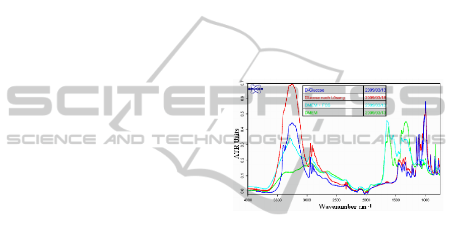
3 RESULTS AND DISCUSSION
The results obtained by applying a simple system
with costs effective components and using DMEM
solutions show that the measurements are
reproducible under the same conditions. Using
different light sources (LASER, LED, IR-emitter)
having wavelengths in the visible and IR and using
photodiodes and thermopile as detectors have shown
the same tendency by measurements. A few
wavelengths have demonstrated more dependency
on the glucose concentrations. As an example of the
measurements, figure 1 shows the high dependency
of the detected light from glucose concentrations in
glucose DMEM solutions by IR around the wave
number 3200 cm
-1
and between 1000 and 1700 cm
-1
.
Figure 1: IR-Spectroscopy by DMEM glucose solution.
The tissue light absorption in the IR-range is too
high due to the high water absorption, so that the
light penetration in tissue is too small. Light emitting
diodes and IR-detectors are also too expensive in
this range. The non-invasive glucose measurement
in this range is then too difficult and very expensive
for home monitoring. Using an IR-emitter as an
example of the results by measurements in DMEM
solution and a thermopile as detector of scattered IR
radiation between 9000 nm and 10000 nm have
shown good results. Instead of IR-Emitter and
thermopile, Laser sources and pyroelectric infrared
sensors can be utilized.
By Raman scattering without enhancement the
resolution of the measurements was not enough for
the glucose concentrations monitoring. Increasing
the measuring time will increase the resolution and
the detection threshold. Figure 2 shows that the
increase of the detection time increases the signal
quality and the resolution; where as the resulting
error will be reduced. The measuring time in Figure
2 by measurements an DMEM-solutions having
different glucose concentrations is five minutes. As
shown in the non-processed signal, the detection
OPTICAL SPECTROSCOPY AND OBSTACLES BY NON-INVASIVE DETECTION OF GLUCOSE
CONCENTRATION BY HOME MONITORING
293
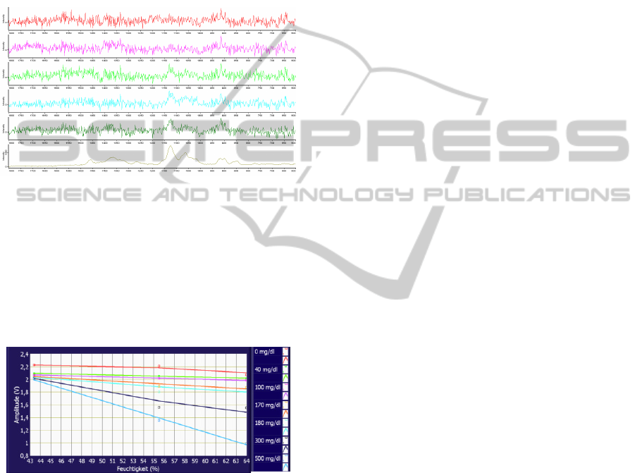
threshold is ca. 30 mg/dl and the resolution seems to
be around 10 mg/dl. Increasing the measuring time
may be possible by in-vivo measurements. The
motion artifact will cause large perturbations to the
detected signals, which can be minimized by
applying an adaptive filter. The enhancement of the
Raman scattering by applying the previous
mentioned methods can produce results having a
high resolution and the detection time may be
reduced.
Figure 2: Raman scattering measured using a LASER
diode with wavelength 785 nm and 80 mW power. From
top: 300 s measuring time; a. 10mg/dl, b. 20 mg/dl, c. 30
mg/dl, d. 40 mg/dl, e. 50 mg/dl, f. reference solution with
a very high glucose concentration.
The realization of a compact simple device for
home monitoring using Raman spectroscopy seems
to be very difficult, but it may be possible using
miniaturized components and intelligent methods.
Figure 3: Scattered measured signal humidity for different
glucose concentrations al parameters.
The relation between the scattered measured
signal and humidity for different glucose
concentrations are shown in Figure 3. The results
obtained by using a fluorescence spectrometer show
the emitted light by stimulation of a DMEM solution
with different glucose concentrations. The detected
signal with 465 nm by the stimulation in the UV
light at the wavelength of 360 nm is not highly
correlated with the glucose concentration. But an
increasing tendency of the emitted light with the
increasing glucose concentration is registered.
The detected signal at 535 nm shows a high
correlation with glucose concentration when
stimulated with a light by the wavelength 485 nm. In
the contrary to the detected signals at 465 nm
mentioned above, a decreasing tendency of the
emitted light with the increasing glucose
concentration is shown. Despite of the variations of
the detected signals around the wavelengths 465 nm
when stimulated with the UV-light at the wavelength
360 nm, non-invasive measurement may deliver
very good results due to other fluorophores in the
skin.
A high correlation of the detected signal at
535 nm with known glucose concentration when
stimulated with 485 nm or 430 nm at different
glucose concentrations was obtained in DMEM-
solutions.
The detected glucose signals are too small and
should be processed carefully. Also the in vivo
measurements are subjected to more noise and
motion artifacts. An adaptive filtering will be needed
for eliminating these perturbations. A noise
reference signal is generated by means of a
Synthesizer or piezoelectric element and will be
adjusted as much as possible to the real noise
contained in the corresponding measurement by the
adaptive filter based on the least mean square
optimization algorithm. This algorithm has delivered
very good results by testing it for the non-invasive
calculations of oxygen saturation by artificial
vibrations of the hand, where a pulse oximeter
sensor is applied at the finger subjected to these
artifacts. We are applying a multisensor technology
that overcomes the obstacles others have faced
trying to measure blood glucose optically through
the skin.
4 CONCLUSIONS
AND FUTURE WORK
Optical methods are valuable and promising for the
non-invasive detection of blood glucose. Raman
scattering can have a very high sensitivity and
resolution, but the development of an in-vivo simple
device till now, seems to be very difficult. IR-
spectroscopy is promising for the development of a
cost effective sensor for home monitoring. Because
of the fact that the detected glucose signals are too
small and subjected to a lot of disturbances from the
surroundings and from the background of the
measured locations due to tissue alteration and
physiological parameter variations, a high signal to
noise ratio measuring system is essential for this
difficult task. Tiny perturbations such as
BIOSIGNALS 2012 - International Conference on Bio-inspired Systems and Signal Processing
294
temperature, humidity and applied pressure
variations can adulterate the measurements. Also
small drift of the characteristics of the electronic and
optical components can cause great disturbances to
the measurements reducing the accuracy or even
yielding invalid measurements. In addition to use a
robust hardware and apply advanced signal
processing methods by the glucose detection, all
these factors have to be taken into account.
The results obtained using a fluorescence
spectrometer having the stimulation/emission wave-
lengths of 360 nm /465 nm, 430 nm / 535 nm and
485 nm / 535 nm (Abdallah, 2011) as well as our
further fluorescence measurements at these and
further wavelengths have shown that fluorescence
spectroscopy is a very promising method. We have
already developed a compact sensible system and
sensor for that aim.
The integration of further parameters can
enhance the accuracy, but the system complexity, its
size and costs have to be minimized to enable the
applying of the device for home monitoring. Also
the number of the measured parameters has to be
minimized in order to reduce the resulting error
caused by the measurements variations..
Also by applying light sources with wavelengths
in IR over 1400 nm the penetration depth of light in
the tissue will be very small because water has a
high absorption of IR-light. This will be important
by detecting glucose or cholesterol using IR-
spectroscopy. The reflective sensors can be applied
proximal in order to avoid the perfusion problem by
applying sensors (in case of transmission) distal to
body extremity like fingers or earlobe. Light
reflected from the tissues and detected by
photodetectors and then the findings can be
interpreted by the software in the sensor. The
reflection sensor can be applied on forehead, back,
breast etc., and hence diagnose the central parts of
the body.
For the detection of glucose concentration
noninvasively using various optical methods, the
interaction between light and definite glucose
solutions was studied. IR-spectroscopy has the
potential for the development of a simple cost
effective sensor for glucose monitoring that can be
used for home care.
Problems with existing methods have
encouraged alternative approaches to glucose
sensing, and those based on multiparameter like
scattering, fluorescence intensity and lifetime have
special advantages, including sensitivity and the
potential for non-invasive measurement when UV,
visible or NIR light is used (Yamakoshi, 2006;
Evans, 2005; Evans, 2003; Pickupa, 2005). The
fluorescence signals using UV light as stimulus and
detection of fluorescence at violet or blue have
shown a very good correlation with the glucose
concentrations in DMEM solution. Light stimulation
with blue light and the detection of fluorescence by
green region shows also a high correlation with the
glucose concentrations. The detected glucose signals
will be subjected to perturbations from the
surroundings and from the background of the
measured locations due to tissue alteration and
physiological parameter variations. All perturbations
such as temperature, humidity and applied pressure
variations have to be considered by the calculations,
as illustrated by Figure 4. The drift of the
characteristics of the system components may cause
high disturbances to the measurements.
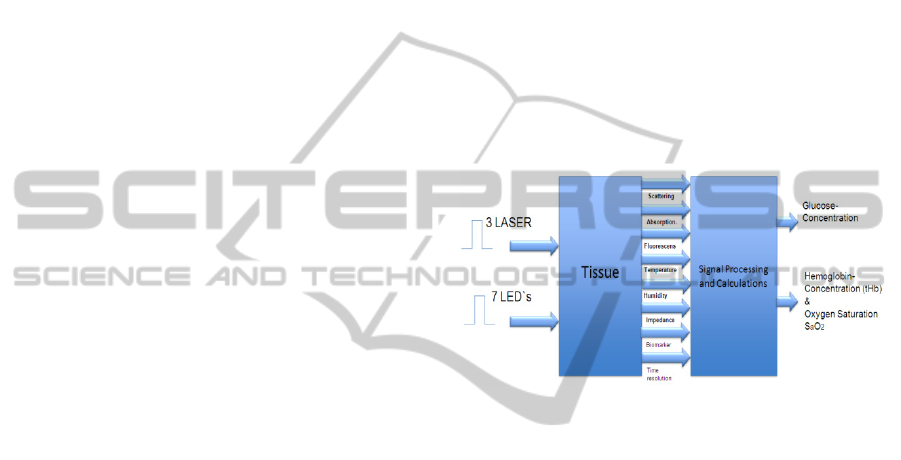
Figure 4: Schematic of a multisensor for non-invasive
detection of blood glucose, hemoglobin concentration, and
fractional oxygen saturation.
There is no doubt that the multiparametic
measurement depending on scattering, absorption
and fluorescence technologies have considerable
promise for glucose sensing.
As a future work, all developed sensors will be
integrated in one system that enables the
simultenous processing of the detected signals
(Caduff, 2009). Other blood components like total
hemoglobin concentrations and fractional oxygen
saturation measured non-invasively have to be taken
as parameters by the glucose calculations. The
suitable locations for measurements may be earlobe
for transmission measurements. For reflection
measurements forehead as well as abdomen or arm
can be chosen. Applying the Twersky theory or
diffusion theory by the calculations are our next
perspectives. After that a clinical study for non-
invasive measurements will be conducted. Applying
the neural fuzzy techniques, the results and the
system will be optimized to obtain the required
resolution and accuracy.
OPTICAL SPECTROSCOPY AND OBSTACLES BY NON-INVASIVE DETECTION OF GLUCOSE
CONCENTRATION BY HOME MONITORING
295

ACKNOWLEDGEMENTS
This work is a part of the project „System for Non-
invasive Detection of Glucose “supported by the
Foundation Baden-Württemberg Stiftung by
Research Program: Microsystem technology for the
life sciences.
REFERENCES
Abdallah O., Qananwah Q., Bolz, A. A., Hansmann J.,
Hinderer S., and Mertsching H., 2011. Fluorescence
Spectroscopy by Detection of Glucose Concentrations
in DMEM-Solutions and its Perspectives for Non-
invasive Measurement BIODEVICES 2011 part of
BIOSTEC, The International Joint Conference on
Biomedical Engineering Systems and Technologies,
Rome, Italy
Abdallah O., Hansmann J., Bolz A. and Mertsching H.,
2010. "IR spectroscopy vs. Raman scattering by
measurement of glucose concentration", Proc. SPIE
7376, 73760B; doi:10.1117/12.871469
Barman Ishan, Chae-Ryon Kong, Gajendra P. Singh,
Ramachandra R. Dasari, and Michael S. Feld, 2010:
Accurate Spectroscopic Calibration for Noninvasive
Glucose Monitoring by Modeling the Physiological
Glucose Dynamics. Analytical Chemistry 2010, Vol.
82, No. 14, pp. 6104–6114
Caduff, A., Talary, M., Mueller, M., Dewarrat, F., Klisic,
J., Donath, M., Heinemann, L., Stahel, W., 2009. Non-
invasive glucose monitoring in patients with Type 1
diabetes: A Multisensor system combining sensors for
dielectric and optical characterisation of skin. Bio-
sensors and Bioelectronics Vol. 24, pp. 2778– 2784.
D’Auria, S., Herman, P., Rossi, M., Lakowicz, J., 1999.
The Fluorescence Emission of the Apo-glucose
Oxidase from Aspergillusniger as Probe to Estimate
Glucose Concentrations. Biochemical and Biophysical
Research Communications, Vol. 263, pp. 550-553.
Evans, N., Gundi, L., Rolinski, O., Birch, D., Pickup, J.,
2003. Non-invasive glucose monitoring by NAD(P)H
autofluorescence spectroscopy in Fibroblasts and
adipocytes: A model for skin glucose sensing.
Diabetes Technology & Therapeutics, Vol. 5, No. 5.
Evans, N.D., Gnudi, L., Rolinski, O.J., Birch, D.J., Pickup,
J.C., 2005. Glucose-dependent changes in NAD(P)H-
related fluorescence lifetime of adipocytes and
fibroblasts in vitro: Potential for non-invasive glucose
sensing in diabetes mellitus. J. of Photochemistry and
Photobiology B: Biology, Vol. 80, pp. 122-129.
Gaster, Barak, Hirsch, MD; Irl B., MD, 1998. The Effects
of Improved Glycemic Controlon Complications in
Type 2 Diabetes, Arch Intern Med/VOL 158
Harman-Boehm Ilana, Avner Gal, Alexander M.
Raykhman, Jeffrey D. Zahn, Eugene Naidis, and
Yulia Mayzel, 2009. Noninvasive Glucose
Monitoring: A Novel Approach, Journal of Diabetes
Science and Technology Volume 3, Issue 2
Khalil, O.S., 2004. Non-invasive glucose measurement
technologies: An update from 1999 to the dawn of the
new millennium. Diabetes Technol. Ther., 6, 660-697
Lakovics, J., 2006. Principles of Fluorescence. Springer.
Maruo K., M. Tsurugi, J.Chin, T. Ota, H. Arimoto, Y.
Yamada, M. Tamura, M. Ishii, and Y. Ozaki, 2003.
"Noninvasive Blood Glucose Assay Using a Newly
Developed Near-Infrared System," IEEE Journal Of
Selected Topics In Quantum Electronics, 9 (2)
Mezzetti Andrea, Cipollone Francesco, Cuccurullo
Franco, 2000. Oxidative stress and cardiovascular
complications in diabetes: Isoprostanesas new markers
on an old paradigm, Cardiovasc. Research 47, 475–
488
Moschou, E., Sharma, P., Deo, S., Daunert, S., 2004.
Fluorescence Glucose Detection: Advances Toward
the Ideal In Vivo Biosensor. Journal of Fluorescence,
Vol. 14, No. 5.
Pambianco Georgia, Tina Costacou, Demetrius Ellis,
Dorothy J. Becker, Ronald Klein, andTrevor J.
Orchard, 2006. The 30-Year Natural History of Type 1
Diabetes Complications: The Pittsburgh Epidemiology
of Diabetes ComplicationsStudy Experience,
DIABETES, VOL. 55
Pickup, C. John, Faeiza Hussain, Nicholas D. Evans, Olaf
J. Rolinski, David J.S. Birch: Review: Fluorescence-
based glucose sensors, Biosensors and Bioelectronics
20, 2555–2565 (2005)
Smith John L., 2011, The Pursuit of Noninvasive Glucose:
“Hunting the Deceitful Turkey” Second Edition
Tura Andrea, Stefano Sbrignadello, Domenico
Cianciavicchia, Giovanni Pacini and Paolo Ravazzani,
2010. A Low Frequency Electromagnetic Sensor for
Indirect Measurement of Glucose Concentration: In
Vitro Experiments in Different Conductive Solutions
Sensors 10, pp. 5346-5358;
Wang Chuji and Sahay Peeyush, 2009, Breath Analysis
Using Laser Spectroscopic Techniques: Breath
Biomarkers, Spectral Fingerprints, and Detection
Limits, Sensors 2009, 9, PP. 8230-8262
Yamakoshi, K. Y. Yamakoshi: Pulse Glucometry, 2006. A
New Approach for Non-invasive Blood Glucose
Measurement Using Instantaneous Differential Near
Infrared Spectrophotometry, Journal of Biomedical
Optics 11(5), 1-11
Zierler, K., 1999. Whole body glucose metabolism. Am J
Physiol, 276(3 Pt 1):E409-E426
BIOSIGNALS 2012 - International Conference on Bio-inspired Systems and Signal Processing
296
