
REGION-BASED SKIN COLOR DETECTION
Rudra P. K. Poudel
1
, Hammadi Nait-Charif
1
, Jian J. Zhang
1
and David Liu
2
1
Media School, Bournemouth University, Weymouth House, Talbot Campus, Poole, Dorset, BH12 5BB, U.K.
2
Siemens Corporate Research, 755 College Road East, Princeton, NJ 08540, U.S.A.
Keywords:
Skin-color Detection, Region-based, Superpixels, Bayes Classifier, Conditional Random Field.
Abstract:
Skin color provides a powerful cue for complex computer vision applications. Although skin color detection
has been an active research area for decades, the mainstream technology is based on the individual pixels.
This paper presents a new region-based technique for skin color detection which outperforms the current
state-of-the-art pixel-based skin color detection method on the popular Compaq dataset (Jones and Rehg,
2002). Color and spatial distance based clustering technique is used to extract the regions from the images,
also known as superpixels. In the first step, our technique uses the state-of-the-art non-parametric pixel-based
skin color classifier (Jones and Rehg, 2002) which we call the basic skin color classifier. The pixel-based skin
color evidence is then aggregated to classify the superpixels. Finally, the Conditional Random Field (CRF)
is applied to further improve the results. As CRF operates over superpixels, the computational overhead is
minimal. Our technique achieves 91.17% true positive rate with 13.12% false negative rate on the Compaq
dataset tested over approximately 14,000 web images.
1 INTRODUCTION
Skin color provides a powerful cue in complex com-
puter vision applications such as hand tracking, face
tracking, and pornography detection. Skin color de-
tection is computationally efficient yet invariant of
rotation, scaling and occlusion. These are the ma-
jor reasons for its popularity. The main challenges of
skin color detection are illumination, ethnicity back-
ground, make-up, hairstyle, eyeglasses, background
color, shadows and motion (Kakumanu et al., 2007).
Many of the skin color detection problems could be
overcome by using infrared (Socolinsky et al., 2003)
and spectral imaging (Pan et al., 2003). However,
such systems are expensive as well as cumbersome
to implement. Moreover, there are many situations
where such systems can not be used such as image
retrieval on the internet.
Most of the skin color detection methods are pixel-
based, which treat each skin or non-skin pixel individ-
ually without considering its neighbours. However,
it is natural to treat skin or non-skin as regions in-
stead of individual pixels. Surprisingly, there are only
few region-based skin detection techniques (Yang
and Ahuja, 1998), (Kruppa et al., 2002), (Jedynak
et al., 2003) and (Sebe et al., 2004). Kruppa (Kruppa
et al., 2002), Yangand Ahuja (Yang and Ahuja, 1998)
searched for elliptical skin color shape to find the
face. Sebe (Sebe et al., 2004) used fixed 3x3 pixel
patches to train a Bayesian network and Jedyank (Je-
dynak et al., 2003) smoothed the results using hidden
Markov model. This paper proposes a new technique
purely based on the concept of regions, irrespective of
the underlying geometrical shape. As such, this tech-
nique can be easily integrated into any skin detection
based system.
Our technique uses a segmentation technique
called superpixel (Moore et al., 2008) and (Ren and
Malik, 2003) to group similar color pixels together.
Then each superpixel is classified as skin or non-skin
by aggregating pixel-based evidence obtained using a
histogram based Bayesian classifier similar to (Jones
and Rehg, 2002). The result is further improved with
CRF, which operate over superpixels instead of pix-
els. Even though the segmentation cost is an overhead
over the pixel-based approach, it greatly reduces the
processing cost further down the line, such as smooth-
ing with CRF. Also, aggregation of pixels into regions
helps to reduce local redundancy and the probability
of merging unrelated pixels (Soatto, 2009). Since
superpixels preserve the boundary of the objects, it
helps to achieve very accurate object segmentations
(Fulkerson et al., 2009).
The presented method not only outperforms the
current state-of-the-art pixel-based skin color detec-
tion methods but also extracts larger skin regions
301
P. K. Poudel R., Nait-Charif H., J. Zhang J. and Liu D..
REGION-BASED SKIN COLOR DETECTION.
DOI: 10.5220/0003801203010306
In Proceedings of the International Conference on Computer Vision Theory and Applications (VISAPP-2012), pages 301-306
ISBN: 978-989-8565-03-7
Copyright
c
2012 SCITEPRESS (Science and Technology Publications, Lda.)

while still keeping the false-positive rate lower, pro-
viding semantically more meaningful results. This
could in turn benefit higher-level vision tasks, such
as face or hand detection. Related work is discussed
in section 2; section 3 presents the proposed region-
based skin color detection technique; experiments and
results are discussed in section 4. Finally, we summa-
rize our work in section 5.
2 RELATED WORK
Skin color detection has two important parts: one is
color space selection and another is color modelling.
RGB: (Jones and Rehg, 2002), HSV: (Zhu et al.,
2004), CIE-Lab: (Kawato and Ohya, 2002), YCbCr:
(Wong et al., 2003), and normalized RGB: (Brown
et al., 2001) are popular color spaces, with RGB and
HSV being the most frequently used. CIE-Lab uni-
formly represents the color based on how two colors
differ to the human observer. HSV shows better re-
sults under varying illumination (Kakumanu et al.,
2007). However, most camera output RGB and il-
lumination variation can be eliminated by increasing
sample size (Jones and Rehg, 2002). Due to this rea-
son the RGB color space is chosen in our experiments.
Skin color modelling falls in three categories: ex-
plicitly defined skin region (Peer et al., 2003), non-
parametric and parametric methods. Histogram based
Bayes classifier is a popular non-parametric mod-
elling approach. Jones and Rehg (Jones and Rehg,
2002) used RGB color space and histograms based
Bayes classifier and obtained 90% true positive rate
with 14.5% false positive rate on unconstrained web
images, a dataset made up of approximately 14,000
images. On parametric skin modelling technique,
mixture of Gaussian has shown the best result (Ter-
rillon et al., 2000). However, Jones and Rehg (Jones
and Rehg, 2002) showed that given enough sam-
ples, the histogram based Bayes classifier technique
is slightly better than mixture of Gaussian. Neural
Network (Phung et al., 2002), self organizing map
(Brown et al., 2001), Bayesian network (Sebe et al.,
2004) and a few other methods have been used for
skin color modelling.
This paper presents a region-based skin color de-
tection method with no prior knowledge on the geo-
metric shape of the regions. The works of Yang and
Ahuja (Yang and Ahuja, 1998), Kruppa (Kruppa
et al., 2002), Jedyank (Jedynak et al., 2003) and Sebe
(Sebe et al., 2004) are the close to ours. However,
Yang and Ahuja (Yang and Ahuja, 1998) used multi-
scale segmentations to find elliptical regions for face
detection. Hence, their model is biased toward ellipti-
(a) Original image (b) After segmentation (c) Segmentation
visualization
Figure 1: An example of superpixel segmentation.
cal objects. Kruppa (Kruppa et al., 2002) also used a
similar concept to find the elliptical region using color
and shape information for face detection. Sebe (Sebe
et al., 2004) used 3x3 fixed size pixel patches. Our
presented technique uses patches with varying sizes,
which is purely based on image evidence, i.e. skin
color in this case. Also, Jedyank (Jedynak et al.,
2003) used hidden Markov model at pixel level, while
we use conditional random fields and operate on su-
perpixel, as described in section 3.4.
3 REGION-BASED APPROACH
We argue that skin is better presented as regions rather
than individual pixels. The proposed region-based ap-
proach has four major components: basic skin classi-
fier (section 3.1), extraction of regions called super-
pixels (section 3.2), superpixels classification (section
3.3), and a smoothing procedure with CRF (section
3.4). Each step is discussed in detail below.
3.1 Basic Skin Color Classifier
Any good skin color classification method can be used
as a basic skin color classifier. This paper uses the
histogram based Bayesian classifier similar to that of
Jones and Rehg (Jones and Rehg, 2002), a state-of-
the-art skin color detection technique.
Learning skin and non-skin histograms: densities
of skin and non-skin color histograms are learned
from the Compaq dataset (Jones and Rehg, 2002).
The Compaq skin color dataset has approximately
4,700 skin images and 9,000 non-skin images col-
lected from free web crawling. It has images from
all ethnic groups with uncontrolled illumination and
background conditions. The number of manually la-
belled pixels is nearly 1 billion. Skin and non-skin
histograms are obtained in RGB color space with 32
bins for each color channel, exactly same to the set-
tings in Jones and Rehg (Jones and Rehg, 2002).
Equal number of skin images are randomly divided
for training and testing. Similarly, equal number of
VISAPP 2012 - International Conference on Computer Vision Theory and Applications
302

non-skin images are randomly divided for training
and testing.
Bayesian skin classifier: Naive Bayes is used to build
the skin and non-skin classifier. The probability of a
color being skin s given a color c, P(s|c), is given by
P(s|c) =
P(c|s)P(s)
P(c)
(1)
where, P(c|s) is the likelihood of a givencolor c being
skin, P(s) is skin color prior and P(c) is color prior.
Similarly, the probability of a color being non-skin ns
given a color c is given by
P(ns|c) =
P(c|ns)P(ns)
P(c)
(2)
where, P(c|ns) is the likelihood of a given color c be-
ing non-skin and P(ns) prior for non-skin color. Fur-
ther P(c) could be calculated as following
P(c) = P(c|s)P(s) + P(c|ns)P(ns) (3)
P(c|s) and P(c|ns) are directly calculated from skin
and non-skin histograms. Prior probabilities: P(s)
and P(ns) can also estimated from the total number
of skin and non-skin samples in the training dataset.
However, for skin and non-skin classification, we can
simply compare P(s|c) to P(ns|c). Using equations
(1) and (2), the ratio of P(s|c) to P(ns|c) can be sim-
plified to
P(s|c)
P(ns|c)
=
P(c|s)P(s)
P(c|ns)P(ns)
(4)
Equation (4) can be thresholded to produce a skin
and non-skin classification rule. Further, P(s) and
P(ns) are also constant so this can be simplified as
follows
P(c|s)
P(c|ns)
> Θ (5)
where Θ is a constant threshold value.
In the experiments, equation (5) is used to find the
skin and non-skin probability for pixels. The values
of P(c|s) and P(c|ns) are directly looked-up from nor-
malized skin and non-skin histograms respectively.
3.2 Superpixels
A region or collection of pixels is called a superpixel.
A five dimensional vector is used to extract the super-
pixels: three RGB color channels and two positional
coordinatesof the pixel, using the quick shift (Vedaldi
and Soatto, 2008) image segmentation algorithm. Su-
perpixels generated from this approach vary in size
and shape, hence the number of superpixels in each
image is highly dependent upon the complexity of the
image. An image with low color variation will have
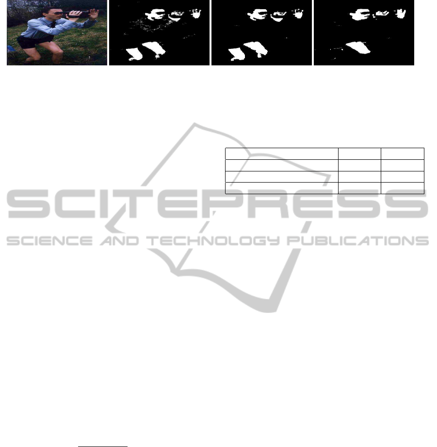
Figure 2: Comparison between pixel-based (Jones and
Rehg, 2002) (middle) and region-based with CRF(right)
skin color classification techniques.
a smaller number of superpixels than an image with
high color variation, as there is no penalty for bound-
ary violation. Generally, the concept of boundary is
not used when extracting the superpixels, however
different objects have different texture or color which
will implicitly act as boundaries. Figure 1 shows the
example of superpixels of an image. In our work we
have used the superpixel extraction library (Vedaldi
and Fulkerson, 2008) for superpixel segmentation.
3.3 Superpixel Classification
First, the pixel based skin color classifier defined on
section 3.1 is used to classify the pixels of the images.
Then the probability of being skin for a given super-
pixel sp with N number of color pixels c is defined as
follows
P(s|sp) =
1
N
N
∑
i
P(s|c
i
) (6)
Similarly the probability of being non-skin for a
given superpixel sp with N number of color pixels c
is defined as follows
P(ns|sp) =
1
N
N
∑
i
P(ns|c
i
) (7)
REGION-BASED SKIN COLOR DETECTION
303
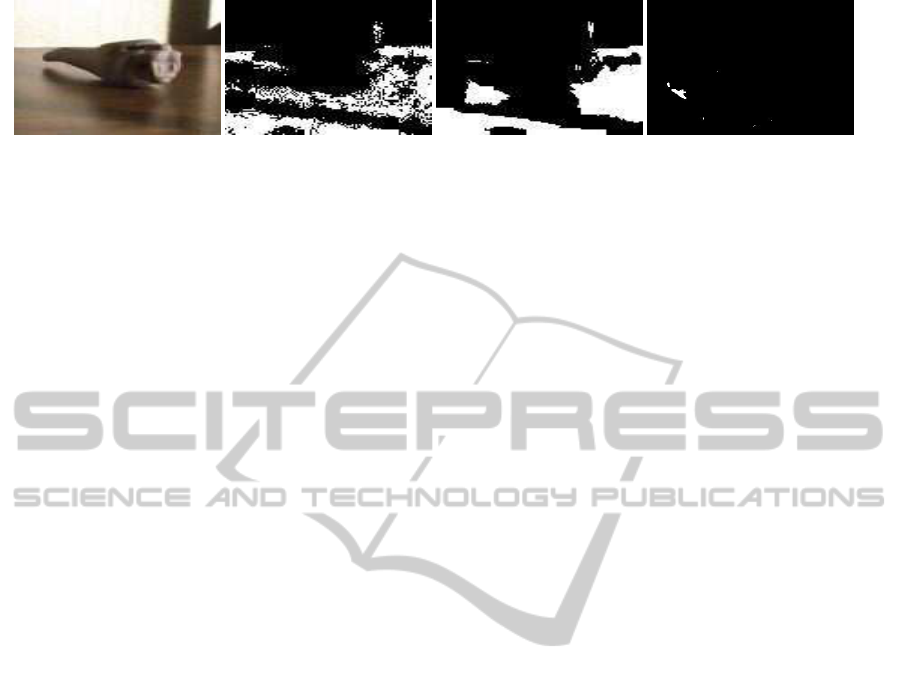
(a) Original image (b) Pixel-based (c) Region-based without CRF (d) Region-based with CRF
Figure 3: This example shows the advantages of the region-based approach even without CRF (see sub figures b and c). Sub
figures c and d show the failure case when CRF is applied.
3.4 Smoothing with CRF
Skin regions have varying size and shape, depending
upon the camera angle, distance from the camera and
human body factors. Hence, to obtain smooth skin re-
gions but still preserve the skin and non-skin bound-
aries, it is necessary to introduce some constraints.
CRF provides a natural way of combining pairwise
constraints. Color difference and length of bound-
ary between adjacent superpixels are used as pairwise
constraints. Optimum skin and non-skin labelling L
of all superpixels S of an image is defined as follows
−log(P(L|S;ω)) = −
∑
s
i
∈S
Ψ(l
i
|s
i
)+ω
∑
(s
i
,s
j
)∈E
Φ(c
i
, c
j
|s
i
, s
j
)
(8)
where ω is the weight of pairwise constraint, E is
the set of edges of superpixel, and i and j are index
nodes in superpixel level graph of an image.
Color potential (Ψ(l
i
|s
i
)): the color potential Ψ cap-
tures the skin and non-skin probability of superpixel
s
i
. We have used skin and non-skin probability for
superpixel directly from superpixel classification de-
fined in section 3.3 for color potential Ψ as follows
Ψ(l
i
|s
i
) = log(P(l
i
|s
i
)) (9)
Edge and boundary potential (Φ(c
i
, c
j
|s
i
, s
j
))):
pairwise edge and boundary potential Φ is defined
similar to those of (Fulkerson et al., 2009)
Φ(c
i
, c
j
|s
i
, s
j
) =
L(s
i
, s
j
)
1+ ||s
i
− s
j
||
, [c
i
6= c
j
] (10)
where L(s
i
, s
j
) is the shared boundary length, and
||s
i
−s
j
|| is the Euclidean norm of the color difference
between s
i
and s
j
superpixels.
Only one pairwise potential is used to make the
system as simple as possible to show that treating skin
color with regions is more effective than with pixels.
This implementation has only one weighting factor ω,
which is optimized using cross validation. We use
the multi-label graph optimization library of (Boykov
et al., 2001), (Boykov and Kolmogorov, 2004) and
(Kolmogorov and Zabih, 2004) for the inference of
skin and non-skin regions. CRF graph is built on the
superpixel level hence CRF optimization is fast.
Table 1: The results of pixel-based and our region-based
technique.
Method TP FP
Jones and Rehg (2002) 90% 14.2%
Our (superpixel only) 91.44% 13.73%
Our (superpixel and CRF) 91.17% 13.12%
4 EXPERIMENTS AND RESULTS
Equal number of training and testing sets are ran-
domly chosen from the Compaq dataset (Jones and
Rehg, 2002) and same training and testing sets are
used for all experiments. The Compaq dataset has ap-
proximately 4,700 skin and 9,000 non-skin images,
freely collected from the web. Basic pixel-based skin
color classifier mentioned in section 3.1 achievessim-
ilar results to those in Jones and Rehg (Jones and
Rehg, 2002). We have used RGB bin size = 32
for each channels, and threshold constant Θ = 1. It
roughly detects 90% skin color with 14.2% false pos-
itive rate.
Superpixel extraction using quick shift is con-
trolled by three parameters: (i) λ controls the trade
off between spatial and color consistency, (ii) σ con-
trols the deviation of density estimator, and (iii) τ
maximum distance in the quick shift tree. We have
used σ = 2, τ = 6, and λ = 0.9 for our experiment.
Which are chosen using grid search as there is no
explicit mechanism to preserve the skin boundaries,
with above selected parameters we have noticed that
97.43% skin pixels are correctly grouped into super-
pixels with 0.35% false positive rate. Average size of
the superpixels increases with the larger valueof τ and
σ and vice versa. Lower values of λ give importance
to spatial factor while higher values give importance
to the color value. Average size of superpixels are
larger when λ is
∼
=
0.5. Skin color detection depends
upon the values of the color channels, hence higher
importance is given to the color consistency in super-
pixel extraction. Also, experiments show that the skin
boundary is not well preserved with higher spatial im-
VISAPP 2012 - International Conference on Computer Vision Theory and Applications
304
(a) Original image (b) Pixel-based (c) Region-based CRF with color in-
formation only
(d) Region-based CRF with color
and border information
Figure 4: Example shows the failure of region-based approach when only a color difference constraint is used with CRF.
portance. The average size of superpixel is 65 in our
experiments. However, the size of superpixels is not
fixed and fully depends on the complexity of the im-
ages.
Table 1 shows the results comparison between
the presented region-based technique and the cur-
rent state-of-the-art pixel-based skin color detection
(Jones and Rehg, 2002) on unconstrained illumina-
tion and background. The region based technique
without CRF has 91.44% true positive rate with
13.73% false positive rate and with CRF has 91.17%
of true positive rate with 13.12% false positive rate.
Simply grouping the pixel-based evidence onto su-
perpixels increased the true positive rate by 1.44%
and decreased the false positive rate by 0.48%. This
shows treating skin as a region yields better results
than using pixels only. Both results from the region-
based techniques are better than the pixel-based tech-
nique.
The results on figure 2 show the effectiveness
of the region-based technique with CRF over pixel-
based method. Region-based technique first groups
the skin and non-skin evidence from each pixels into
superpixels level using basic skin color classifier,
which helps to remove noise. This is the main rea-
son why only grouping the pixel-based evidence into
superpixels increases the true positive rate by 1.44%
and reduces the false positive rate by 0.5% (see table
1). Also, CRF helps further extract larger smooth skin
regions by exploiting neighbouring color information
and boundary sharing between superpixels.
However, there are also some cases where region-
based technique performs worse than pixel-based
technique when we apply the CRF. Figure 3 are such
examples. Skin-like looking pixels and high bound-
ary sharing between skin and non-skin regions are the
main reason of the failure. However, we also exper-
imented using the color difference constraint only on
CRF instead of both color difference and boundary
sharing constraints and found that it performs better
when skin regions are very small and narrow. But
overall CRF with both neighbour color difference and
length of boundary sharing constraints performed bet-
ter. Figure 4 shows an example where CRF with
both neighbours color difference and length of bound-
ary sharing performs better than only with neighbours
color difference.
Skin regions do not have the same color values,
even the closest skin color pixels within superpixels
have different color values. Also, other skin-like ob-
jects exist. Hence, results can be further improved
using texture information. This is left for our future
work.
5 CONCLUSIONS
This paper presents a region-based skin color detec-
tion technique, which outperforms the current state-
of-the-art pixel-based technique. Color and spatial
distance based clustering technique is used to extract
the regions from the images, also known as superpix-
els. In the first step, our technique uses the state-of-
the-art non-parametric pixel-based skin color classi-
fier (Jones and Rehg, 2002) which we call the basic
skin color classifier. The pixel-based skin color evi-
dence is then aggregated to classify the superpixels.
Finally, the CRF is applied to further improve the re-
sults. As CRF operates over superpixels, the compu-
tational overhead is minimal.
The proposed region-based technique achieved
91.44% true positive rate with 13.73% false positive
rate without CRF optimization and 91.17% true pos-
itive rate with 13.12% false positive rate with CRF
optimization. Grouping the pixel-based evidence into
superpixels increased the true positive rate by 1.44%
and reduced the false positive rate by 0.48%. More-
over, the region-based approach produced smoother
results than the pixel-based methods. Skin commonly
appears as regions of similar pixels, so treating skin as
a region is advantageous over treating it as an individ-
ual pixel. Due to the illumination, background reflec-
tion and other noise factors, pixel values vary greatly
and groupingthem into a region helps to removenoise
by collecting evidence from neighbouring pixels.
These results suggest that skin color detection
REGION-BASED SKIN COLOR DETECTION
305

should be region-based rather than pixel-based. Also,
by adding more constraints on the CRF similar to
(Shotton et al., 2006) , the detection rate can be im-
proved. Moreover, any better skin color classification
method can be used as our basic skin color classifi-
cation module and can be easily combined with our
region-based skin color detection framework defined
in section 3 to improve the results.
REFERENCES
Boykov, Y. and Kolmogorov, V. (2004). An experi-
mental comparison of min-cut/max- flow algorithms
for energy minimization in vision. IEEE Transac-
tions on Pattern Analysis and Machine Intelligence,
26(9):1124–1137.
Boykov, Y., Veksler, O., and Zabih, R. (2001). Fast ap-
proximate energy minimization via graph cuts. IEEE
Transactions on Pattern Analysis and Machine Intel-
ligence, pages 1222–1239.
Brown, D., Craw, I., and Lewthwaite, J. (2001). A som
based approach to skin detection with application in
real time systems. In Proceedings of the British Ma-
chine Vision Conference, volume 2, pages 491–500.
Fulkerson, B., Vedaldi, A., and Soatto, S. (2009). Class
segmentation and object localization with superpixel
neighborhoods. In Proceedings International Confer-
ence on Computer Vision, volume 5.
Jedynak, B., Zheng, H., and Daoudi, M. (2003). Maxi-
mum entropy models for skin detection. In Energy
Minimization Methods in Computer Vision and Pat-
tern Recognition, pages 180–193.
Jones, M. J. and Rehg, J. M. (2002). Statistical color mod-
els with application to skin detection. International
Journal of Computer Vision, 46(1):81–96.
Kakumanu, P., Makrogiannis, S., and Bourbakis, N. (2007).
A survey of skin-color modeling and detection meth-
ods. Pattern Recognition, 40(3):1106–1122.
Kawato, S. and Ohya, J. (2002). Automatic skin-color dis-
tribution extraction for face detection and tracking. In
International Conference on Signal Processing, vol-
ume 2, pages 1415–1418. IEEE.
Kolmogorov, V. and Zabih, R. (2004). What energy func-
tions can be minimized via graph cuts? IEEE Trans-
actions on Pattern Analysis and Machine Intelligence,
26(2):147–159.
Kruppa, H., Bauer, M., and Schiele, B. (2002). Skin patch
detection in real-world images. In Van Gool, L.,
editor, Pattern Recognition, volume 2449 of Lecture
Notes in Computer Science, pages 109–116. Springer
Berlin / Heidelberg.
Moore, A. P., Prince, S., Warrell, J., Mohammed, U., and
Jones, G. (2008). Superpixel lattices. In IEEE Con-
ference on Computer Vision and Pattern Recognition.
Pan, Z., Healey, G., Prasad, M., and Tromberg, B. (2003).
Face recognition in hyperspectral images. IEEE
Transactions on Pattern Analysis and Machine Intel-
ligence, 25(12):1552–1560.
Peer, P., Kovac, J., and Solina, F. (2003). Human skin colour
clustering for face detection. In International Confer-
ence on Computer as a Tool.
Phung, S. L., Chai, D., and Bouzerdoum, A. (2002). A uni-
versal and robust human skin color model using neural
networks. In Proceedings of International Joint Con-
ference on Neural Networks, volume 4, pages 2844–
2849.
Ren, X. and Malik, J. (2003). Learning a classification
model for segmentation. In IEEE International Con-
ference on Computer Vision, volume 1.
Sebe, N., Cohen, I., Huang, T., and Gevers, T. (2004). Skin
detection: A bayesian network approach. In Proceed-
ings of the 17th International Conference on Pattern
Recognition, pages 903–906, Cambridge, UK.
Shotton, J., Winn, J., Rother, C., and Criminisi, A. (2006).
Textonboost: Joint appearance, shape and context
modeling for multi-class object recognition and seg-
mentation. Proceedings of European Conference on
Computer Vision, pages 1–15.
Soatto, S. (2009). Actionable information in vision. In Pro-
ceedings of the International Conference on Computer
Vision, volume 25.
Socolinsky, D. A., Selinger, A., and Neuheisel, J. D. (2003).
Face recognition with visible and thermal infrared im-
agery. Computer Vision and Image Understanding,
91(1-2):72–114.
Terrillon, J. C., Fukamachi, H., Akamatsu, S., and Shirazi,
M. N. (2000). Comparative performance of different
skin chrominance models and chrominance spaces for
the automatic detection of human faces in color im-
ages. In Fourth IEEE International Conference on
Automatic Face and Gesture Recognition, page 54.
Vedaldi, A. and Fulkerson, B. (2008). VLFeat: An open
and portable library of computer vision algorithms.
http://www.vlfeat.org.
Vedaldi, A. and Soatto, S. (2008). Quick shift and kernel
methods for mode seeking. Proceedings of European
Conference on Computer Vision, pages 705–718.
Wong, K. W., Lam, K. M., and Siu, W. C. (2003). A ro-
bust scheme for live detection of human faces in color
images. Signal Processing: Image Communication,
18(2):103–114.
Yang, M. H. and Ahuja, N. (1998). Detecting human faces
in color images. In International Conference on Image
Processing, 1998, volume 1, pages 127–130.
Zhu, Q., Cheng, K. T., Wu, C. T., and Wu, Y. L. (2004).
Adaptive learning of an accurate skin-color model.
In Sixth IEEE International Conference on Automatic
Face and Gesture Recognition, pages 37–42.
VISAPP 2012 - International Conference on Computer Vision Theory and Applications
306
