
A Single Electrical Acupuncture Needle with Bipolar Electrodes for
Biotissue Discrimination
Giseok Kang
1
, Jae-Cheon Kim
1
, Sohee Kim
1,2
and Jong-Hyun Lee
1,2
1
Department of Medical System Engineering, Gwangju Institute of Science and Technology, Gwangju, Republic of Korea
2
Department of Mechatronics, Gwangju Institute of Science and Technology, Gwangju, Republic of Korea
Keywords: Bipolar Electrodes, Acupuncture Needle, Biotissue, Electrical Impedance, MEMS, Flexible Photomask,
Parylene C.
Abstract: In oriental medicine, acupuncture is an essential treatment for the muscle tissue relaxation. For treatment,
electrical stimulations to the tissue conducted with multiple electrical acupuncture needles have generally
been used. However, the sting depth of a needle can be handled only by the sense of the oriental medicine
doctor. Moreover, it is difficult to use multiple needles to focus the electrical stimulation on a tissue of small
volume, and, likewise difficult to distinguish various tissues. In order to overcome the aforementioned
shortcomings, we developed a single acupuncture needle that has bipolar electrodes on the surface of the
needle tip by using a novel flexible parylene C film photomask. The interdigitated electrodes, 31.25 m in
width and 32.00 m in gap, were passivated by parylene C film to prevent metal debris from spreading into
the tissue. The electrical acupuncture needle was developed based on the conventional acupuncture needle
(400 m in diameter), so that the needle will give a familiar sensation to patients. We demonstrate the metal
patterning technique with a high resolution that has less than 2.95 % dimensional error compared to the
designed metal pattern dimensions. The biotissues were well distinguished by phase angle at 1 MHz of
14.6°, -32.7°, and 43.6° for skin, muscle, and ligament of a chicken, respectively.
1 INTRODUCTION
Acupuncture is an Asian alternative medicine
methodology that uses metal needles inserted into
the skin for patient treatment. The treatment, based
on traditional medicine theory, insists that human
body imbalances, due to unstable flow of the qi, can
be controlled by acupuncture (Langevin and
Yandow, 2002). Though current scientific research
has reported the physiological efficacy of the
acupuncture, some studies have still concluded that
the efficacy of acupuncture can be explained by the
placebo effect (White et al., 2003). In order to clear
up this controversy, acupuncture needs more
research from the scientific point of view.
Most of acupuncture treatments are focused on
muscle tissue relaxation, additionally supported by
electrical stimulations (Sandberg et al., 2003). In this
conventional stimulation, multiple needles are
needed because a single needle acts as only one
electrode. When the volume of tissue to be treated is
small, it is difficult to use a conventional electrical
acupuncture needle to locate the designated tissue in
a one shot. Therefore, it is difficult to distinguish
between various tissues by means of electrical
signals. In addition, the single conventional needle is
not efficient in stimulation of the target tissue
because the electrical signal tends to disperse into
the surrounding tissue. For effective acupuncture
treatment, thus, accuracy of detection and
localization of electrical stimulation are
indispensable.
In this paper, we have developed bipolar
electrodes on a single acupuncture needle surface
using micro-electromechanical systems (MEMS)
technology. In order to fabricate the acupuncture
needle, a novel flexible photomask made of parylene
C film is used. The flexible photomask is designed
to make direct contact with a curved substrate,
which makes it possible to pattern the electrodes on
the surface of conventional acupuncture needles
with high resolution. Parylene C is a material
featuring high biocompatibility and good electrical
insulation for the electrode passivation.
To validate the developed electrical acupuncture
needle, various biotissues (skin, muscle, and
47
Kang G., Kim J., Kim S. and Lee J..
A Single Electrical Acupuncture Needle with Bipolar Electrodes for Biotissue Discrimination.
DOI: 10.5220/0004237700470050
In Proceedings of the International Conference on Biomedical Electronics and Devices (BIODEVICES-2013), pages 47-50
ISBN: 978-989-8565-34-1
Copyright
c
2013 SCITEPRESS (Science and Technology Publications, Lda.)
ligament of a chicken) were characterized in terms
of electrical impedance. For steady and precise
results, every test was conducted using an automated
real-time measurement system under identical
conditions.
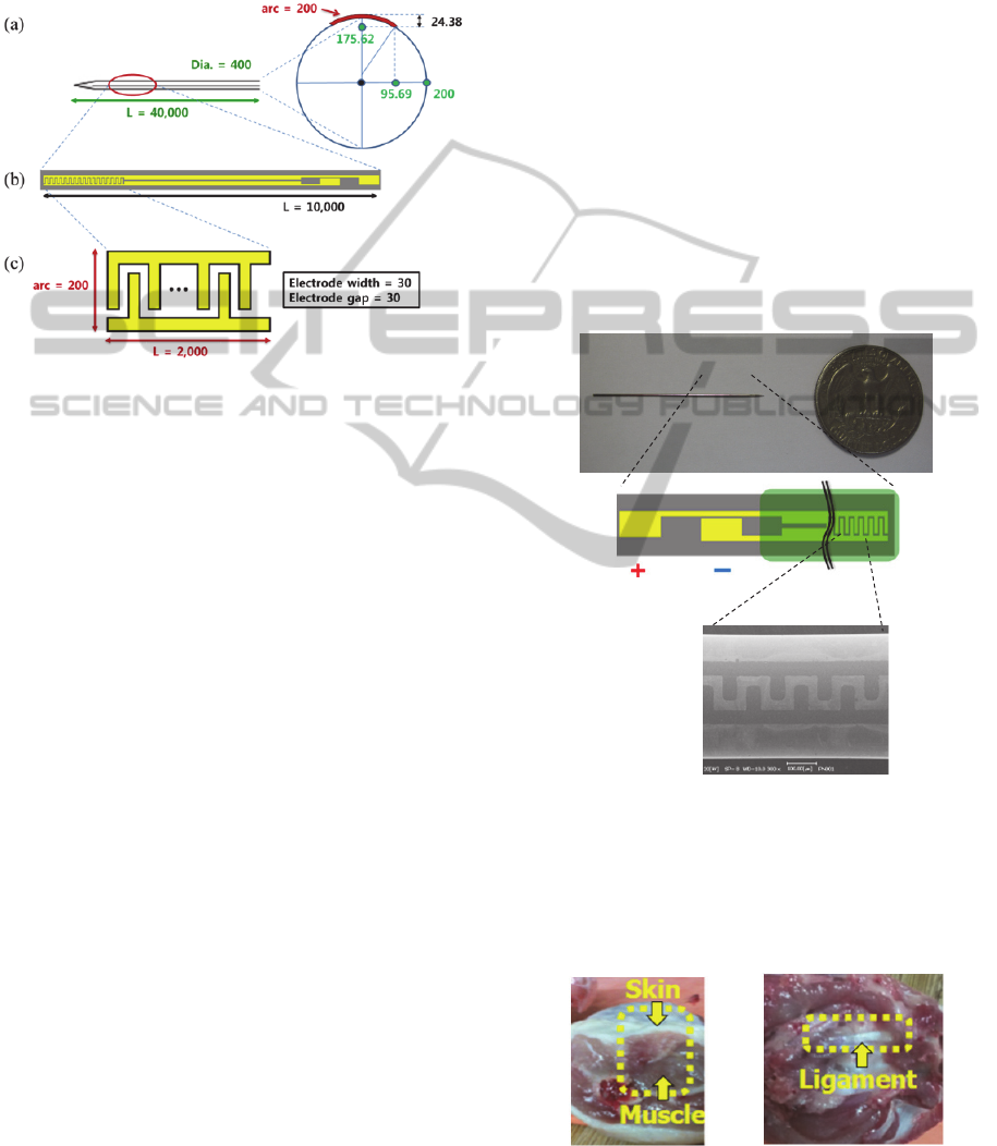
Figure 1: Schematics of the conventional acupuncture
needle and bipolar electrodes pattern [dimensions in m];
(a) overall view of the conventional acupuncture needle
(400 m in diameter, 30 mm in length) and cross-section
view of the electrodes on the needle surface, (b) overall
view of the bipolar electrode pattern made of Ti/Au (200
m in width, 10 mm in length), and (c) detailed view of
the interdigitated electrode (2 mm in length; both width
and gap are 30 m).
2 DEVICE CONFIGURATION
A flexible parylene C film photomask has been
developed to fabricate high resolution patterns on
the curved surface of the conventional acupuncture
needle, whose diameter can be as small as 400 um.
Compared to the previous flexible polydimethly-
siloxane (PDMS) photomask (Kim et al., 2009), the
patterning capability in this study was greatly
improved, showing a smaller pattern size and no
cracks in the metal layer. This improved patterning
was achieved by locating the parylene masking layer
on the neutral axis (geometric centroid of the beam
or membrane) that contains no longitudinal stresses
or strains (Bedford and Liechti, 2000). The Cr layer
(around 150 nm thick) can be located in the centroid
of the parylene C layers with a very low eccentricity.
This is because the parylene C is deposited by
chemical vapor deposition (CVD), which shows a
few tens of nm error in thickness (Yang et al., 1998).
The dimensions of the designed electrical
acupuncture needle are illustrated in Figure 1.
Considering the dimensions of the biotissue under
testing the interdigitated electrode (IDE), the
electrical sensing part on the acupuncture needle,
was designed to have 30
m
both in width and gap,
and to have 2 mm in length. The acupuncture needle
was electrically insulated by the biocompatible
polymer, parylene C (Schmidt et al., 1988). This will
also prevent metal debris from spreading into the
living tissue during operation. Figure 2 shows the
electrical acupuncture needle passivated with
parylene C.
3 BIOTISSUE EXPERIMENTS
To evaluate the fabricated electrical acupuncture
needle, an ex-vivo efficiency test of the biotissues
was conducted. Arrows in Figure 3 indicate the
biotissues (skin,
muscle, and ligament) of the chicken.
(a)
(b)
(c)
Figure 2: Configuration of the electrical acupuncture
needle passivated by parylene C: (a) fabricated electrical
acupuncture needle, (b) schematic of the Ti/Au bipolar
electrodes (bright yellow), the conventional acupuncture
needle (dark gray) and parylene C electrical insulation
layer (translucent green), and (c) SEM image of a part of
the IDE.
(a) (b)
Figure 3: Chicken biotissues: (a) skin (white) and muscle
(pink), and (b) ligament (white line).
BIODEVICES2013-InternationalConferenceonBiomedicalElectronicsandDevices
48
The test maintained identical environment
conditions (room temperature of 26 °C, room
humidity of 50%) in a clean booth. The bipolar
electrodes of the electrical acupuncture needle were
connected to an impedance analyzer (HP4294A,
USA) and a laptop in order to automatically measure
the electrical signals of the biotissues. Constant
voltage (0.5 V
pp
) and current (0.1 mA) were applied
to the biotissues from the impedance analyzer at 7
frequencies from 1 kHz to 1 MHz, as shown in
Figure 4. The sinusoidal electrical signal was
applied to the electrodes located on the needle
through the impedance probe kit (HP42941A, USA).
The probe kit performs a 4-wire (or Kelvin)
measurement method which automatically
compensates the lead and contact resistance to
measure impedance characteristics accurately
(Siegal and Galloway, 2008). Visual basic for
application (VBA) in Excel (Microsoft, USA) was
used to control the impedance analyzer and to
collect raw data into text files.
To verify the effectiveness of the developed
needle for the biotissue discrimination, a
conventional method using two electrical
acupuncture needles (separation distance 5 mm) was
evaluated as well. The tip of the conventional needle,
whose diameter is identical to that of the developed
needle, was not electrically passivated
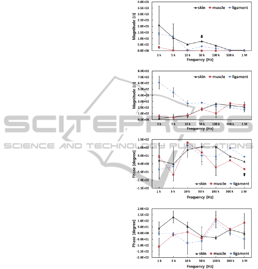
The validation of the electrical acupuncture
needles as a sensor was performed by measuring the
electrical impedances of various biotissues (skin,
muscle, and ligament of a chicken). The electrical
impedances of the tissues, the magnitude, and the
phase angle were measured, with results as shown in
Figure 4. All the data points corresponding to each
frequency indicate the average values calculated
with 200 data points of electrical response data for
each biotissue. Furthermore, the data points,
including the error bar (a standard deviation value),
are clearly differentiated between the biotissues at
the particular frequencies of the components of the
electrical impedance.
As shown in Figure 4, only the developed needle
can distinguish the biotissues both in the magnitude
and phase. We believe that the high performance of
the developed needle as an electrical sensor is
attributed to the electrodes in micro scale. In the test
using the bipolar needle, impedance magnitude of
the skin is larger than that of the muscle. That can be
explained by the fact that electrical signals is easily
conducted through each fiber (very large individual
cells) of muscle, but the skin is highly
inhomogeneous resulting in the most resistive tissue
in the human body (Miklavcic et al., 2006).
(a) magnitude in the developed needle test.
(b) magnitude in the conventional needle test.
(c) phase angle in the developed needle test.
(d) phase in the conventional needle test.
Figure 4: Electrical impedance responses as a function of
the frequency for the skin, muscle, and ligament of the
chicken: (a) magnitude in the developed needle test, (b)
magnitude in the conventional needle test, (c) phase in the
developed needle test, and (d) phase in the conventional
needle test. The vertical bars represent the error, defined
by the standard deviation. The arrows indicate the
frequency at which the differentiation index reaches the
maximum value.
In order to quantitatively evaluate the
effectiveness of the differentiation, differentiation
index (D), was used, as in our previous work:
ASingleElectricalAcupunctureNeedlewithBipolarElectrodesforBiotissueDiscrimination
49

D = G/D
A
, (1)
where G and D
A
are the gap and the average
difference, respectively (Kang, Yoo, Kim and Lee,
2012). A negative value of D means that the data
points overlapped, while a positive value of D close
to 1.0 (the maximum value of D) means that the data
points are well distinguished with a low-variance.
To validate the difference of the electrical
impedance between the biotissues, the D values
were estimated following the sequence of tissues as
the needle penetrated the tissues; D
SM
corresponds to
the differentiation index between the skin and
muscle; D
ML
corresponds to the differentiation index
between muscle and ligament. The maximum D
values of the magnitude and phase angle for the
biotissues are summarized in Table 1. The best case
for the biotissue discrimination took place for the
phase angle at 1 MHz; 0.97 for both D
SM
and D
ML
.
The differentiation indices, based on the
electrical impedances, were confirmed to have
efficiently distinguished the biotissues. This electro-
thermal acupuncture needle, integrated with a
microheater to focus heat-effects on a localized area,
can be a good medical appliance for a precision
treatment.
Table 1: Maximum differentiation index values of
magnitude and phase angle for biotissue discrimination.
Biotissue
Differentiation
index
Magnitude Phase angle
at 50 kHz at 1 MHz
Skin & muscle D
SM
0.95 0.97
Muscle &
ligament
D
ML
0.93 0.97
4 CONCLUSIONS
We designed and fabricated a novel electrical
acupuncture needle with bipolar electrodes for
biotissue discrimination. With the developed
acupuncture needle, various biotissues were
electrically characterized, and were definitely
distinguished at a particular frequency in real-time.
It is expected that the developed electrical
acupuncture needle with enhanced sensing accuracy
will be greatly beneficial to patients.
ACKNOWLEDGEMENTS
The research was supported by a grant from the
Institute of Medical System Engineering (iMSE) in
the GIST, Korea.
REFERENCES
Bedford, A. and Liechti, K. M. (2000). Chap 8, Mechanics
of Materials, Prentice-Hall. New Jersey.
Siegal, S., and Galloway, J. (2008). Thermal Test Chip
Design and Performance Considerations. 24
th
IEEE
Semi-Therm Symposium, 59-62.
Kang, G., Yoo, S., Kim, H., and Lee, J. (2012).
Differentiation between Normal and Cancerous Cells
at the Single Cell Level using 3-D Electrode Electrical
Impedance Spectroscopy. IEEE Sensors Journal. 12,
1084-1089.
Kim, J., Takama, N., Kim, B. and Fujita, H. (2009).
Optical–Softlithographic Technology for Patterning on
Curved Surfaces. J. Micromech. Microeng, 19, 1-8.
Miklavcic, D., Pavselj, N. and Hart, F. X. (2006). Electric
Properties of Tissues, John Wiley & Sons.
Langevin, H. M. and Yandow, J. A. (2002). Relationship
of Acupuncture Points and Meridians to Connective
Tissue Planes. The Anatomical Record (New Aant.),
269, 257-265.
Sandberg, M., Lundeberg, T., Lindberg, L. and Gerdle, B.
(2003). Effects of Acupuncture on Skin and Muscle
Blood Flow in Healthy Subjects. Eur J Appl Physiol,
90, 114-119.
Schmidt, E. M., McIntosh, J. S. and Bak, M. J. (1988).
Long-term Implants of Parylene-C Coated
Microelectrodes. Med. Biol. Eng. Comput., 26, 96-101.
White, P., Lewith, G., Hopwood, V. and Prescott, P.
(2003). The Placebo Needle, Is it a Valid and
Convincing Placebo for Use in Acupuncture Trials? A
Randomized, Single-blind, Cross-over Pilot Trial.
PAIN, 106, 401-409.
Yang, G. R., Ganguli, S., Karcz, J., Gill, W. N. and Lu, T.
M. (1998). High Deposition Rate Parylene Films.
Journal of Crystal Growth, 183, 385-390.
BIODEVICES2013-InternationalConferenceonBiomedicalElectronicsandDevices
50
