
Comparison of Two Techniques for Lifting Low-lying Objects on a Table
Part II: EMG and Psychological Measurement
Angelina Thiers, Harald Loose, Katja Orlowski, Mildred Bl
¨
asing and Marco Wallmann
Department of Informatics and Media, Brandenburg University of Applied Sciences,
Magdeburger Str. 50, 14770 Brandenburg, Germany
Keywords:
Back Pain, Load Lifting, Stoop Lifting, Squat Lifting, Electromyography, Psychological Criteria.
Abstract:
The purpose of this study was to determine differences in health benefits and fatigue when using various
lifting techniques. Worldwide, back pain is a common disease. In this context, muscular tension in shoulder
and neck areas as well as tension-type headaches are the most common side effects. One frequent cause for
this pain is connected with the wrong lifting and carrying of loads. To avoid these types of back pain numerous
recommendations concerning the right lifting technique already exist. The most common recommendation is
that one should use squat lifting instead of stoop lifting. By means of this technique a relief for the back
should be obtained. However, these benefits have not been proven yet. For this study eight healthy subjects
were evaluated. The test persons had to lift a load for ten minutes. During the lifting task the muscle activity of
nine muscles was documented. At the same time, psychological data were collected in a questionnaire. Both,
the physiological and psychological data revealed differences between the lifting techniques. During the stoop
lifting, a higher burdening of the back muscles was measured. In addition, following the exercise, a greater
and prolonged discomfort in the back muscles was documented.
1 INTRODUCTION
People who had not at least once in their lifetime
low back pain are the minority. A survey of German
health insurances companies showed that back pain is
one of the five most frequent illnesses. Almost 80%
of all people in Central Europe have reported at least
once in their lifetime about back problems. Espe-
cially, the number of children involved highlights the
increasing importance of prevention programs (Lez-
ius, 2004).
Frequently, the affected persons do not only suffer
from low back pain but also from muscular tensions
in the spine, shoulder and neck areas. Additionally,
back pain often leads to tension-type headache. Com-
mon causes of these pain patterns are incorrect or ex-
cessive pressure to the back as well as psychological
factors like stress. In everyday life, heavy lifting and
moving of loads are equally as responsible for an in-
creased stress of the back as wrong postures during
lifting (Kempf, 2009; Buhr, 2011).
The current recommendations for avoiding back
pains consist primarily of guidelines for a correct lift-
ing technique. The often suggested squat lifting is
done by bent knees and erected back. In contrast, the
stoop lifting is characterized by straight legs and a
bent back. However, the advantages of squat lifting
in terms of effectiveness and positive health benefits
have not been sufficiently proven (Dietrich, 2009).
To proof the suspected superiority of the squat lift-
ing technique different studies have been made. Re-
vuelta et al. (Revuelta et al., 2000) as well as Straker
and Duncan (Straker and Duncan, 2000) were able
to show that using the squat lifting leads to a higher
heart rate response than the stoop lifting. A different
physical strain of the lifting techniques was also con-
firmed by analyzing the muscle activity. Besides dif-
ferent activity and fatigue patterns, recorded datasets
also showed that a heavy pressure caused a changed in
the lifting technique from squat to stoop (Troup et al.,
1983; Hagen et al., 1994; Dietrich, 2009).
The aim of the current study was to show that there
is a difference in effectiveness, health benefits and fa-
tigue between both techniques. For the verification
of these assumptions a loading test was conducted.
Analyzing psychological data as well as physiologi-
cal data should show the differences between squat
and stoop lifting.
248
Thiers A., Loose H., Orlowski K., Bläsing M. and Wallmann M..
Comparison of Two Techniques for Lifting Low-lying Objects on a Table - Part II: EMG and Psychological Measurement.
DOI: 10.5220/0004327802480253
In Proceedings of the International Conference on Bio-inspired Systems and Signal Processing (BIOSIGNALS-2013), pages 248-253
ISBN: 978-989-8565-36-5
Copyright
c
2013 SCITEPRESS (Science and Technology Publications, Lda.)

2 EXPERIMENTAL SETUP
2.1 Subjects
Eight students were recruited through personal con-
tact. An equal distribution of the sexes was given. The
ages ranged from 18 to 27 years. The average body
height of the test persons was 173.5 (SD 8.6) cm.
All were healthy, without back pain symptoms and
gave their written consent to participate after being
informed about the experiment.
2.2 Test Procedure
On the first day of the experiment the subjects had to
execute the stoop lifting. Test persons returned one
week later to perform the squat technique.
Initially, the test persons were briefed about the
selected lifting technique (Loose et al., 2013). A max-
imum voluntary contraction (MVC) test for all ana-
lyzed muscles followed. Afterwards, the subjects did
a test lift with the aim to practice the correct motion
sequence while it was being made sure that the test
person is not restricted. The lifting task itself con-
sisted of three different stages. Firstly, the test person
had to remain still for one minute. During the second
phase, the subject performed the lifting of the load
for ten minutes. The requirements included both, the
correct motion sequence and a lifting frequency of at
least five lifts per minute. The final stage was equal
to the initial phase, one minute rest. A different load
weight for the sexes was given (male: 15 kg , female:
8.4 kg) (Loose et al., 2013). For the post-processing
of the lifting task an immediate survey about the sub-
jective effort and discomfort was conducted. Further-
more, a questionnaire about the same topics was filled
out on each of the seven following days.
2.3 Measurement
The used Shimmer
TM
sensors are characterized by
their small form and their low power consump-
tion. The Bluetooth technology offers real-time data
streaming. The basic module consists of an on-board
micro-controller, wireless communication modules, a
microSD slot and an integrated three-axis accelerom-
eter. In this study the used daughterboard was the
electromyography (EMG). The EMG module records
the electrical activity of a muscle with a sampling fre-
quency of 1024 Hz. The sensor disposes of three con-
nectors which allow to measure two or three chan-
nel pre-amplificated EMG-signal (Shimmer Research
Support, 2012).
2.4 Sensor Placement
Muscular contraction data were collected for nine
muscles. The EMG was recorded from the follow-
ing trunk and extremities muscles: M. gastrocnemius
medialis and lateralis, M. biceps femoris, M. vastus
lateralis, M. rectus femoris, M. gluteus maximus, M.
erector spinae, M. trapezius ascendes and M. biceps
brachii. The muscles were chosen based on their
function to carry out the defined motion sequences.
Ag/AgCl surface electrodes were applied after a stan-
dard skin preparation considering the recommenda-
tions from the SENIAM project (SENIAM project,
2012).
2.5 Data Analysis
The first questionnaire was completed immediately
after the lifting task. The second questionnaire was
completed on a daily basis at home over a period of
seven days. The content included questions regard-
ing the effort and discomfort in general and in spe-
cial parts of the body. Using an eleven-point answer
scale, the test persons had to judge from 0 (no ef-
fort/discomfort) to 10 (very high effort/discomfort).
The statistical evaluation was performed by using
mean values to compare the two lifting techniques.
The raw EMG signal was band-pass filtered from
15 to 500 Hz (Merletti and Parker, 2004). Addition-
ally, a notch filter with a blocking frequency of 50 Hz
was applied.
For analyzing the signal in the time domain a full-
wave rectification was carried out. Statistical parame-
ters like maximal or mean amplitude were calculated
over the time. They were calculated over an inter-
val of seven seconds, which is the average length of
a lifting cycle. Additionally, the parameters were cal-
culated for ten detected liftings. Using the MVC data
for the standardization of the EMG an inter-proband
analysis was possible. A second standardization was
made by using time normalization.
Transformation of the EMG signal from the
time to the frequency domain was achieved by the
Fast Fourier Transformation over signal segments of
512 ms (Grimshaw et al., 2006; Kaplanis et al., 2009).
The Frequency-Analysis included the computation of
the median frequency as well as the computation of
the total power. Both parameters were used as indi-
cators for muscle fatigue (Merletti and Parker, 2004;
Lukas, 2000).
The accumulation of the power density spectrum
for all frequencies is defined as the total power, fig-
ured in equation 1 (Kaplanis et al., 2009).
ComparisonofTwoTechniquesforLiftingLow-lyingObjectsonaTable-PartII:EMGandPsychologicalMeasurement
249
E
totalPower
=
Z
∞
0
S
PD
( f )d f (1)
The median frequency is the frequency where the
accumulated power spectrum energy is 50 % of the
total power spectrum, figured in equation 2 (Chang
et al., 2012; Kaplanis et al., 2009).
F
medianFrequency
=
1
2
Z
∞
0
S
PD
( f )d f (2)
3 RESULTS
3.1 Physiological Data
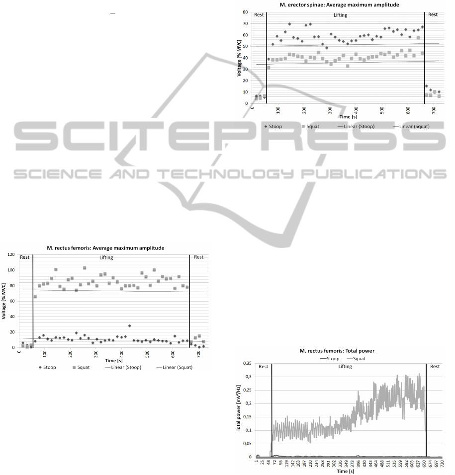
For the comparison of the course of the maximum am-
plitude of the M. rectus femoris in stoop and squat the
measured values of all subjects were averaged. Fig-
ure 1 shows the changes in the maximum amplitude
(normalized values) of the M. rectus femoris through-
out the lifting task. A comparison of the stoop and
squat values illustrates the higher strain of the M. rec-
tus femoris in squat lifting. On average, using squat
technique produces threefold greater voltage values
than using stoop technique. That corresponds to the
characteristic that in squat the load is lifted from the
knees and not from the back.
Figure 1: M. rectus femoris - Comparison of the maximum
amplitude in stoop and squat.
As in the previous calculation, the maximum am-
plitudes of all subjects were averaged. The visualiza-
tion of the averaged values (normalized values) of the
M. erector spinae is shown in figure 2. Both tech-
niques have in common that on average the values
rise over time. This observation supports the asser-
tion that load lifting leads to a fatigue in the back
muscle. The figure further indicates that the stoop
technique involves a higher participation of the back
muscle. The consideration of figure 2 together with
figure 1 points out for both techniques that the mea-
sured values reflect the strain in the suspected body
areas. The relative measurements in stoop show a
threefold higher effort in M. erector spinae than in M.
rectus femoris. The measured values in squat tech-
nique show a strongly contrasting distribution. M.
rectus femoris is producing twice as high values as
M. erector spinae.
Figure 2: M. erector spinae - Comparison of the maximum
amplitude in stoop and squat.
Fatigue is defined in muscle physiology as a state
when a subject can no longer maintain a required
force (Merletti and Parker, 2004). Hence the main-
tenance demands an increasing recruitment of motor
units (Lukas, 2000). Figure 3 shows the course of
the total power of one test person during the load lift.
The difference between the absolute values for both
techniques are clearly visible. Furthermore, there is
a noticeable course difference. For the total power in
stoop nearly static values were recorded. In contrast,
the calculated total power when using squat technique
shows a steady increase. Both observations indicate a
higher strain in M. rectus femoris during squat lifting.
Additionally, the course of the total power illustrates
the fatigue of the muscle when using squat lifting.
Figure 3: M. rectus femoris - Comparison of the total power
in stoop and squat.
One of the most widely applied indexes for muscle
fatigue is the median frequency. In the state of mus-
cle fatigue the EMG spectrum is compressed towards
BIOSIGNALS2013-InternationalConferenceonBio-inspiredSystemsandSignalProcessing
250
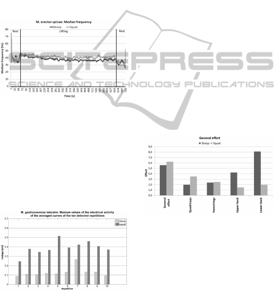
lower frequencies (Merletti and Parker, 2004). The
development of the median frequency of M. erector
spinae for both lifting techniques is represented in fig-
ure 4. Both graphs show descending median frequen-
cies. Hence, squat lifting is as well as stoop lifting
fatiguing for the back muscles. However, a close-up
look at the values shows that the decrease of the fre-
quency in stoop lifting is higher than in squat. The al-
legation that stoop lifting causes a higher strain on the
back is supported by figure 4. The course of the me-
dian frequency of this subject is similar to the curves
of the other test persons.
Figure 4: M. erector spinae - Comparison of the median
frequency in stoop and squat.
To assess the fatigue of the M. gastrocnemius lat-
eralis during the execution of both techniques the
maximum amplitude (time normalized) of ten de-
tected repetitions was considered. According to
Werner (Werner, 2006), the fatigue of a muscle ac-
crues as a result of strain and is recognizable by the
increase of the electrical activity. On average, the test
persons showed higher maximum values during the
squat than the stoop technique. Figure 5 illustrates the
behavior of the maximum amplitude of the M. gas-
trocnemius lateralis for both techniques over the ten
detected repetitions. Because of the higher electrical
Figure 5: Maximum values of the electrical activity of the
averaged curves of the ten repetitions, M. gastrocnemius lat-
eralis.
activity during the squat technique it can be assumed
that this technique led to a higher strain. However,
if the behavior of the maximum values is considered
over the ten repetitions, no continuous increase or de-
crease is visible. Referring to the first and the last de-
tected repetition, there is an increase of the values in
both techniques. Hence, a fatigue of the M. gastroc-
nemius lateralis could not be proven. This could be
due to either none of the techniques causing a fatigue
of the M. gastrocnemius lateralis or too many factors
being present that influence EMG signals.
3.2 Psychological Data
The immediately conducted survey of all test persons
after the lifting task (fig. 6) revealed as part of the
comparison of stoop and squat lifting that the sub-
jective general effort in squat lifting is higher than in
stoop. A detailed look at the distribution in figure 6
shows that both, the effort in quadriceps and biceps
femoris in squat lifting are higher than in stoop lift-
ing. A contrary result is shown in terms of subjec-
tive exertion in low and upper back. In both cases
the perceived effort in stoop lifting is several times
higher than in squat lifting. The information gath-
ered from the test persons consultation presents the
expected results in dependency to the described mo-
tion sequences of the lifting techniques.
Figure 6: Comparison of the general effort in stoop and
squat
In addition, the surveys sought information about
the actual discomfort. In general, all test persons suf-
fered a higher discomfort after using the squat tech-
nique. However, a more detailed analysis of this
point revealed that while some body areas led to a
higher discomfort when using the squat technique
other body areas had a higher discomfort following
the stoop technique. Firstly, the look at discomfort of
the thigh in figure 7 shows that using the squat tech-
nique caused higher values, especially in the quadri-
ceps. The discomfort is nearly seven times higher
ComparisonofTwoTechniquesforLiftingLow-lyingObjectsonaTable-PartII:EMGandPsychologicalMeasurement
251
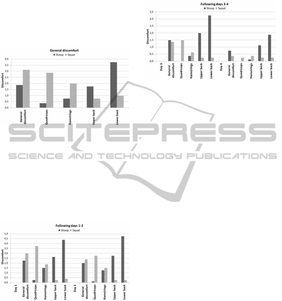
than using the stoop technique. Therefore, the use of
the stoop technique can be expected to have a higher
discomfort in the back. Figure 7 confirms the assump-
tion that the subjective pain after using stoop is in the
lower as well as in the upper back many times higher
than after using the squat lifting.
Figure 7: Comparison of the general discomfort in stoop
and squat.
The assessment of general discomfort one day af-
ter the lifting tasks showed similar values to those ob-
tained immediately after the lifting. The comparison
of the course of the complaints differs (fig. 8). The
discomfort on the first days after using the squat tech-
nique shows a decreasing level. In contrast, the com-
plaints in the back after using stoop are still growing
on the second day. Compared to the squat technique,
the pain level in the lower back is in stoop over ten
times higher.
Figure 8: Following day 1 and 2 - Comparison of the gen-
eral discomfort in stoop and squat.
The course of the general pain levels shows a
faster regeneration after using squat technique. On
the third day after the lifting task the comparison of
the general pain shows for the first time lower values
for the squat lifting. Already on the fourth day af-
ter using the squat technique, the test person reported
scarcely any complaints. The values of the subjective
pain in the back after using stoop are comparatively
high.
Figure 9: Following day 3 and 4 - Comparison of the gen-
eral discomfort in stoop and squat.
4 DISCUSSION
The main finding in this experimental study is that the
EMG patterns of stoop and squat lifting differ. On
one hand, high values in muscle activity were sus-
pected for the M. rectus femoris. On the other hand,
the presumption for the squat lifting was a high per-
formance of the M. erector spinae. Both assumptions
are proven with the analyzed EMG courses. More-
over, in the frequency domain the typical properties
of the lifting techniques are visible. The comparison
of the course of total power of the M. rectus femoris
in stoop and squat showed a great difference in the ef-
fort of the muscle. The increase of the total power in
squat indicates the fatigue of the muscle. The higher
voltage values as well as the fatigue point out that the
effort in squat is higher than in stoop. The detailed
observation of the maximum amplitudes and the fa-
tigue of the M. erector spinae show lower differences.
Although stoop technique creates higher voltage val-
ues and a steeper decrease of the median frequency of
the M. erector spinae, the differences are not as pow-
erful as the differences in squat technique. This can
be caused by the definition of the motion sequence of
Loose et al. (Loose et al., 2013). A lifting and lower-
ing of weights without placing the weights on a table
might be useful for a more conclusive result. Addi-
tionally, the placement of the sensors can be more ef-
fectively. As the investigation of the muscle activity
of the M. gastrocnemius lateralis shows that this mus-
cle is less important for the evaluation of the lifting
techniques. Instead, the placement of additional sen-
sors at the back can be useful.
The analysis of the subjective pain impressions
recorded via the questionnaires support the statement
that squat lifting is healthier than stoop lifting. Es-
pecially the course of the discomfort in the four days
following the lifting shows that complaints after using
stoop are more intensive and the regeneration takes
BIOSIGNALS2013-InternationalConferenceonBio-inspiredSystemsandSignalProcessing
252

longer. Furthermore, the observation of the general
effort and general discomfort immediately after the
lifting task confirms the thesis that squat is more fa-
tiguing than stoop lifting.
5 CONCLUSIONS
This paper investigated the hypothesis that the squat
lifting technique is more ergonomic, healthy and less
exhausting on a real life example of a combined mo-
tion of lifting and putting a beer crate into a car trunk.
The analysis of the physiological and psychological
data indicates on the one hand that squat causes a
higher effort in the upper legs and on the other hand
that the effort of the back is higher in stoop. Fur-
thermore, the investigation of the discomfort in the
back shows that the intensity and duration of the com-
plaints are higher when using stoop. In contrary, the
ECG analyzed in Loose et al. (Loose et al., 2013)
showed a higher physical stress when using squat. In
summary, it can be concluded that no general assump-
tion for an optimal lifting technique can be made.
Further assumptions about the lifting techniques
can be reached by an individual analysis of the lifting
cycles. This can be realized by an observation of the
EMG patterns or by the use of the Kinect data (Loose
et al., 2013).
The feature of the characterized motion sequence
is that it represents an everyday situation like lifting a
beer (or similarly shopping) crate into a car. However,
the experimental study was not able to show a signifi-
cant benefit of the squat lifting with regards to the fa-
tigue of the M. erector spinae. Due to that reason the
validity of the statement that squat lifting is healthier
has to be proven in everyday situations. Another con-
tinuation of the study could focus on the behavior of
the muscle activity when lifting different loads using
various lifting techniques.
Further measurements (Kinect and ECG data), re-
sults, discussions and finalizing conclusions are in-
cluded in Loose et al. (Loose et al., 2013).
REFERENCES
Buhr, M. (2011). Der gesunde R
¨
ucken: R
¨
uckenschmerzen
vorbeugen und heilen. humboldt.
Chang, K.-M., Liu, S.-H., and Wu, X.-H. (2012). A wireless
semg recording system and its application to muscle
fatigue detection. Sensors, pages 489–499.
Dietrich, R. (2009). Zeitaufgel
¨
oste Frequenzanalyse
von EMG-Signalen bei dynamischen Hebevorg
¨
angen
mit zunehmenden Lasten. PhD thesis, Humboldt-
Universit
¨
at zu Berlin.
Grimshaw, P., Lees, A., and Fowler, N. (2006). Sport and
Exercise Biomechanics (BIOS Instant Notes). Bios
Scientific Publ.
Hagen, K. B., Harms-Ringdahl, K., and Hall
´
en, J. (1994).
Influence of lifting technique on perceptual and car-
diovascular responses to submaximal repetitive lift-
ing. European Journal of Applied Physiology, pages
477–482.
Kaplanis, P., Pattichis, C., Hadjileontiadis, L., and Roberts,
V. (2009). Surface emg analysis on normal subjects
based on isometric voluntary contraction. Journal of
Electromyography & Kinesiology, pages 157–171.
Kempf, H.-D. (2009). Die Neue R
¨
uckenschule. Springer.
Lezius, T. (2004). Volkskrankheit r
¨
uckenschmerzen - ein
forschungsprojekt unter gesundheitspsychologischen
aspekten. Master’s thesis, Evangelische Hochschule
N
¨
urnberg.
Loose, H., Orlowski, K., Thiers, A., and Tezlaff, L. (2013).
Comparison of two techniques for lifting low-lying
objects on a table: Part i: Setup, ecg and motion mea-
surement. BIOSIGNALS 2013.
Lukas, C. M. (2000). Kraftverhalten und elektromyo-
graphische Untersuchungen an der Unterschenkel-
muskulatur bei Patienten nach operativ versorgter
Achillessehnenruptur. PhD thesis, Eberhard-Karls-
Universit
¨
at zu T
¨
ubingen.
Merletti, R. and Parker, P. A. (2004). Electromyography.
John Wiley & Sons.
Revuelta, N., Dauphin, A., Kowslowski, O., Dubois, D.,
and Thevenon, A. (2000). Heart rate response to two
lifting techniques. Archives of Physical Medicine and
Rehabilitation, 81:958–959.
SENIAM project (2012). Sensor placement. Website.
Available online at http://www.seniam.org; visited on
October 25th 2012.
Shimmer Research Support (2012). EMG User Guide Rev
1.2. Shimmer Research.
Straker, L. and Duncan, P. (2000). Psychophysical and psy-
chological comparison of squat and stoop lifting by
young females. Australian Journal of Physiotherapy,
pages 27–32.
Troup, J., Leskinen, T. P. J., Stalhammar, H. R., and
Kuorinka, I. A. A. (1983). A comparison of intraab-
dominal pressure increases, hip torque, and lumbar
vertebral compression in different lifting techniques.
Human Factors: The Journal of the Human Factors
and Ergonomics Society, pages 517–525.
Werner, F. (2006). Auswirkungen differenzieller
Erm
¨
udungsprogramme auf ausgew
¨
ahlte Bewe-
gungsparameter und die Muskelaktionspotenziale von
Triceps surae und Quadriceps femoris. PhD thesis,
Friedrich-Schiller-Universit
¨
at Jena.
ComparisonofTwoTechniquesforLiftingLow-lyingObjectsonaTable-PartII:EMGandPsychologicalMeasurement
253
