
A Physiological Evaluation of Immersive Experience of a View Control
Method using Eyelid EMG
Masaki Omata
1
, Satoshi Kagoshima
2
and Yasunari Suzuki
2
1
Interdisciplinary Graduate School of Medicine and Engineering, University of Yamanashi, Takeda, Kofu, Japan
2
Department of Computer Science and Media Engineering, University of Yamanashi, Takeda, Kofu, Japan
Keywords:
physiological Evaluation, Immersive Impression, Eyelid-Movement, Electromyogram, Virtual Environment.
Abstract:
This paper describes that the number of blood-volume pulses (BVP) and the level of skin conductance (SC)
increased more with increasing immersive impression with a view control method using eyelid electromyo-
graphy in virtual environment (VE) than those with a mouse control method. We have developed the view
control method and the visual feedback associated with electromyography (EMG) signals of movements of
user’s eyelids. The method provides a user with more immersive experiences in a virtual environment because
of strong relationship between eyelid movement and visual feedback. This paper reports a physiological evalu-
ation experiment to compare it with a common mouse input method by measuring subjects’ physiological data
of their fear of an open high place in a virtual environment. Based on the results, we find the eyelid-movement
input method improves the user’s immersive impression more significantly than the mouse input method.
1 INTRODUCTION
In recent years, some devices have been developed on
how to provide a user with more immersive impres-
sion in a virtual environment (VE)(Nagahara et al.,
2005), (Meehan et al., 2002). In the research field,
some visual displays have been developed as visually-
immersive devices such as a head-mounted display
that covers the user’s view and peripheral vision, a
wall-sized screen that covers the user’s whole body
and 3D display that give a user a sense of depth. On
the other hand, some interaction techniques in a vir-
tual environment have been designed (Touyama et al.,
2006), (Ries et al., 2008), (Asai et al., 2002), (Man-
ders et al., 2008), (Steinicke et al., 2009), (Miyashita
et al., 2008), (Haffegee et al., 2007), (Kikuya et al.,
2011). Particularly, it is important to provide a user
with embodiment interaction techniques in a virtual
environment, just like the same as natural embodi-
ment motion in the real world. Therefore, several ges-
tural interaction techniques and several walk-through
environments have been developed. By using the in-
teraction techniques and the displays, users can more
naturally interact with a virtual environment in where
visual feedback to the users corresponds with motor
skills of the users.
However, these existing embodied interaction
techniques have some shortcomings of the correspon-
dence between motor skills and visual feedback. For
instance, people usually scrunch up their eyes to look
carefully at an important point. Conversely, people
usually open their eyes widely with to get a wide view
of surrounding area. Existing virtual reality systems
do not incorporate the basic movement like looking.
They usually provide users with control devices such
as a mouse, a joystick and a location sensor to control
user’s view. Because there is no real-world-like rela-
tionship (eye movement and view) between the device
operations and the visual feedback, the device opera-
tions may make the user aware of the gap between
the real world in where the user controls the device
and the virtual world in where the user sees and may
reduce the user’s immersive impression.
We, therefore, have developed a view control
method and the visual feedback associated with elec-
tromyographic (EMG) signals of movements of user’s
eyelids (TheAuthors, 2011). The method provides
users with more immersive experiences in a virtual
environment because of the strong relationships be-
tween the eyelid movements and the visual feedback.
Moreover, the visual feedback enhances a user’s vi-
sual functions in a virtual world, such as zoom-in/out
and see-through, by following the relationship be-
tween motor skills of seeing and views in the real
world. We think that the method will be used for
a natural and direct view control system in a three-
224
Omata M., Kagoshima S. and Suzuki Y..
A Physiological Evaluation of Immersive Experience of a View Control Method using Eyelid EMG.
DOI: 10.5220/0004719102240231
In Proceedings of the International Conference on Physiological Computing Systems (PhyCS-2014), pages 224-231
ISBN: 978-989-758-006-2
Copyright
c
2014 SCITEPRESS (Science and Technology Publications, Lda.)

dimensional virtual environment.
Another research issue of VE interaction is how
to evaluate user’s immersive experience. A question-
naire, such as Likert scale or semantic differential
method, is one of conventional evaluation techniques,
but it is not continuous and not objective. Addition-
ally, it is difficult to measure and evaluate sensation
by conducting a performance evaluation such as task
completion time and error ratio. We therefore con-
ducted a physiological evaluation test to compare the
immersive impression with the eyelid-movement in-
put method with that with a mouse input method.
This paper describes related work, the develop-
ment summary of the system, the physiological eval-
uation test about the users’ immersive impression and
improvement of the recognition algorithm of EMG of
user’s eyelid movements.
2 RELATED WORK
Asai et al. developed a vision-based interface based
on body position for viewpoint control in an immer-
sive projection display by tracking 3D positions of
the arms and head of the user by using image pro-
cessing without attached devices (Asai et al., 2002).
They evaluated the utility of the interface by com-
paring the interface with a joystick. The results in-
dicate that the performance of the interface is com-
parable to that of the joystick in terms of viewpoint
control, but enhances the sensation of speed. Man-
ders et al. presented a method for interacting with 3D
objects in an immersed 3D virtual environment with a
head-mounted display (HMD) by tracking user’s hand
gestures with a stereo camera (Manders et al., 2008).
The system allows the user to manipulate a 3D object
with five degrees of freedom using a series of intuitive
hand gestures. Steinicke et al. developed a VR-based
user interface for presence-enhancing gameplay with
which players can explore the game environment in
the most natural way, i. e., by real walking (Steinicke
et al., 2009). While the player walked through the
virtual game environment by wearing a HMD, they
analyzed the usage of transitional environments via
which the player could enter a virtual world. The re-
sults of the psychophysical experiments have shown
that players can be guided on circular arc with a ra-
dius of 22.03m whereas they believe themselves to be
walking straight. Oshita proposed a motion-capture-
based control framework for third-person view virtual
environments with a large screen (Oshita, 2006). The
framework can generate seamless transitions between
user controlled motion and system generated reactive
motions.
Because these systems use user’s body motions to
control view in virtual environment, there are a few
fundamental problems. A problem is that the view
control is rough by using body motions. Another
problem is that the device input makes the user feel
the seam between the real and virtual worlds. There-
fore, some systems to adopt eye movement for view
control have been developed.
Miyashita et al. proposed an electrooculography
(EOG)-based gaze interface that was implemented by
mounting EOG sensors on a HMD with a head-tracker
and proposed a gaze estimation method on the HMD
screen(Miyashita et al., 2008). The accuracy of gaze
estimation was experimentally determined to be 68.9
%. The system solves the problems of eye-tracker
devices’ block to a HMD. Haffegee et al. focusses
on methods and algorithms for using an eye-tracker
to takes the eye-tracker output and converts it into a
virtual world gaze vector in an immersive VE by us-
ing an eye camera and a head tracker (Haffegee et al.,
2007).
These systems mainly adopt the user’s line of
sight. However, people usually use their eye move-
ments to not only control line of sight but also several
controls for sight such as focus, gaze, interest and af-
fect. For instance, people usually tighten their eyelids
to look carefully. People also widen their eyes to look
at someone with interest or in surprise. Additionally,
because a person with x-ray vision in a science fic-
tion movie tightens his/her eyelids to look through ob-
jects, we proposed that it is possible to enhance user’s
visual functions in a virtual environment by tracking
eye movements in detail.
3 A VIEW CONTROL METHOD
USING EMG OF EYELIDS
We proposed a view control method associated to
movements of use’s eyelids to enhance user’s vi-
sual functions in a virtual environment (TheAuthors,
2011). The basic method deals with three types
of eyelid movements: staring, neutral and widely-
opening. The neutral is a state of looking at some-
thing naturally without straining or widely-opening
(see figure 1a). The staring is a state of looking care-
fully with gathering eyebrows (see figure 1b). The
widely-opening is a state of looking at whole with
raising eyebrows (see figure 1c). Figure 2 shows the
state transition diagram of the three states.
By using the states, we designed two types
of view control techniques in a virtual environ-
ment: the zoom in/out control technique and the see
through/annotation control technique. By using the
APhysiologicalEvaluationofImmersiveExperienceofaViewControlMethodusingEyelidEMG
225
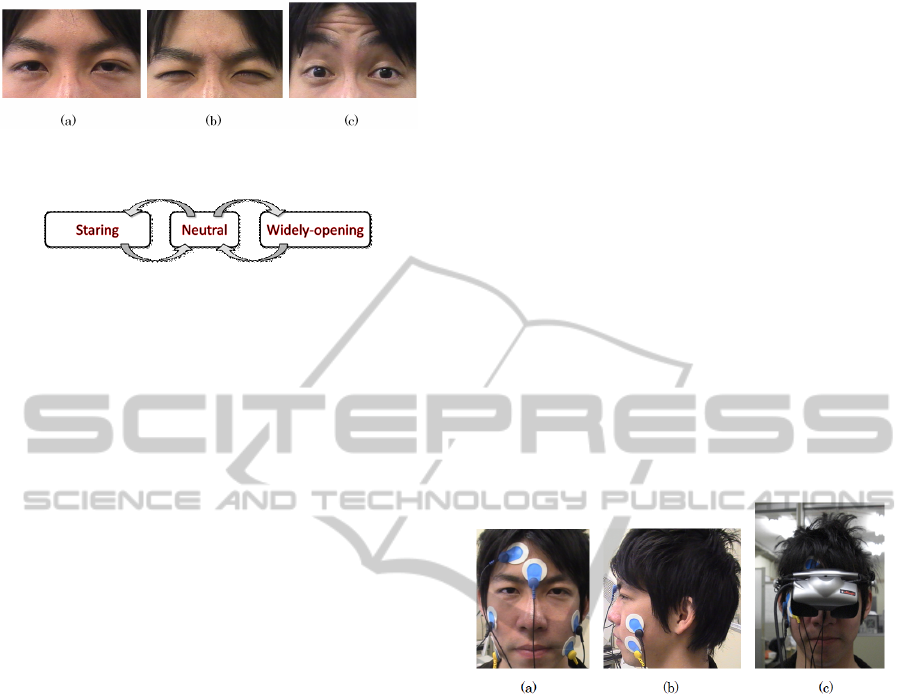
Figure 1: Three states of eyelid movements: (a) neutral, (b)
staring, and (c) widely-opening.
Figure 2: State-transition diagram of the three eyelid states.
zoom in/out technique, a user can look at a distant
object as the zoomed-in view by looking at it with
gathering eyebrows. Moreover, the user can look at
objects, which are outside of the user’s neutral view,
as the zoomed-out view by looking at the center of the
view with raising eyebrows. The view of zoom in/out
becomes the normal view when the user repositions
the eyebrows and relaxes the palpebral muscles.
On the other hand, by using see-
through/annotation technique, a user can look
inside an object such as an engine of a car by looking
at it with gathering eyebrows. Moreover, the user can
read annotation text, such as spec of a car around it
by looking at it with raising eyebrows. The views
become the initial view when the user repositions the
eyebrows and relaxes the palpebral muscles.
We think that the system improves the user’s im-
mersive experience because of the relationship be-
tween the actual eye movements and the visual feed-
back. This advantage solves the problem of reducing
a user’s immersive impression because of the tenuous
relationship between visual feedback and the move-
ment of conventional input devices, such as a mouse,
a joystick, or a location sensor.
Additionally, the system has a possibility to pro-
vide a user with controlling the view in multiple steps
by adjusting the strength of his/her eyelid muscle. In
other words, the stronger the user gathers his/her eye-
brows, the larger the zoom is, and the higher the trans-
parency is.
3.1 Implementation
We use a Z800 3D Visor (eMagin Co.) as a
head-mounted display (HMD), a ProComp Infiniti
(Thought Technology Ltd.) as a biofeedback device,
two MyoScan sensors (Thought Technology Ltd.) as
pre-amplified surface electromyography (EMG) sen-
sors, and a PC to implement an eyelid-movement rec-
ognizer and a 3 D virtual environment . The speci-
fications of the HMD are as follows: a 105-inch vir-
tual screen at a distance of 3.6 m, 40-degree angle
of view, horizontal 360-degree angle and vertical 60-
degree angle head tracking, 800 600 pixels. The
biofeedback device and the EMG sensors are used to
record the EMG signals on a user’s facial surface and
to send the data to the PC via USB. The recording
frequency is 20 Hz, and the recorded signals are raw
voltage values. The PC is used to process the EMG
data and to render the scene of a virtual environment
with Microsoft Visual C++ and OpenGL. The three
dimensional view of the scene is controlled with the 6
degrees of freedom data of the head-tracking HMD.
Figures 3 shows the HMD and the electrodes of
the EMG sensors attached to a user’s face. An EMG
sensor consists of three electrodes: positive, negative,
and reference. We use an EMG sensor to detect eyelid
movements because it is difficult to detect it by image
data processing with a camera, such as a study of Val-
star et.al(Valstar et al., 2006), because of the narrow
space between user’s face and the HMD (Figure 3c).
Figure 3: State of wearing the HMD and attaching the EMG
sensors in our proposed system.
3.2 Measuring EMG and Detecting
Eyelid Movements
EMG is a technique for detecting and amplifying tiny
electrical impulses that are generated by muscle fibers
when they contract. An EMG sensor can record the
signals from all the muscle fibers within the recording
area of the sensor contact. Some research studies use
EMG to develop human interfaces. Manabe et al. pro-
posed the use of an EMG of facial muscles to control
an electric-powered wheelchair(Manabe et al., 2009).
Agustin et al. presented a low-cost gaze-pointing and
EMG-clicking device, which a user employs with an
EMG headband on his/her forehead (Agustin et al.,
2009). Clark et al. used the EMG on a user’s arm
movement to control a robotic arm (Jr. et al., 2010).
Also, Costanza st al. presented a formal evaluation
of subtle motionless gestures based on EMG signals
from the upper arm for interaction in a mobile con-
text(Costanza et al., 2007) , and Gibert et al. de-
PhyCS2014-InternationalConferenceonPhysiologicalComputingSystems
226
veloped describe a light expression recognition sys-
tem based on 8 facial EMG sensors placed on specific
muscles able to discriminate 6 expressions to enhance
human computer interaction (Gibert et al., 2009). It is
practical to use EMG data in order to enable a com-
puter to intuitively interact with body movements.
Our system measures the EMG data between the
eyebrows of a user and between the upper and lower
right side of a user’s right eye to detect three types of
eyelid movements: staring, neutral, and wide open-
ing (Figure 1). This means that the system measures
the EMG signals of the frontalis muscle to recognize a
wide opening state, and the EMG signals of the corru-
gator muscle to recognize a staring state. This is based
on our preliminary experiment (TheAuthors, 2011).
3.3 Calibration and Recognition
Typically, the values of the electric potential of the
EMG signals are positive and negative. Therefore,
our system converts all values into absolute values for
simple statistical processing for calibration and recog-
nition.
Our system calibrates the thresholds of the EMG
data to recognize the three types of eyelid movements
for an individual user, before the user uses the view
control method. After a user puts the EMG sensors
on his/her face, the system records the EMG data five
times among 20 frames at the two places while the
user maintains each of the eyelid movements. Next,
the system calculates the average and the standard de-
viation (SD) of the recorded EMG data of each eyelid
movement state of the user. Based on the calculations,
the threshold of the neutral state is the sum of the aver-
age and the SD of the state. The threshold of the star-
ing state is the difference between the average and the
SD of the state, and the threshold of the wide opening
state is the difference between the average and the SD
of the state.
While a user uses the eyelid-view-control method,
the system continuously records the EMG data among
four frames at the two places, calculates two averages
and two SDs from the data and compares the averages
and the SDs with the thresholds of the calibration data
in order to recognize the eyelid movements.
4 PHYSIOLOGICAL
EVALUATION
We conducted a physiological evaluation experiment
to compare the visually immersive experience of
the eyelid-movement input method with that of a
mouse input method as a conventional common in-
put method. We measured skin conductance (SC) of
a subject’s hand and blood-volume pulse (BVP) on
a subject’s finger and analyzing them. The reason is
that physiological data continuously and objectively
reflect emotions of users (subjects) who are experi-
encing an immersive virtual environment.
4.1 Experimental Task
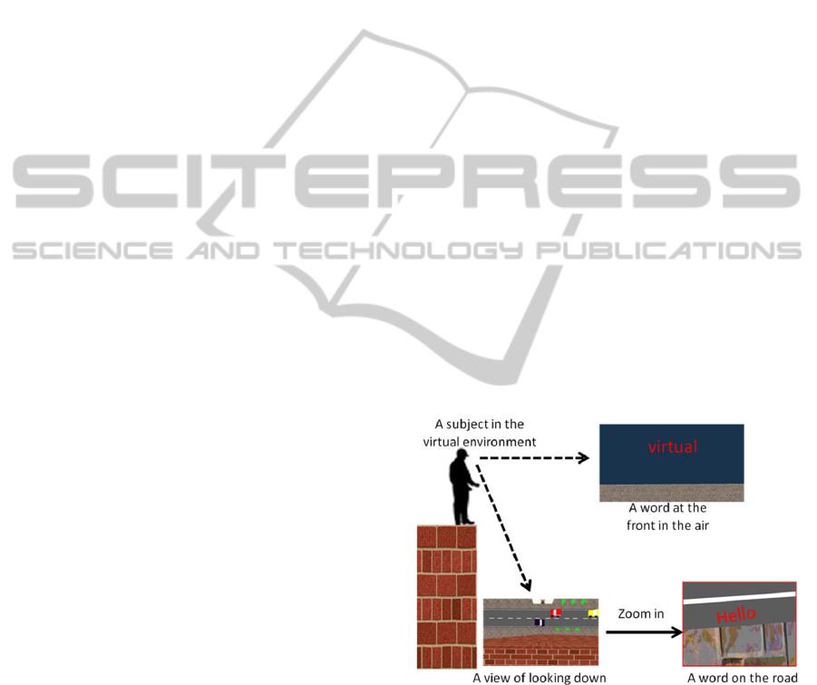
The experimental task is to read a word and say it in
the air in front of the subject and on the road under the
subject’s foot from the edge of the top of a tall build-
ing in a 3D virtual environment. The subjects perform
the task by using our system or a mouse while wear-
ing the HMD for a first person’s view with head track-
ing. Figure 4 illustrates the task. A subject stands at
the edge of the top of a tall building in a virtual en-
vironment. First, the subject reads a word and says it
on the road after looking down at the road by bending
his/her neck down and zooming in on the word (for
example, Figure 4 shows gHelloh). We set the font
size of the word to small so that it is impossible for
the subjects to read it without zooming it in. Next, the
subject reads a word and says it in the air in front of
him/her by raising his/her head (for example, Figure 4
shows gvirtualh). After that, the subject reads another
word and says it on the road again by zooming it in.
The subject repeats the task for five words.
Figure 4: Schematic representation of the experimental
task.
We set the task of reading a word from a high alti-
tude because we could provide the subjects with a fear
of the open high place in the 3D virtual environment,
and hypothesize that the more immersive feelings a
subject had, the more he/she would experience a fear
of the place. For the purpose of the more immersive
experience, we asked the subjects to stand at the edge
of the board that was approximately 4 cm thick (Fig-
ure 5).
APhysiologicalEvaluationofImmersiveExperienceofaViewControlMethodusingEyelidEMG
227

Figure 5: State of standing on a board to provide the subject
with the feeling of standing on the edge of a building.
4.2 Procedure
We covered the HMD with a black thick cloth to block
natural light from entering between the HMD and the
subject’s face, because the light in the real world de-
creases the subject’s immersive feelings in a virtual
world.
All subjects performed the task once using each
method (within subjects); the eyelid-movement input
method and a mouse input method. For the mouse in-
put method, a subject held a mouse on his/her thigh
while standing on the board. The left click button was
used to zoom in and the right click button was used
to zoom out. To avoid an ordering effect between
the two methods, half of the subjects performed the
task of eyelid movements first and the other half per-
formed the task of mouse clicking first.
The subjects practiced how to use each of the
methods for approximately 1 min before they started
to perform the task by using the assigned method.
During practice, the subjects confirmed the head-
tracking function of the HMD and how to zoom in
and out by standing on the ground in the virtual en-
vironment, not by standing on the top of a building.
The subjects performed the experimental task at the
rate of approximately 2 min per method. We recorded
the subject’s physiological data of the time in order
to validate the differences in the physiological data of
a heightened sense of fear of the high place between
the eyelid-movement input method and the mouse in-
put method. We assumed these differences to reflect
the differences in immersive feelings between the in-
put methods.
After completing the tasks, the subjects answered
a questionnaire consisting of five-point scale pair
comparisons about the differences they perceived be-
tween the input methods. Table 1 shows the question-
naire.
4.3 Measurement of Physiological Data
We analyzed how the subjects immersed in the vir-
tual environment when using each of the methods by
measuring the skin conductance (SC) of a subject’s
hand and the blood-volume pulse (BVP) on a sub-
Table 1: Questionnaire for user’s experience.
Questions and indexes
Q1DFor which did you feel more immersive impression?
1:mouse ——— 5: eyelid movement
Q2DFor which did you become more aware?
1:mouse ——— 5: eyelid movement
Q3DWhich was more intuitive?
1:mouse ——— 5: eyelid movement
Q4DWhich was easier to control as intended?
1:mouse ——— 5: eyelid movement
Q5DFor which did you feel more tired?
1:mouse ——— 5: eyelid movement
Q6DWhich do you like to use?
1:mouse ——— 5: eyelid movement
ject’s finger (Figure 6) with a ProComp infiniti en-
coder. We measured the physiological data because
they are known to increase according to the degree of
tension of the fear of high place (Meehan et al., 2002)
and to estimating arousal and valence levels of emo-
tions (Soleymani et al., 2008), (Lin et al., 2005). We
recorded the SC and the BVP of a subject during prac-
tice and the experimental tasks, and calculated the dif-
ferences of the average of the data between practice
and the tasks. We hypothesized that the differences of
the average between the practice and the experimental
task with the eyelid-movement input method is more
than that between the practice and the mouse input
method, because the subjects experienced the fear of
high place by the eyelid-movement method to a larger
extent than that by the mouse method. We think the
high embodiment of the eyelid movements makes feel
more immersive to the user.
Figure 6: State of attaching an SC sensor and a BVP sensor.
4.4 Results
Ten subjects (men aged between 21 and 25 years
old) participated in the experiment. Figure 7 shows
a box plot of the average differences of SC between
the practice (on the ground) and the experiment (at
the top of the building) in each of the input meth-
ods (mouse input and eyelid movement), and Fig-
ure 8 shows a box plot of the average differences of
the number of BVP pulses between the practice and
the experiment in each of the input methods. On
the basis of the results, we observe a significant dif-
PhyCS2014-InternationalConferenceonPhysiologicalComputingSystems
228
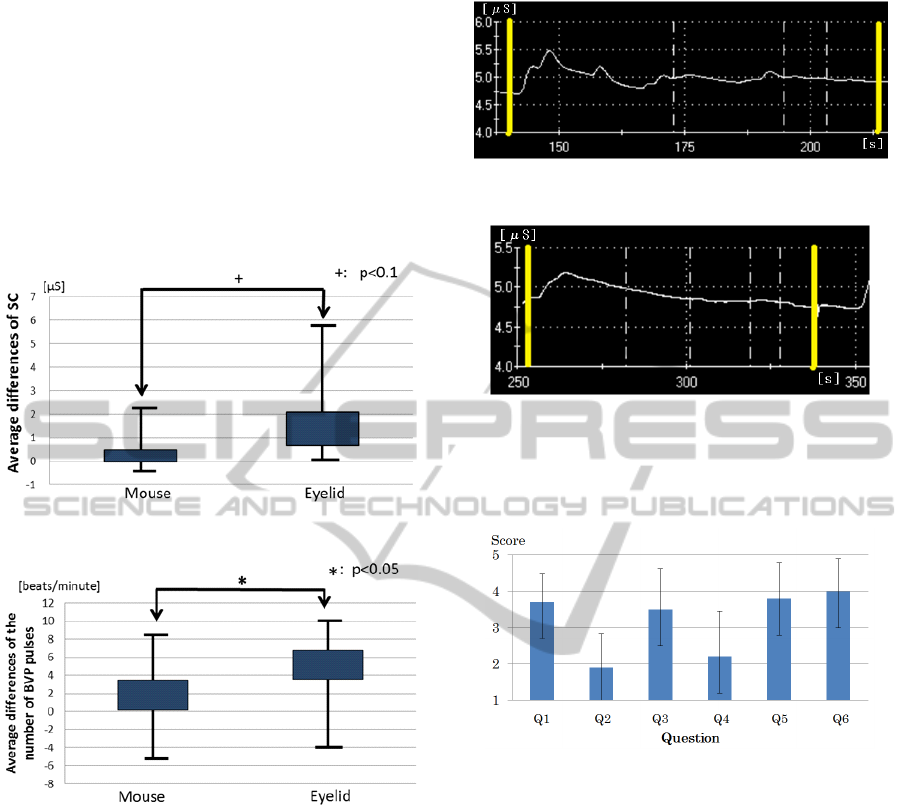
ference in SC between the input methods (ANOVA,
F = 5.130,d f = 1/9, p < 0.1). Therefore, we find
that the SC of the eyelid-movement input method
is significantly higher than that of the mouse input
method. In addition, there is a significant differ-
ence in the BVP between the input methods (ANOVA,
F = 3.943,d f = 1/9, p < 0.05). Therefore, we also
find that the number of BVP pulses of the eyelid-
movement input method is significantly higher than
that of the mouse input method.
Figure 7: Average differences of SC of the input methods.
Figure 8: Average differences of the number of BVP pulses
of the input methods.
As observed from the typical signals of SC in both
methods (Figures 9 and 10), the values of SC of most
of the subjects increased soon after the subject saw the
high altitude. Then, the values gradually decreased.
However, in the eyelid-movement input method, there
were typically some waves on the SC signals (Figures
9). The solid perpendicular lines of Figure 9 and Fig-
ure 10 show start and finish of the task.
Figure 11 shows the averages and the standard de-
viations of the answers to the questionnaire (Table
1). In terms of results, we find the subjects consid-
ered that the eyelid-movement input method is more
immersive than the mouse input method (Q1). Also,
they considered the eyelid-movement input method is
intuitive (Q3) because of marginal awareness of the
boundary between the real and virtual worlds (Q2);
they also would like to use it again (Q6). However,
Figure 9: Typical SC signals in eyelid-movement input
method.
Figure 10: Typical SC signals in mouse input method.
they observed that it was not as controllable as in-
tended (Q4), and they felt tired by using it (Q5).
Figure 11: Average and SD scores of the answers.
4.5 Discussion
Based on the results, our proposed eyelid-movement
input method improves the users’ immersive experi-
ences more effectively than the mouse input method.
We find that the eyelid-movement input method has a
significant fear effect on the users’ immersive experi-
ence because of the increase in SC and BVP pulses.
Additionally, the differences of the physiological data
between the eyelid-movement input method and the
mouse input method denote the same tendency of the
results of the questionnaire. We think that the physi-
ological data show not only the differences but also
the time series variation of subjects’ sense of fear.
Therefore, we consider that the waves of the SC in
eyelid-movement input method (Figure 9) show the
noticeable changes in the sense of fear with the phys-
ical and intuitive operation. We also consider that this
is due to the fact that the eyelid-movement input is
APhysiologicalEvaluationofImmersiveExperienceofaViewControlMethodusingEyelidEMG
229
associated with the act of looking at something, but
the mouse input is not associated with any such act.
Therefore, the subjects performed the task intuitively,
without being aware of the boundary between the real
and virtual worlds, and immersed themselves in the
virtual world.
On the other hand, based on the results of the
questionnaire, the eyelid-movement input did not per-
form as the users intended a few times, as compared
with the mouse input, and imposed a physical load on
the subjects. We consider that this is due to the fact
that the system failed to recognize some eyelid move-
ments a few times, although the system recognized
all the mouse clicks correctly. Actually, some sub-
jects experienced cognitive loads because of recogni-
tion errors between the zoom-in and zoom-out move-
ments, and some subjects experienced physical loads,
because a user of the eyelid-movement input method
was required to maintain a state, such as wide open-
ing of the eyelids. In addition, the average time to
complete the task by using the mouse input was 83
s, which was shorter than the time of the eyelid-
movement input, which was 160 s. For these rea-
sons, we have improved the recognition algorithm to
recognize each eyelid movement more accuracy, and
we have enhanced the system to recognize not only
the three types of eyelid movements but also multi-
degrees of eyelid state based on the present three
states.
5 IMPROVEMENT
We improved the recognition algorithm by adapting
an algorithm to remove outliers of EMG data because
there had been a lot of variation in the EMG data. We
have used the Smirnov-Grubbs’ test that detects out-
liers (p < 0.05) not included a normal distribution of
recorded EMG data, removes them and repeats the de-
tection and the remove until an outlier is undetectable.
Expression (1) shows the Smirnov-Grubbs’ test.
T =
|X
max
−X |
√
U
(1)
where X is mean of data, X
max
is the farthest value
from X, U is dispersion and T is test statistic. If a
value is less than T , the value is an outlier.
We conducted an experiment to verify the recogni-
tion rate of the improved system. Ten subjects (eight
men and two women, aged between 21 and 22 years
old) participated in the experiment. They did three
types of eyelid movements ten times each type in a
random order (within-subjects design) by using the
conventional system and the improved system.
Table 2: Recognition rates of the three states of the conven-
tional system (%).
Neutral Staring Widly-Opening
Average 82.0 78.0 84.0
SD 14.0 24.4 20.0
Table 3: Recognition rates of the three states of the im-
proved system (%).
Neutral Staring Widely-Opening
Average 90.0 90.0 92.0
SD 7.7 8.9 8.7
Table 3 and table 4 show the recognition rates of
each movement of each system. The average recogni-
tion rate of the conventional system is 80 % and that
of the improved system is 90 %. Additionally, the de-
viation of the improved system is smaller than that of
the conventional system. We therefore find that the
improved system is more accuracy than the conven-
tional system.
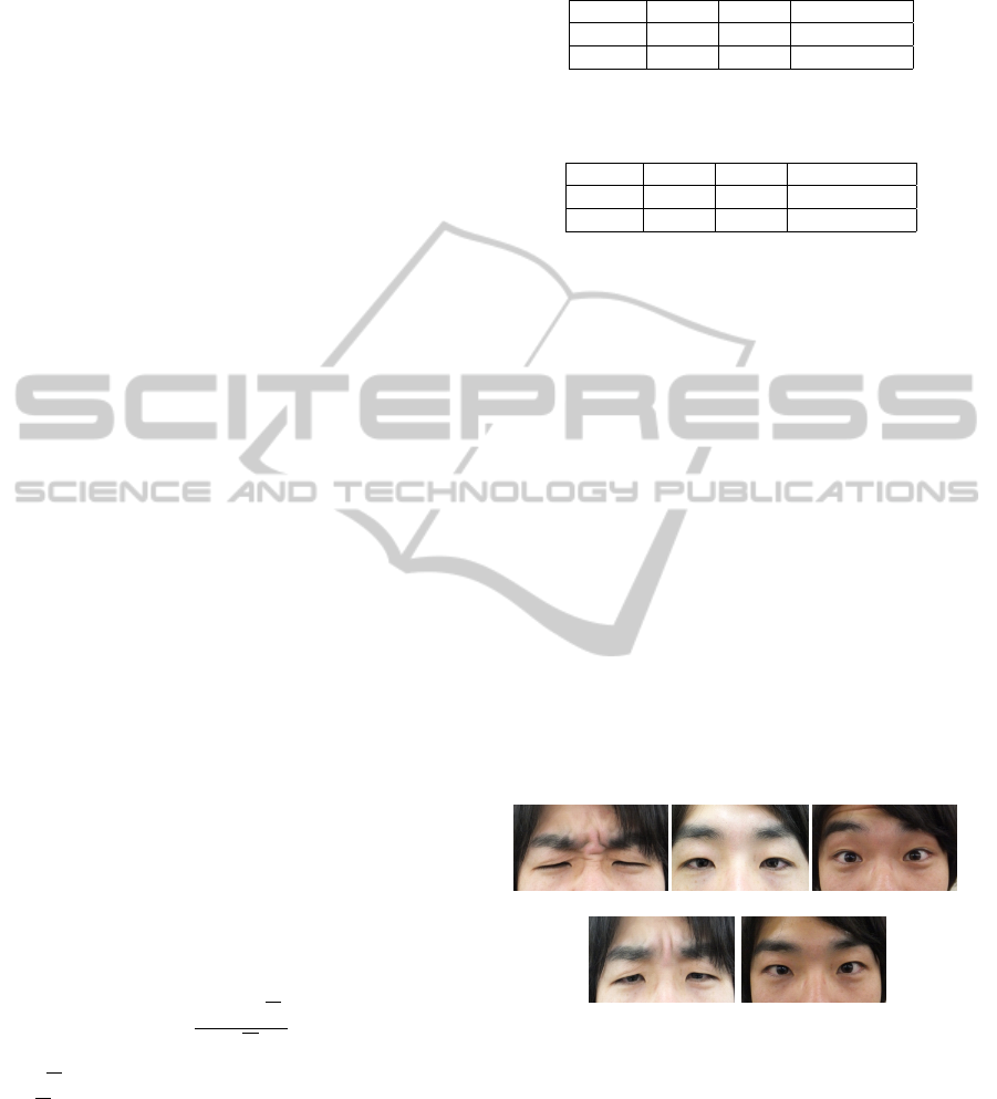
Moreover, we enhanced the number of states to
recognize eyelid movements from three types to five
types; strongly staring, softly staring, neutral, softly
opening, and widely-opening (see figure 12). As the
result of verifying the recognition rates of the five
movements like the experiment described above, table
4 shows the recognition rates of the five types (within-
subjects design) of ten subjects (eight men and two
women, aged between 21 and 22 years old). The rate
of the softly staring and the rate of the softly-opening
are lower than other rates.
(a) (c) (e)
(b) (d)
Figure 12: Three states of eyelid movements: (a) strongly
staring, (b) softly staring, (c) neutral, (d) softly opening, and
(e) widely-opening.
6 CONCLUSIONS
We conducted an experiment to compare our pro-
posed method with the mouse input method by mea-
suring the subjects’ physiological data of the fear of
the open high place in the virtual environment. On the
PhyCS2014-InternationalConferenceonPhysiologicalComputingSystems
230

Table 4: Recognition rates of the three states of the im-
proved system (%).
Neutral
Strongly-
staring
Softly-
staring
Widely-
Opening
Softly-
opening
Average 93.0 91.0 81.0 94.0 74.0
SD 6.4 7.0 9.4 6.6 6.6
basis of the results, we find that the eyelid-movement
input method improves the user’s immersive impres-
sion more significantly than the mouse input method.
Therefore, we conclude that it is important to immerse
an input method in an immersive virtual environment
because the user can use it intuitively and does not
have to be concerned about the seam between the real
and the virtual worlds. We also conclude that physio-
logical data are useful in continuously and objectively
evaluating usability of an input method.
For future work, we plan to further improve
the user’s immersive experience by applying real-
world embodiment movements and emotions esti-
mated from a user’s physiological data, such as EMG,
SC, BVP, brain waves and cerebral blood flow.
REFERENCES
Agustin, J. S., Hansen, J. P., Hansen, D. W., and Skovs-
gaard, H. H. T. (2009). Low-cost gaze pointing and
emg clicking. chi extended abstracts. CHI Extended
Abstracts 2009, pages 3247–3252.
Asai, K., Osawa, N., Sugimoto, Y., and Tanaka, Y. (2002).
Viewpoint motion control by body position in im-
mersive projection display. Proc. of SAC2002, pages
1074–1079.
Costanza, E., Inverso, S. A., Allen, R., and Maes, P. (2007).
Intimate interfaces in action: assessing the usability
and subtlety of emg-based motionless gestures. Proc.
of CHI 2007, pages 819–828.
Gibert, G., Pruzinec, M., Schultz, T., and Stevens, C.
(2009). Enhancement of human computer interac-
tion with facial electromyographic sensors. Proc. of
OZCHI 2009, pages 421–424.
Haffegee, A., Alexandrov, V. N., and Barrow, R. (2007).
Eye tracking and gaze vector calculation within im-
mersive virtual environments. Proc. of VRSTACM,
pages 225–226.
Jr., F. C., Nguyen, D., Guerra-Filho, G., and Huber, M.
(2010). Identification of static and dynamic muscle
activation patterns for intuitive human/computer inter-
faces. Proc. of PETRA 2010.
Kikuya, K., Hara, S., Shinzato, Y., Ijichi, K., Abe, H., Mat-
suno, T., Shikata, K., and Ohshima, T. (2011). Hyak-
ki men: Development of a mixed reality attraction
with gestural user interface. INTERACTION 2011,
2011(3):469–472.
Lin, T., Hu, W., Omata, M., and Imamiya, A. (2005). Do
physiological data relate to traditional usability in-
dexes? Proc. of OZCHI ’05.
Manabe, T., Tamura, H., and Tanno, K. (2009). The control
experiments of the electric wheelchair using s-emg of
facial muscles. Proc. FIT2009, pages 541–542.
Manders, C., Farbiz, F., Tang, K. Y., Yuan, M., Chong, B.,
and Chua, G. G. (2008). Interacting with 3d objects
in a virtual environment using an intuitive gesture sys-
tem. Proc. of VRCAI08.
Meehan, M., Insko, B., Whitton, M., and Brooks, F. P.
(2002). Physiological measures of presence in stress-
ful virtual environments. ACM Trans. Graph. In SIG-
GRAPH ’02, 21(3):645–652.
Miyashita, H., Hayashi, M., and ichi Okada, K. (2008).
Implementation of eog-based gaze estimation in hmd
with head-tracker. Proc. of ICAT2008, pages 20–27.
Nagahara, H., Yagi, Y., and Yachida, M. (2005). A
wide-field-of-view catadioptrical head-mounted dis-
play. Electronics and Communications in Japan, J88-
D-II(1):95–104.
Oshita, M. (2006). Motion-capture-based avatar control
framework in third-person view virtual environments.
Proc. of ACE ’06.
Ries, B., Interrante, V., Kaeding, M., and Anderson, L.
(2008). The effect of self-embodiment on distance
perception in immersive virtual environments. Proc.
of VRST 2008, 15(5):167–170.
Soleymani, M., Chanel, G., Kierkels, J. J. M., and Pun,
T. (2008). Affective ranking of movie scenes using
physiological signals and content analysis. Proc. of
MSACM, pages 32–39.
Steinicke, F., Bruder, G., Hinrichs, K. H., and Steed, A.
(2009). Presence-enhancing real walking user inter-
face for first-person video games. Proc. of ACM SIG-
GRAPH Symposium on Video Games, pages 111–118.
TheAuthors (2011). *****. *****.
Touyama, H., Hirota, K., and Hirose, M. (2006). Imple-
mentation of electromyogram interface in cabin im-
mersive multiscreen display. Proc. of IEEE Virtual
Reality 2006, pages 273–276.
Valstar, M., Pantic, M., Ambadar, Z., and Cohn, J. F. (2006).
Spontaneous vs. posed facial behavior: Automatic
analysis of brow actions. Proc. of ICMI ’06, pages
162–170.
APhysiologicalEvaluationofImmersiveExperienceofaViewControlMethodusingEyelidEMG
231
