
Removal of Gradient Artefacts during Transient Head Movements
for Continuous EEG-fMRI
José L. Ferreira
1
, Ronald M. Aarts
1,2
and Pierre J. M. Cluitmans
1,3
1
Department of Electrical Engineering, Eindhoven University of Technology, Eindhoven, The Netherlands
2
Philips Research Laboratories Eindhoven, Eindhoven, The Netherlands
3
Kempenhaeghe Epilepsy Center, Heeze, The Netherlands
Keywords: Combined EEG-fMRI, Gradient Artefact Correction, Transient Head Movements, Cubic Spline
Interpolation.
Abstract: This paper presents a novel approach for removing gradient artefacts from the EEG signal recorded during
continuous EEG-fMRI, which are influenced by transient head movements of the subject within the
magnetic scanner. Transient head movements provoke abrupt changes in the gradient artefact waveform, in
such a way that they compromise the estimation of an artefact waveform to be subtracted and achieve the
EEG correction. According to our proposed methodology, a cubic spline waveform is used to model and
represent the signal transitions components. This model is then used to change and approximate the shape of
the EEG signal as homogeneous data, in order to improve the performance of the gradient artefact
correction technique. The proposed approach also makes use of the signal slope adaption (SSD) method,
combined with sum-of-sinusoids modelling for correction of the gradient artefact. Our methodology reveals
to perform a robust and satisfactory removal of gradient artefacts under the occurrence of abrupt transient
head movements.
1 INTRODUCTION
Albeit combination of EEG-fMRI constitutes a
powerful and promising tool for brain activity
mapping as well as cognitive studies and research,
the occurrence of artefacts in the EEG signal still
represents a challenge to be overcome in order to
consolidate and broaden the range of application of
such a technique (Moosmann et al., 2009; Ritter et
al., 2010; De Munck et al., 2013). It is the case of
the gradient or imaging acquisition artefact, which is
induced in the electroencephalogram by the rapidly
varying gradient magnetic fields of the fMRI
equipment (Ritter et al., 2010). The gradient artefact
has amplitudes (up to 10
4
µV) that can be much
larger than those of the clinical EEG (up to 300 µV).
It possesses a characteristic waveform, which is
approximately the differential waveform of the
gradient magnetic fields that originate the artefact
(Anami et al., 2003;Ritter et al., 2010; Olson, 2010).
Because of the periodic and stationary nature of
the gradient artefact waveform, correction methods
based upon subtraction in time-domain and its
variants have been proposed and successfully
employed for artefact correction and subsequent
EEG restoration (Allen, et al. 2000; Garreffa et al.
2003; Gonçalves et al., 2007; De Munck et al.,
2013). In this way, the established average artefact
subtraction (AAS) methodology proposed by Allen
et al. (2000) has proven to be very effective for
cleaning up imaging artefacts. However, as
discussed by Yan et al. (2009), head motions of the
subject within the fMRI scanner compromise its
efficacy because of the alterations and transients that
are inserted in the artefact waveform. Thereby, the
averaging process results in an inaccurate estimation
of the artefact template (Sun and Hinrichs, 2009;
Koskinen and Vartiainen, 2009), which leads to
arising residual artefacts in the corrected EEG.
The usage of a sliding average artefact template
achieves to minimize this problem, decreasing the
probability of movement within a particular
averaging window. Nevertheless, in addition to
increasing the risk of subtraction of a clinical event
of interest of the EEG signal, the windows which
coincide with the movement continue locally altered
by using this approach (Yan et al., 2009). In order to
circumvent that problem, Moosmann et al. (2009)
213
L. Ferreira J., Aarts R. and J. M. Cluitmans P..
Removal of Gradient Artefacts during Transient Head Movements for Continuous EEG-fMRI.
DOI: 10.5220/0004802002130220
In Proceedings of the International Conference on Bio-inspired Systems and Signal Processing (BIOSIGNALS-2014), pages 213-220
ISBN: 978-989-758-011-6
Copyright
c
2014 SCITEPRESS (Science and Technology Publications, Lda.)

propose a correction procedure that uses information
related to head movement parameters from the fMRI
to improve the accuracy of the artefact template. In
the same way, Sun and Hinrichs (2009) describe a
method whereby the considered epochs for template
averaging are selected by weighting factors that
account the influence of the head position and
movement on the artefact shape. However, under the
occurrence of abrupt head movements, the artefact
correction obtained by those approaches could not
achieve accurate artefact template estimation as well
(Moosmann et al., 2009; Sun and Hinrichs, 2009).
Moosmann et al. (2009) even mention the strong
need for development of other correction methods
which take the occurrence of abrupt head motions
into account during simultaneous EEG-fMRI.
2 OBJECTIVES
Instead of using an artefact average template,
Ferreira et al. (2013a) propose a gradient artefact
correction methodology whereby the gradient
artefact waveform is approximated by the sum of a
set of sinusoids. According to Niazy et al. (2005),
artefact frequencies overlap the EEG bandwidth in
discrete harmonic frequency intervals (or frequency
bins) whose fundamental corresponds to the inverse
of the MR echo-planar slice time (ST) parameter.
Such frequency components can be modelled as a
sum of sinusoids waveforms which are then
subtracted to obtaining the EEG restoration (El-
Tatar and Fokapu, 2011; Ferreira et al., 2013a). An
advantage of using such a modelling approach is that
it does not require extensive calculation of MR
parameters as well as time-alignment of the internal
clocks of the EEG and fMRI equipments.
Furthermore, estimation of the average waveform is
not based upon an averaging process, in addition to
predicting the artefact waveform variability over the
time (Ferreira et al. 2013a).
The objectives of this paper are to investigate
and adapt the methodology proposed by Ferreira et
al. (2013a) for correction of gradient artefacts
affected by abrupt signal transients provoked by
head movements. In this way, we have proposed to
model and represent those signal transients as cubic
splines curves, which are used to modify and
approximate the shape of the EEG as homogeneous
data. This approach shows to improve the
performance of the gradient artefact correction, as
described in the sections Materials and Methods and
Results. Moreover, the proposed methodology
reveals itself to be robust to such signal transitions,
as shown in the section Results.
3 MATERIALS AND METHODS
3.1 Subjects
The EEG recordings were collected simultaneously
with the fMRI data for a research focused on
epilepsy and post-traumatic stress disorder (PTSD)
(Van Liempt et al., 2011; Ferreira et al., 2012;
Ferreira et al., 2013a), jointly developed by the
department of Psychiatry of Universiteit Medisch
Centrum Utrecht, the Research Centre Military
Mental Health Care in the Dutch Central Military
Hospital in Utrecht, and the Department of Research
and Development of the Kempenhaeghe Epilepsy
Center in Heeze, The Netherlands.
The data were recorded from military veterans
with PTSD which were in mission abroad through
the outpatient clinic of the Military Mental Health
Care. All participants were male and aged between
18 and 60 years.
3.2 Characteristics of the Used Data
Functional magnetic resonance imaging scanning
was carried out using a 3 T Scanner (Philips,
Eindhoven, The Netherlands) at Kempenhaeghe
Epilepsy Center. An MRI-compatible 64 channel
polysomnograph (MRI 64, MicroMed, Treviso,
Italy) was used to collect one ECG channel, two
EOG channels, one EMG channel and 60 EEG
channels.
The subjects were scanned using a functional
echo-planar imaging sequence with 33 transversal
slices (thickness 3 mm, TE 30 ms, TR 2500 ms).
EEG electrodes positioning was in accordance with
the international 10-20 system electrodes placement.
The sampling rate for signal acquiring was 2048 Hz
(Ferreira et al., 2012).
3.3 Proposed Methodology for Signal
Transients Modelling and Gradient
Artefact Removal
Representative raw EEG excerpts containing abrupt
transients caused by subject head movements were
selected and processed in accordance with the
algorithm block diagram of figure 1. Each step of
the algorithm was implemented and applied to the
EEG data in MATLAB environment.
BIOSIGNALS2014-InternationalConferenceonBio-inspiredSystemsandSignalProcessing
214
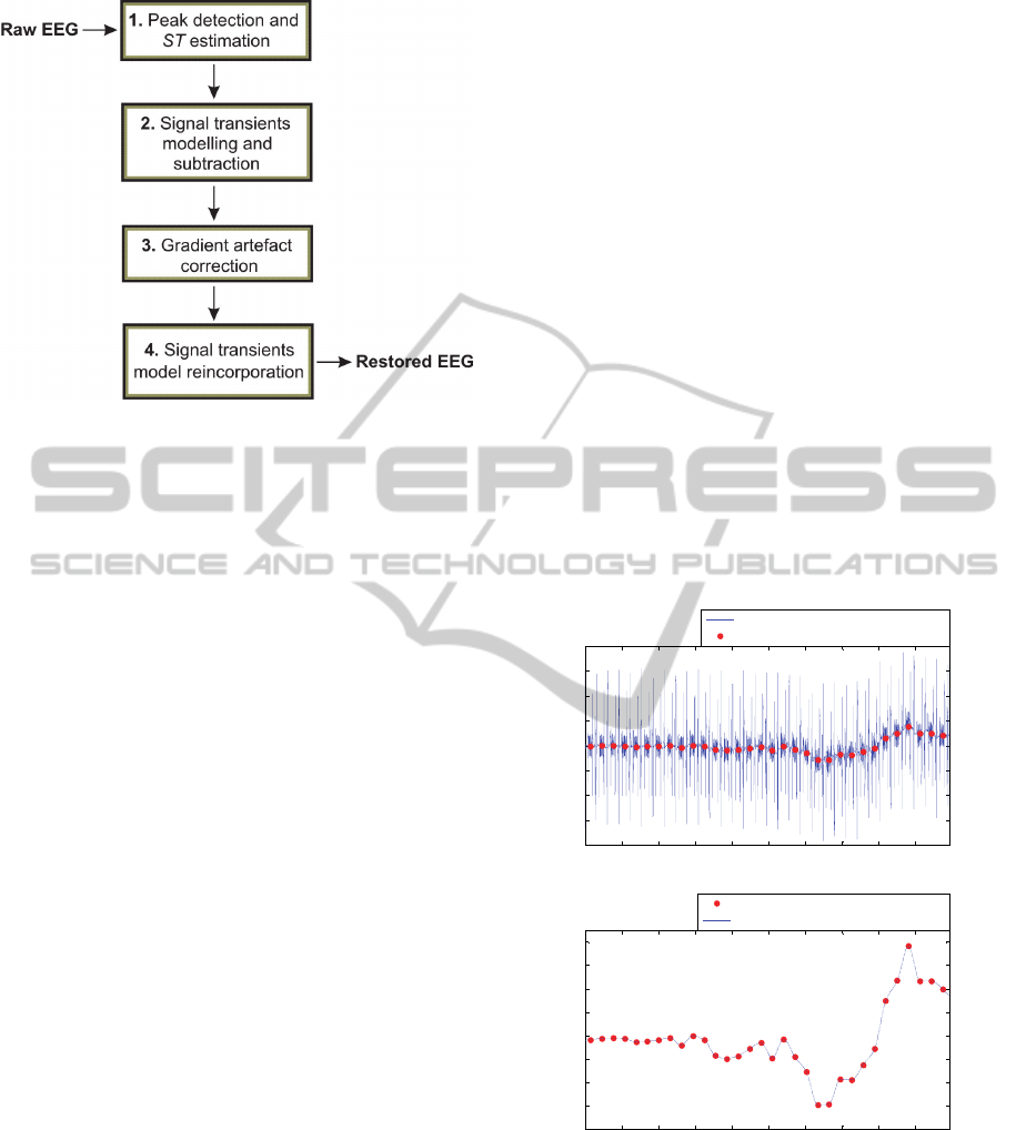
Figure 1: Block diagram structure of the proposed
methodology for gradient artefact correction under
transient head movements.
3.3.1 Peak Detection and ST Estimation
Implementation of the methodology illustrated in
figure 1 requires the initial detection of the typical
gradient artefact peaks, which are observed in the
raw EEG data recorded within the MR scanner. Such
detection is necessary for implementation of the
signal transients modelling (step 2 of figure 1).
Localization of those peaks are important for
estimation of the echo-planar slice time (ST) as well,
parameter used during the gradient artefact
correction methodology proposed by Ferreira et al.
(2013a) and applied in the step 3 of figure 1.
In order to detect the peaks, we used the peak
detection algorithms proposed by Garreffa et al.
(2003). Because of the EEG excerpts under analysis
were contaminated with transients, making difficult
the correct localization of the peaks, we have used
the ECG signal recorded simultaneously with the
EEG channels to perform the peak detection, as
performed by Ferreira et al. (2012).
For the data under analysis, the value of ST was
estimated at 155 ± 1 samples, which corresponds to
the time interval of 75.68 ± 0.50 ms (Ferreira et al.,
2012). The length of the raw EEG window was set
as 32 × 155 samples (Ferreira et al., 2013a).
3.3.2 Signal Transients Modelling and
Subtraction
The following equation was taken into account for
representation of the raw EEG signal (EEG
raw
):
arttranstrueraw
SSEEGEEG
,
(1)
where EEG
true
corresponds to the true EEG signal;
S
trans
corresponds to the signal transients introduced
by the head movement; and S
art
is the gradient
artefact.
In order to model the signal transients, the EEG
excerpts were divided into epochs (slices) whose
length is equal to the time between the gradient
artefacts peaks, ST (155 ± 1 samples), observed in
the raw EEG. Afterwards, we have taken into
account to average all samples of each epoch
separately. Making the assumption that the gradient
artefact waveform is stationary (i.e., it can be
considered a slowly varying process from epoch to
epoch) and has zero mean, that average only would
run over values associated with the EEG signal and
the signal transients. Thereby, the resulting average
values associated with each epoch would correspond
to the mean variation of the signal transients and
low-frequency components related to the true EEG
signal, from epoch to epoch. Figure 2a illustrates the
implementation of such a procedure. The illustrative
raw EEG excerpt shown in this figure was extracted
from the recordings of one subject, electrode
position F8:
0 500 1000 1500 2000 2500 3000 3500 4000 4500
-4000
-3000
-2000
-1000
0
1000
2000
3000
4000
Signal (
V)
Sample
0 500 1000 1500 2000 2500 3000 3500 4000 4500
-800
-600
-400
-200
0
200
400
600
800
Signal (
V)
Sample
Raw EEG data
Resulting points from epoch average
Resulting points from epoch average
Interpolated cubic spline
b
a
Figure 2: (a) Raw EEG data (blue trace) and resulting
values from averaging of each epoch (red points); (b)
resulting values from averaging each epoch (red points),
approximated by a cubic spline curve (blue trace).
The average values were plotted in the middle of
each epoch (red points). Such points have been
plotted in figure 2b as well, together with a cubic
spline curve which was used to fit those points in a
RemovalofGradientArtefactsduringTransientHeadMovementsforContinuousEEG-fMRI
215
time continuous sense. According to Wolberg and
Alfy (1999), cubic splines are very useful to fit a
smooth continuous curve to discrete data. The usage
of cubic splines as interpolants is especially
attractive because they make use of piecewise
polynomials with low-order to interpolate the data.
Moreover, the data can be modelled by respecting
constraints of smoothness and monotonicity. The
usage of cubic splines was proposed by Koskinen
and Vartiainen (2009) to improve the artefact
template estimation during application of the AAS
method (Allen et al., 2000).
Therefore, the fitted spline also corresponds to
the mean variation of the signal transients and low-
frequency components associated with the true EEG
signal from epoch to epoch. In turn, the frequency
activity associated with the gradient artefact and the
true EEG high-frequency components are contained
in the signal resulting from the subtraction of the
spline from the raw EEG of figure 2a. Such
characteristics can be observed in figure 3.
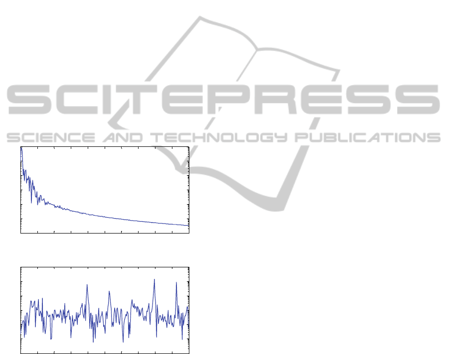
0 10 20 30 40 50 60 70 80 90 100
10
2
10
3
10
4
10
5
10
6
10
7
10
8
Frequency (Hz)
Power/frequency (
V
2
/Hz)
0 10 20 30 40 50 60 70 80 90 100
10
2
10
3
10
4
10
5
10
6
10
7
10
8
Frequency (Hz)
Power/frequency (
V
2
/Hz)
a
b
Figure 3: (a) Power spectrum of the fitted spline curve
shown in figure 2b; (b) power spectrum of the subtraction
of such a spline from the raw EEG of figure 2a. The
gradient artefact frequencies are contained in (b).
Thereby, although transients in the EEG signal
caused by abrupt head movements possess strong
high-frequency components, they can be
characterized as low-frequency activity in
comparison with the gradient artefact frequency
components. Hence, equation (1) was changed to
equation (2):
hpraw
SSEEG
,
(2)
where S
p
is the fitted spline, which corresponds to
the sum of the signal transients S
trans
and the low-
frequency components of the EEG
true
; and S
h
corresponds to the sum of the gradient artefact S
art
and the high-frequency components of the EEG
true
.
Therefore, according to equation (2) and figure
3, the gradient artefact correction should be applied
uniquely in the signal S
h
, which is the component of
the raw EEG that, in fact, contains the artefact
activity. Thus, there is no need to apply such
correction in the S
p
, in such a way that the
inaccuracies introduced by the signal transients
caused by the subject head movements during the
application of the gradient artefact approach can be
minimized.
To fit the cubic spline curve shown in figure 2,
we have used the piecewise cubic Hermite
interpolation method (Kreyszig, 2011; Fritsch and
Carlson, 1980). According to the Hermite
interpolation setup, given two points (x
j
, y
j
) and (x
j+1
,
y
j+1
), they are linked by the cubic interpolating
polynomial H
j
(x) with the following constraints:
jjj
yxH
)(
,
11
)(
jjj
yxH
,
jjj
y'xH'
)(
,
11
)(
jjj
y'xH'
.
(3)
H
j
(x) is described as (x
j
≤ x ≤ x
j+1
):
2
)()()(
jjjjjj
xxcxxbaxH
)()(
1
2
jjj
xxxxd
,
(4)
where the coefficients a
j
, b
j
, c
j
, and d
j
are calculated
by taking into account the values of x
j
, x
j+1
, y
j
, y
j+1
,
and certain slopes y’
j
and y’
j+1
at the two segment
endpoints. These slopes are chosen in such a way
that the shape and monotonicity within the data are
respected. Finally, the piecewise interpolant is found
by joining the J local cubic interpolants:
J
j
j
xHxH
1
)()(
.
(5)
In MATLAB, we have implemented the cubic
Hermite interpolation method using the routine
‘pchip’.
3.3.3 Gradient Artefact Correction
As mentioned above, we used the gradient artefact
BIOSIGNALS2014-InternationalConferenceonBio-inspiredSystemsandSignalProcessing
216
correction methodology proposed by Ferreira et al.
(2013a).
According to this method, initially a non-linear
filter based upon the signal slope adaption (SSD)
approach (Ferreira et al., 2013b; Ferreira et al.,
2013c) is applied to the raw EEG in order to remove
artefact high-frequency components. Here, we have
applied this filter directly to the signal S
h
, as
suggested earlier.
Because of such a filter is based upon the
difference between consecutive samples of the
signal, we observed that large signal slopes
associated with abrupt signal transients affect the
computational performance of the filtering
processing. In similar way, estimation of the
frequency components associated with the sum-of-
sinusoids model (Ferreira et al., 2013a) can be
affected by undesirable frequency activities inserted
by those transients. We noticed that such drawbacks
are minimized by using the signal S
h
instead of the
raw EEG, during carrying out the gradient artefact
correction.
The resulting signal after removal of the gradient
artefact constitutes the signal EEG
corct
.
3.3.4 Signal Transients Model
Reincorporation
As the fitted spline model contains low-frequency
components associated with the EEG signal, the
signal S
p
cannot be left out of the estimation of the
restored EEG, EEG
rest
, but it must be
reincorporated, as follows:
pcorctrest
SEEGEEG
.
(6)
Therefore, the proposed methodology is specifically
addressed to remove the gradient artefacts from the
raw EEG. Thus, the baseline associated with the
signal transients still remains in the restored EEG.
4 RESULTS
Figures 4 and 5 illustrate the application of the
proposed methodology to remove the gradient
artefact from the raw EEG excerpt of figure 2. In
figure 4a, the raw EEG was reproduced from figure
2. It can be noticed that the beginning of the abrupt
transient in this signal occurs around 188.5 s.
Figure 4b shows the signal S
h
, resulting from the
subtraction of the fitted spline from the raw EEG. It
can be noticed that S
h
approximately possesses the
shape of homogeneous data (i.e., data without abrupt
transients caused by head movements) in
comparison with the raw EEG signal. This fact,
therefore, enables minimization of the influence of
the signal transient components on estimation of the
artefact waveform. In consequence, application of
the gradient correction technique is performed in a
more accurate way.
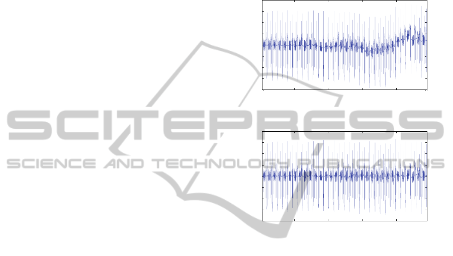
187.5 188 188.5 189
-4000
-3000
-2000
-1000
0
1000
2000
3000
4000
Signal (
V)
Time (s)
Raw EEG
187.5 188 188.5 189
-4000
-3000
-2000
-1000
0
1000
2000
3000
4000
Signal (
V)
Time (s)
Subtraction between the raw EEG and the fitted spline
b
a
Figure 4: (a) Raw EEG data; (b) signal S
h
resulting from
the subtraction of the fitted spline curve depicted in figure
2b from the raw EEG. The signal S
h
possesses an
approximated homogeneous data shape in comparison
with the raw EEG of (a).
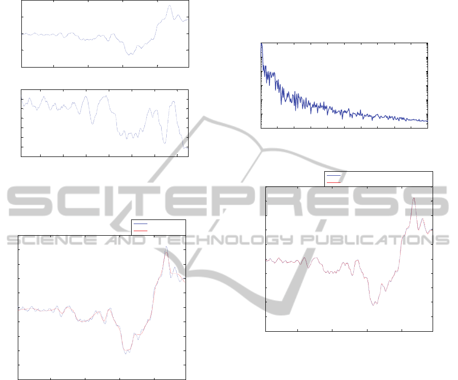
In figure 5, the restored EEG after application of the
proposed methodology is depicted. It can be
observed that the gradient artefact was cleaned up.
In figure 6, the restored EEG signal and the fitted
spline are superimposed for comparison purposes.
As can be observed, such curves are quite similar,
with a cross-correlation equal to 0.995. This fact
indicates that the restored EEG signal contains weak
frequency activity associated with S
h
. It also
confirms the idea that the points resulting from the
average of each epoch (and the respective fitted
spline) correspond to the mean variation of the
signal transients and low-frequency components
associated with the EEG signal, from epoch to
epoch, as assumed during implementation of our
approach.
Figure 7 depicts the power spectrum of the
restored EEG. The frequency activity associated
with the gradient artefact (figure 3b) was effectively
attenuated. Finally, figure 8 reveals that the usage of
RemovalofGradientArtefactsduringTransientHeadMovementsforContinuousEEG-fMRI
217
187.5 188 188.5 189
-1000
-500
0
500
1000
Signal (
V)
Time (s)
Restored EEG
187.2 187.4 187.6 187.8 188 188.2 188.4
-300
-250
-200
-150
-100
-50
0
50
Signal (
V)
Time (s)
b
a
Figure 5: (a) Restored EEG data after application of the
methodology depicted in figure 1; (b) Zooming in the
signal (a) around 187.7 s.
187.5 188 188.5 189
-1000
-800
-600
-400
-200
0
200
400
600
800
1000
Time (s)
Signal (
V)
Restored EEG
Fitted spline
Figure 6: Restored EEG signal (blue trace) and fitted cubic
spline curve (red trace). The cross-correlation between
these curves is equal to 0.995.
the cubic spline for representation of the signal
transitions can be employed to improve the gradient
artefact correction obtained by the AAS method
(Allen et al., 2000, Moosmann et al., 2009). For
evaluation of this case scenario, we used this
approach in the step 3 of figure 1 as well, after
application of the non-linear filter by SSD (Ferreira
et al., 2013b; Ferreira et al., 2013c) in the signals of
figure 4. Taking into account the signal of figure 4b,
it can be noticed that the EEG restoration obtained
by both correction methods are quite similar (figure
8). In turn, considering only the AAS method, the
artefact interference without subtraction of the spline
was estimated at 21 µV pk-pk, whereas such
interference was reduced to 13 µV pk-pk by
performing the subtraction of the S
p
. Therefore, the
subtraction of the spline proves to attenuate
alterations or inaccuracies introduced in the artefact
waveform estimative by the signal transients.
0 10 20 30 40 50 60 70 80 90 100
10
2
10
3
10
4
10
5
10
6
10
7
10
8
Frequency (Hz)
Power/frequency (
V
2
/Hz)
Figure 7: Power spectrum of the restored EEG signal.
187.5 188 188.5 189
-1000
-800
-600
-400
-200
0
200
400
600
800
1000
Time (s)
Signal (
V)
Restored EEG of figure 5
Restored EEG using the AAS method
Figure 8: Restored EEG signal of figure 5 (blue trace) and
restored EEG signal by the AAS method (red trace).
5 DISCUSSION
A number of approaches have achieved a
satisfactory removal of gradient artefacts from the
EEG signal recorded within the fMRI scanner during
the occurrence of subject head movements.
Nevertheless, the development of further correction
techniques is still demanded to improve the quality
of the EEG restoration in case of abrupt transients
caused by the head motions (Moosmann et al., 2009;
Sun and Hinrichs, 2009).
In this sense, we have proposed to model those
signal transients by averaging the samples of each
epoch into which the raw EEG was divided (figure
2). Such procedure also represents a moving-average
filtering process of ST samples, which removes
high-frequency components from the raw EEG,
including those ones related to the gradient artefact
BIOSIGNALS2014-InternationalConferenceonBio-inspiredSystemsandSignalProcessing
218

activity. Hence, the cubic spline used to fit the
resulting values from epoch averaging contains low-
frequency components associated with the EEG
signal and the signal transients per se, as depicted in
figures 3 and 6.
As shown in figure 4, the representation of the
signal transitions by cubic splines and the
subtraction of the respective model from the raw
EEG allow the data obtaining a homogeneous shape.
This fact yields to improve the performance of the
gradient artefact correction method by its application
in the signal S
h
, instead of the raw EEG, which
makes it robust to the signal transients. Hence, as
shown in figure 5 and 7, application of the proposed
approach achieves an effective removal of the
gradient artefact under the occurrence of signal
transitions caused by head movements.
Therefore, the subtraction and reincorporation of
the spline model from the raw EEG data, according
to the methodology depicted in figure 1, act as an
artifice to preserve the signal transients and EEG
low-frequency signal components from an
unnecessary processing by the artefact correction
method. The transients are responsible to introduce
inaccuracies and alterations in the artefact waveform
estimative and, in consequence, in the restored EEG
(Yan et al., 2009). We have noticed that the
proposed procedure is even useful during the
occurrence of longer lasting head movements and
homogeneous data correction. An additional
advantage associated with using the cubic spline
modelling is that eventual outliers which could be
obscured in the epoch averaging process can be
inserted in the interpolated curve as well, depending
on the need, in order to obtaining a better
representation of the signal transients. Those
characteristics shall be better evaluated in future
work.
It is noteworthy that instead of using splines,
application of a low-pass filter in the raw EEG does
not show to be adequate during implementation of
the proposed methodology. The higher the cut-off
frequency of the filter, the more is the amount of
gradient artefact frequency components which
remains in the signal S
p
, in case of using low-pass
filtering. Thereby, the gradient correction method
should be applied in S
p
as well. On the other hand, a
low cut-off frequency provokes insertion of
frequency components associated with the transients
in the signal S
h
, in such a way that those
inaccuracies could continue being introduced in the
restored EEG during the application of the gradient
artefact correction approach.
Another advantage observed within application
of the methodology described in figure 1 is that it
does not require additional information associated
with the head movements, quantified by using
sensors or related to the fMRI equipment. Rather,
according to the approach proposed in this work,
such information is directly inferred from the EEG
data during the signal transients modelling (step 2 of
figure 1).
As shown in figure 8, the restored EEG of figure
5 and the restoration obtained by the application of
the AAS method (Allen et al., 2000) in the step 3 of
figure 1 are quite similar. Therefore, it indicates that
the cubic spline can be employed for a satisfactory
EEG restoration by the AAS method as well, during
the occurrence of abrupt subject head motions. As a
further suggestion for future work, the usage of the
cubic spline for signal transients modelling shall be
assessed within the application of other artefact
correction methodologies.
6 CONCLUSIONS
In this work, we have proposed a novel method for
removing gradient artefacts from the EEG signal
recorded within the fMRI scanner under the
occurrence of abrupt subject head movements.
The proposed approach makes use of a cubic
spline curve to model signal transients caused by the
head motions. The subtraction and reincorporation
of such a model is used to change the EEG data
shape, which reveals to improve the performance of
the employed gradient artefact correction method.
Our methodology shows to perform an effective
and robust removal of the gradient artefact from the
EEG signal during the occurrence of abrupt signal
transients caused by head movements. Therefore,
such an approach constitutes a promising tool for a
satisfactory EEG correction within studies and
patients in scenarios in which it is difficult to
prevent those types of movements.
ACKNOWLEDGEMENTS
We are grateful to Saskia van Liempt, M.D., and
Col. Eric Vermetten, M.D., Ph.D. from the
University Medical Center/Central Military
Hospital, Utrecht, for providing the data presented in
this work. This work has been made possible by a
grant from the European Union and Erasmus
Mundus – EBW II Project, and by a grant from
CNPq – Science without Borders Program.
RemovalofGradientArtefactsduringTransientHeadMovementsforContinuousEEG-fMRI
219

REFERENCES
Allen, P., Josephs, O., Turner, R., 2000. A method for
removing imaging artifact from continuous EEG
recorded during functional MRI. NeuroImage 12, 230-
239.
Anami, K., Mori, T., Tanaka, F., Kawagoe, Y., Okamoto,
J., Yarita, M., Ohnishi, T., Yumoto, M., Matsuda, H.,
Saitoh, O., 2003. Stepping stone sampling for
retrieving artifact-free electroencephalogram during
functional magnetic resonance imaging. NeuroImage
19, 281–295.
De Munck, J., Van Houdt, P., Gonçalves, S., Van Wegen,
E., Ossenblok, P., 2013. Novel artefact removal
algorithms for co-registered EEG/fMRI based on
selective averaging and subtraction. NeuroImage 64,
407-415.
El-Tatar, A., Fokapu, O., 2011. Modeling MR induced
artifacts contaminating electrophysiological signals
recorded during MRI. Proceedings of the 33
rd
Annual
International Conference of the IEEE EMBS 2011,
Boston, Massachusetts, August 30 – September 3,
2011. 7135-7138.
Ferreira, J., Cluitmans, P., Aarts, R.M., 2012. Gradient
artefact correction in the EEG signal recorded within
the fMRI scanner. Proceedings of the 5
th
International
Conference on Bio-inspired Systems and Signal
Processing, BIOSIGNALS 2012, Vilamoura, Portugal,
February 1 – 4, 2012. 110-117.
Ferreira, J., Cluitmans, P., Aarts, R.M., 2013a. Gradient
artefact modelling using a set of sinusoidal waveforms
for EEG correction during continuous fMRI. Signal
Processing Research 2, 39-48.
Ferreira, J., Cluitmans, P., Aarts, R.M., 2013b. Non-linear
filter for gradient artefact correction during
simultaneous EEG-fMRI. Signal Processing Research
2, 55-63.
Ferreira, J., Cluitmans, P., Aarts, R.M., 2013c. Detection
of sharp wave activity in biological signals using
differentiation between consecutive samples.
Proceedings of the 6
th
International Conference on
Bio-inspired Systems and Signal Processing,
BIOSIGNALS 2013, Barcelona, Spain, February 11 –
14, 2013. 327-332.
Fritsch, F., Carlson, R., 1980. Monotone piecewise cubic
interpolation. SIAM. J. Numer. Anal. 17, 238-246.
Garreffa, G., Carnì, M., Gualniera, G., Ricci, G.B.,
Bozzao, L., De Carli, D., Morasso, P., Pantano, P.,
Colonnese, C., Roma, V., Maraviglia, B., 2003. Real-
time MR artifacts filtering during continuous
EEG/fMRI acquisition. Magn. Res. Imaging. 21, 1175-
1189.
Gonçalves, S., Pouwels, P., Kuijer, J., Heethaar, R., De
Munck, J., 2007. Artifact removal in co-registered
EEG/fMRI by selective average subtraction. Clin.
Neurophysiol. 118, 823-838.
Koskinen, M., Vartiainen, N., 2009. Removal of imaging
artifacts in EEG during simultaneous EEG/fMRI
recording: reconstruction of a high-precision artifact
template. NeuroImage 46, 160-167.
Kreyszig, E., 2011. Advanced engineering mathematics.
10
th
e. Wiley: New York.
Moosmann, M., Schönfelder, V., Specht, K., Scheeringa,
R., Nordby, H., Hugdahl, K., 2009. Realignment
parameter-informed artefact correction for
simultaneous EEG-fMRI recordings. NeuroImage 45,
1144-1150.
Niazy, R., Beckmann, C., Iannetti, G., Brady, J., Smith, S.,
2005. Removal of FMRI environment artifacts from
EEG data using optimal basis sets. NeuroImage 28,
720-737.
Olson, W., 2010. Basic concepts of medical
instrumentation. In J. Webster (ed.), Medical
instrumentation: application and design. 4
th
ed. Wiley:
New York.
Ritter, P., Becker, R., Freyer, F., Villringer, A., 2010. EEG
quality: the image acquisition artifact. In C. Mulert, L.
Limieux (eds.), EEG-fMRI: Physiological basis,
technique and applications. Springer: Verlag, Berlin,
Heidelberg.
Sun, L., Hinrichs, H., 2009. Simultaneously recorded
EEG-fMRI: removal of gradient artifacts by
subtraction of head movement related average artifact
waveforms. Hum. Brain Mapp. 30, 3361–3377.
Van Liempt, S., Vermetten, E., Lentjes, E., Arends, J.,
Westenberg, H., 2011. Decreased nocturnal growth
hormone secretion and sleep fragmentation in combat-
related posttraumatic stress disorder; potential
predictors of impaired memory consolidation.
Psychoneuroendocrino. 36, 1361-1369.
Wolberg, G., Alfy, I., 1999. Monotonic cubic spline
interpolation. Proceedings of the Computer Graphics
International Conference, CGI 1999, Canmore,
Canada, June 7 – 11, 1999. 188-195.
Yan, W., Mullinger, K., Brookes, M., Bowtell, R., 2009.
Understanding gradient artefacts in simultaneous
EEG/fMRI. NeuroImage 46, 459-471.
BIOSIGNALS2014-InternationalConferenceonBio-inspiredSystemsandSignalProcessing
220
