
Electrical Stimulation of the Transcutaneous Posterior Tibial Nerve
for the Treatment of Patients with Detrusor Overactivity
Due to Neurogenic Hiperactive Bladder in Multiple Sclerosis
A Case Study
Fabiana S. B. Perez
1
, Luciana R. Tenório Peixoto
1
, Fabiano Soares
2
,
Cristiano Jacques Miosso
2
and Adson F. da Rocha
2
1
School of Medicine, University of Brasilía, Brasília, DF, Brazil
2
University of Brasília at Gama, Gama, DF, Brazil
Keywords: Electrical Stimulation, Multiple Sclerosis, Neurogenic Bladder Incontinence and Posterior Tibial Nerve.
Abstract: This case study evaluates the therapeutic use of Transcutaneous Electrical Nerve Stimulation (TENS) of the
posterior tibial nerve for treating one patient with multiple sclerosis (MS) showing signs of urinary
incontinence (UI) due to detrusor overactivity (DO). Patient: MS with UI and sensory loss. Method: Using
the current therapy twice a week for 20 minutes in 10 sessions and monitoring electrodes during electrical
stimulation. Results: We observed an improvement in urge incontinence with reduced trips to the bathroom
during the day and night. Both the post voiding sense of desire and pain during urination disappeared.
Conclusions: This study shows an indication that the use of TENS in the current technique of posterior tibial
nerve can reduce the uninhibited detrusor contractions and improve the quality of life of patients with MS
due to a reduction of urinary incontinence and also reduce the number of times that the patient's urinates,
thus providing better quality of sleep, humor, personal relationship, less embarrassment and reduction of
stress. In this way, this study justifies a wide investigation with multiple subjects.
1 INTRODUCTION
Multiple sclerosis (MS) is a neurological disease of
high incidence in young adults with a picture of
multifocal demyelination in the central nervous
system (Coelho, 2009; Poser , 1986; Misulis, 2008).
The urgency or urinary incontinence (UI) may occur
as the initial manifestation in most patients with MS
(Stephen, 1995).
Involuntary loss of urine is a problem of social
order and hygiene, causing embarrassment and
changes in behavior such as social isolation, low
self-esteem and psychosocial disorders (Oliveira,
2010). The most common etiology of urinary
incontinence is neurogenic (Monteiro, 2009).
The four handles or neurological pathways in the
control of urination, and which are related to each
other, are: the core trunk detrusor cerebral cortex
(loop I), the core detrusor muscle spinal / sacral
brainstem (loop II)-sacral urethral sphincter of the
bladder (loop III), and the sacral-brain (loop IV).
Pathways I and IV are responsible for voluntary
control of urination. Pathways II and III, on the other
hand, regulate the contractions of the detrusor
bladder emptying to promote and coordinate efforts
between the detrusor and urethra (Stephenson and
O’Connor, 2004).
The neurogenic bladder dysfunction is defined as
a neurological disease produced by nerve damage
that interferes with the mechanisms of voluntary and
involuntary urination, thus causing changes in
normal bladder function
. The neurogenic bladder
corresponds to the overactive and/or underactivity of
the detrusor (Azevedo et al, 1990).
The
underactivebladderretentionoroverflowis
characterized by
urinary loss that occurs when
intravesical pressure exceeds urethral pressure. This
is associated with bladder distention, but in the
absence of detrusor activity. This overflow happens
when one reaches the limits of distensibility or
compliance of the bladder (Miltrano, 2009).
According to the International Continence
Society, the overactive bladder is defined as a
neurogenic injury due to the presence of involuntary
154
S. B. Perez F., R. Tenório Peixoto L., Soares F., Jacques Miosso C. and F. da Rocha A..
Electrical Stimulation of the Transcutaneous Posterior Tibial Nerve for the Treatment of Patients with Detrusor Overactivity Due to Neurogenic Hiperactive
Bladder in Multiple Sclerosis - A Case Study.
DOI: 10.5220/0004806101540158
In Proceedings of the International Conference on Biomedical Electronics and Devices (BIODEVICES-2014), pages 154-158
ISBN: 978-989-758-013-0
Copyright
c
2014 SCITEPRESS (Science and Technology Publications, Lda.)

detrusor contractions during the filling phase
(Coelho, 2009). This is characterized by urinary
incontinence, urinary frequency, nocturia and
urgency (Fischer-Sgrott et al, 2009).
The existent techniques of treatment for UI are
electrostimulation applications directed to the
perineum muscle, using either internal anal
electrodes for men or surface electrodes in the
region. These two techniques are embarrassing,
invasive (in the first case) and may cause discomfort
and burns if the patient has abnormal sensibility
(Marques, 2008).
Treatments with transcutaneous electrical
stimulation in the posterior tibial nerve aim at
reducing UI and assume that the path of the nerve
there are neuronais projections of the bladder
(Fischer-Sgrott et al, 2009).
The TENS current is used for the treatment of
urinary incontinence by bladder hyperactive (BH).
The electrodes are placed bilaterally in the medial
region of the legs, causing motor and sensory
stimulation as the current is applied. During each
session, the patient’s neurological physiotherapist
observes the stimulation caused by the motor
current, and the sensory way is not changed to
modulate the current flow. This technique promotes
the reduction of involuntary detrusor contractions
(Marques, 2008). Regarding the TENS current for
the treatment of BH, some researchers propose a
sequence of pulses with a frequency of around 20 Hz
and with a duration of around 200 miliseconds per
pulse (Amarenco, 2003).
The therapies using electric currents can be used
in neurological patients with abnormal sensitivity,
because applying electrical stimulation displays
rhythmic flexion of the hallux, thus indicating the
correct placement of electrodes and confirming this
to be intact innervation (Maciel and Souto, 2009).
However, in individuals with Babinski's reflex, it is
difficult to dispense the current therapeutic
modulation due to incorrect motor response they
have, so that it becomes impossible to control the
intensity offered by the device and electro-motor
response.
In the case of hyposensitivity, the dose should be
applied until it causes rhythmic inflections of the big
toe, and it should then be reduced until the motor
action disappears. The provided dose agrees with
several studies arguing that the ideal intensity must
be maintained according to the threshold of each
patient and below the motor threshold (Fischer-
Sgrott et al, 2008; Maciel and Souto, 2009;
Amarenco et al, 2003; Kabay et al, 2009).
2 CLINICAL CASE
A 36 year-old black male, married and with one son.
At the age of 28, after suffering a crisis caused by
the disease, the patient was diagnosed with MS.
After a few years, another MS crisis occurred, with
the same characteristics aforementioned, and the
patient again recovered later. The patient reported
having suffered from UI starting 6 years after the
MS was found, and that the UI was diagnosed as
caused by neurogenic overactive bladder, as
confirmed by urodynamic examination.
During the evaluation, the patient was lethargic,
with initiative and responsive. In neurological
evaluation, we noted sensory deficit and the
presence of cutaneous reflex - planting. In the
evaluation of urogynecologic history, we noted that
the main complaint consisted of dysuria, urinary
frequency and incontinence urge. We found that the
frequency of urination during the day corresponded
to 15 trips to the bathroom to urinate and during the
night corresponded to 9 trips to the bathroom, which
confirmed the picture of urinary urgency and
nocturia. The patient reported pain and post-voiding
desire during the act of micturition. He also reported
the presence of active sexual activity with urination,
as well as hypertension. He denied making use of
liners even with urinary incontinence in clothes.
3 METHOD
On the first evaluation day, we applied the
physiotherapy assessment protocol forms, which the
patient completed in the Urogynecology laboratory
at Unifesp. We instructed the patient to complete a
voiding diary for three days after treatment.
During the neurological assessment, a physical
examination showed no sensory deficit and bilateral
Babinski's reflex. Given that no pathological reflex
existed, we positioned the electrodes in the path of
the posterior tibial nerve, to detect whether
innervation was intact, by using electrical
stimulation (we used the TENS NEURODYN/FES
portable device, by Ibramed Ltda). This stimulation
was based on a sequence of 200-milisecond puses,
with a frequency of 20 Hz, following the
recommendation by (Amarenco, 2003). Since the
patient had reported hypertension before the
treatment, his blood pressure (BP) was monitored in
all the sessions.
The treatment protocol consisted of 10 sessions,
twice a week and lasting 20 minutes each. We
ElectricalStimulationoftheTranscutaneousPosteriorTibialNervefortheTreatmentofPatientswithDetrusor
OveractivityDuetoNeurogenicHiperactiveBladderinMultipleSclerosis-ACaseStudy
155
applied the TENS current through two channels,
using four electrodes positioned transcutaneously
and bilaterally in the lower limb (2 electrodes per
channel). For each channel, one electrode was fixed
to the posterior medial malleolus and the other
10 cm above. The intensity parameter due to
hyposensitivity was measured through the signal
engine rhythmic inflections of hallux. A maximum
intensity of 30 mA was applied, for safety reasons.
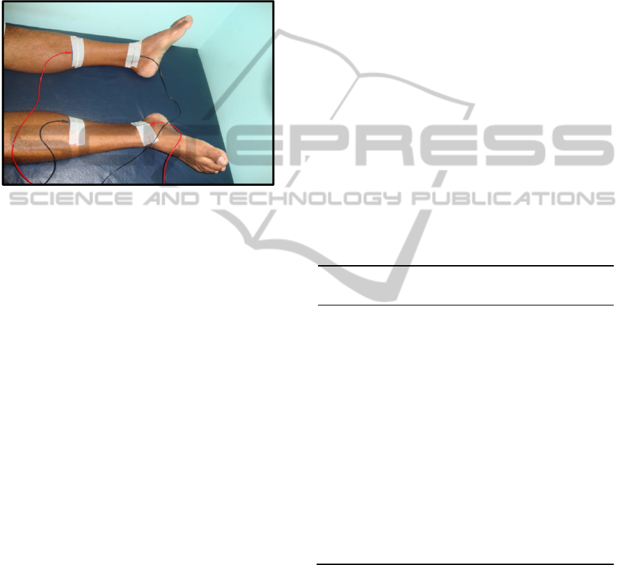
Figure 1: Positioning of the 4 eletrodes, two for each
channel, used to apply the electric currents during the
TENS sessions.
4 RESULTS AND DISCUSSION
This paper presents results of a case study monitored
by descriptive assessments from the Unifesp
Physiotherapeutic Protocol in Urogynecology and
the voiding diary for three days. The patient was
submitted to physiotherapeutic treatment with
transcutaneous electrical stimulation in order to
attenuate urological clinical complaints. This
justifies the choice of a treatment by elective
electrostimulation – separated from other techniques
such as kinesiotherapy. Both evaluation procedures
were applied i) before treatment, ii) after 20 TENS
sessions, iii) within a year after being treated with
TENS. It should be noted that the patient was still
followed and evaluated after discharge from
physiotherapeutic urological attendance and the
same clinical signs obtained after 20 TENS sessions
were preserved.
This case study of urinary incontinence treatment
in a MS patient was made possible because the same
therapist followed the patient throughout the study,
even though the latter was also attended for
treatment of other neurological symptoms such as
unsteadiness and difficulties with static and dynamic
coordination, aggravated during crises. MS patients
commonly face periods of illness aggravation and
remission. Nevertheless, clinical urological results
obtained in this case study remained constant even
after a year.
According to Kabay et al (2009), when applied
to people suffering from multiple sclerosis, the
technique noticeably decreased nocturia in 75% of
patients. Marques (2008) reports decrease in
nocturia with 38% of symptoms relief. Another
study found improvement in nighttime urination in
21% of cases (Govier et al, 2001). Table 1 shows
that after 10 sessions of electrical stimulation of the
posterior tibial nerve, there was decrease in the signs
of urinating discomfort, as well as reduction in the
number of urinary frequency during the day,
nocturia, and urgency incontinence episodes. We
can justify improvement of urinary urgency
conditions through a study in which urodynamic
evaluation with electrical stimulation of the posterior
tibial nerve revealed maximum bladder capacity can
increase together with a decrease of involuntary
detrusor contractions during standard cystometry
(Amarenco, 2003).
Table 1: Urologic evaluation of signs frequency before
treatment (f
before
), after 10 sessions (f
10
) and after one year
of treatment (f
1year
) with electrical stimulation sessions.
Main
complaint
F
before
F
10 sessions
F
20sessions
1
F1year
Nocturia
9 5 0 0
Void desire
during the
day
15 3 a 4 normal normal
Sensation
act voing
pain and
desire
after
voing
burning confortable Confortable
Urge
incontinence
2.7 1.3 normal normal
Cause of
urinary loss
urge
incon-
tinence
urge
incontinence
normal normal
1
after 20 sessions
In general, urinary incontinence treatment was
considered effective given the observation that
urinary loss episodes were reduced by 50%. With
regard to this parameter, involuntary loss of urine
onto clothes was reported 5 times by the patient
before treatment due urge incontinence and 2 times
after 10 electrical stimulation sessions. This
reduction may be explained in terms of possible
neuromodulation provoked by the TENS current.
BIODEVICES2014-InternationalConferenceonBiomedicalElectronicsandDevices
156

This research found a limitation in the loss of
patient sensibility caused by MS.
According to Tilbery (2006), it is common for
patients with MS to present paraparesis of varying
intensity, especially in the lower limbs. Therefore,
the difficulty of using electrotherapy with these
patients consists in adjusting the intensities of the
current. We propose to perform the sensitivity test
before applying the therapeutic current. During
electrical stimulation it is important to carefully
observe the threshold of individuals with
hypersensibility. For that reason, the current
intensity should be slowly graded until reaching an
individualized parameter below the stimulation
threshold of motor innervation (Kabay et al, 2009;
Amarenco, 2003; Fischer-Sgrott et al, 2009; Maciel
and Souto, 2009).
5 CONCLUSIONS
The transcutaneous electrical stimulation in the
posterior tibial nerve can be considered as an
alternative in the treatment of urinary incontinence
in detrusor hyperactivity. Our results suggest that the
technique may be effective in reducing the
uninhibited detrusor contractions after 10 sessions of
electrostimulation.
This technique is also relatively inexpensive,
non-invasive, non-embarrassing, comfortable,
painless, effective, with targeted action on the
detrusor muscle, easy to apply and free of side
effects of medications. Also, we believe that it can
have high acceptance and adherence by the patients,
since it does not result in great discomfort and each
session takes 20 minutes.
We emphasize that, in this study, the technique
resulted in a reduction in daytime and nighttime
urinary frequency and in the number of episodes of
urge incontinence. It can then have an impact in
reducing stress and embarrassment and the number
of urinary tract infections, as well as improving
sleep quality and mood and thus providing a better
quality of life for individuals with MS. It is then a
good alternative in the treatment of lower urinary
tract dysfunction, as suggested by the patient in this
study.
ACKNOWLEDGEMENTS
We leave to express our sincere thanks to Doctor
Denise Sistorelli Diniz, a neurologist at the Hospital
das Clínicas, Federal University of Goiás, for
supporting our research. The authors thank the
Alfredo Nasser Scholl (Unifan).
REFERENCES
Amarenco, G. et al. Urodynamic Effect of Acute
Transcutaneous Posterior Tibial Nerve Stimulation in
Overactive Bladder. American Urological Association.
v. 109, p. 2215-2215, 2003.
Azevedo, M. A. J.; Maria, M. L. S.; SOLER, L. M. A.
Promovendo o auto-cuidado: treinamento e assistência
de enfermagem a pacientes portadores de bexiga
neurogênica. Revista Brasileira de Enfermagem,
Brasília, v. 43, n. 1/4, p. 52-57, jan./dez. 1990.
Coelho, M. M. Avaliação Urodinâmica na Esclerose
Múltipla. Acta Urológica, v. 26, n. 3, 2009.
Fischer-Sgrott, F. O.; Manffra, E. F.; Junior, W. F. S. B.
Qualidade de vida de mulheres com bexiga hiperativa
refratária tratadas com estimulação elétrica do nervo
tibial posterior. Revista Brasileira de Fisioterapia. São
Carlos, v. 13, n. 6, p. 480-6, nov./dez. 2009.
Govier, F. E. et al. Percutaneous afferent neuromodulation
for the refractory overactive bladder results of a
multicenter study. The Journal of Urology, United
States, v. 165. 2001.
Kabay, S. et al. The clinical and urodynamic results of a 3
–month percutaneous posterior tibial nerve stimulation
treatment in patients with with multiple sclerosis-
related neurogenic bladder dysfunction. Neurourology
and Urodynamics, United States, v. 28 , p. 964-968,
abr.. 2009.
Maciel, L. C.; Souto, S. Estimulação do Nervo Tibial
Posterior (Ptns) no Tratamento da Bexiga Hiperativa.
In: Palma, P. (Ed.). Urofisioterapia: Aplicações
Clínicas das Técnicas Fisioterapêuticas nas Disfunções
Miccionais e do Assoalho Pélvico. 1ª ed. São Paulo:
Unicamp, 2009, Cap. 20, p. 223-227.
Marques, A. A. A eletroestimulação do nervo tibial
posterior no tratamento da bexiga hiperativa.
Unicamp. Campinas, SP: [s.n.], 2008.
Miltrano, P. Fisiopatologia e classificação da
incontinência urinária. In: Moreno. (Org.). Fisioterapia
em Uroginecologia. Barueri: Manole, 2009. p. 29-37.
Misulis, K. E. Head, C. T. In: Netter. Distúrbios Auto-
imunes. Neurologia Essencial. São Paulo: Saunders
Elsevier, 2008. p. 297-309.
Monteiro, E.S. et al. Queixas Urinárias em Mulheres com
Infarto Cerebral. Revista Neurociências. V. 17, p. 103-
7, 2009.
Moritz, J. E. et al. Bexiga Neurogênica. 2005. 39p.
Trabalho de conclusão de curso de medicina,
Universidade Federal de Santa Catarina, UFSC,
Florianópolis, 2005.
Oliveira, K. A. C et al. Técnicas Fisioterapêuticas no
Tratamento e Prevenção da Incontinência Urinária de
Esforço na Mulher. Revista Eletrônica F@pciência.
Apucarana, v.1, n.1, p. 31-49, 2007.
ElectricalStimulationoftheTranscutaneousPosteriorTibialNervefortheTreatmentofPatientswithDetrusor
OveractivityDuetoNeurogenicHiperactiveBladderinMultipleSclerosis-ACaseStudy
157

Poser, C. M. et al. Doenças desmielinizantes. In:
ROWLAND, L. P.; MERRITT. Tratado de
Neurologia. Rio de Janeiro: Guanabara Koogan, 1986.
p. 603-621.
Souza, R. O.; Figueiredo, W. M. O. Reflexo Cutâneo
Plantar em Extensão. Arquivos de Neuropsiquiatria.
Rio de Janeiro, p. 318-323, 1995.
Stephen, H. L. Esclerose Múltipla e outras doenças
desmielinizantes. In: HARRISON. Medicina Interna.
São Paulo: Nueva Editorial Inter Americana, 1995. p.
2401-2411.
Stephenson, R. G.; O’connor, L. J. Fisioterapia Aplicada à
Ginecologia e Obstetrícia. 2ª ed. São Paulo: Manole,
2004, 515 p.
Tilbery, C. P. Esclerose Múltipla e Outras Doenças
Desmielinizantes. In: LOPES, A. C. Tratado de
Clínica Médica. São Paulo: Roca, v. 2, 2006, cap. 201,
p. 2317-2322.
BIODEVICES2014-InternationalConferenceonBiomedicalElectronicsandDevices
158
