
Mechanically Flexiable Biosensor for Detection
of Photoplethysography
Kian Davoudi,
Moein Shayeganyan, Kouhyar Tavakolian and Bozena Kamisnka
Simon Fraser University, 8888 University Drive, Burnabyr, Canada
Keywords: Photoplethysmography, Heart Rate, Inkjet Printing, Polymer Metallization, Flexible Sensor.
Abstract: Acute cardiovascular failure could be detected by continuous monitoring of electrocardiogram (ECG).
While electrode allocation on skin challenges quality of electrocardiograph, beat-to-beat heart rate obtained
from photoplethysmography (PPG) could be used to indicate cardiovascular activity as an alternative to
heart rate from ECG. In this paper we proposed a mechanically flexible PPG sensor integrated on thin layer
of polymer. Mean and standard deviation of beat-to-beat heart rate was obtained from a flexible PPG sensor
and compared to the beat-to-beat heart rate obtained from a commercial ECG and PPG devices. The
standard deviation of beat-to-beat heart rate from ECG and PPG intervals were analysed by the Bland and
Altman analysis. The corresponding 95% limits of agreement were estimated as 0.034 to -0.01 for PPG
flexible sensor compared to ECG and as 0.0081 to 0.0037 for PPG flexible sensor compared to PPG
commercial device. The good correlation between the measurement results demonstrated capability of our
proposed mechanical flexible PPG sensor to be used as practical alternative to ECG for heart rate variability
(HRV) analysis.
1 INTRODUCTION
Increasing cost of healthcare and accelerated aging
of society incite hospitals and other medical
caregivers to look for solutions that are inexpensive,
yet maintain proper quality of care. The prospects of
efficient remote health and activity monitoring using
biosensors have recently gained a lot of interest and
stimulate research in the area of wearable
electronics. Technological advances in sensors,
wireless communication and integrated circuits have
brought about small, inexpensive wearable
physiological monitors. These devices are usually
capable of sensing one or more vital signs, e.g. heart
rate, body temperature and blood pressure, then
communicating the acquired data to a local or
remote processing and interpretation centre.
The conventional wet adhesive Ag/AgCI
Electrocardiography (ECG) electrodes are used
almost universally in clinical applications. They
provide an excellent signal but are cumbersome and
irritating for mobile users. The chief advantage of
the standard clinical wet electrodes is their strong
adhesion to skin. However, their main issue is long-
term use problematic for a patient comfort (Chi,
2010). Adhesive wet electrodes stay fixed to
specific, clinical standard locations on the body.
These standard electrodes not only adhere well to a
body, but also are robust, inexpensive and simple.
As an alternative, dry and noncontact electrodes
without gel have been introduced to address the
comfort issues with the adhesive electrodes. These
electrodes offer few advantages for patients with
extremely sensitive skin burn units (Griffith, 1979)
and neonatal care (Bouwstra, 2009). However the
dry electrodes are much more difficult to secure
against the skin and they have yet to achieve the
acceptance for medical use. They also add cost and
complexity in active electrode circuitry. For these
technologies to be useful mechanical solutions must
be devised to place the dry electrodes in the proper
position or an alternative application must be found.
Photoplethysmography (PPG) is a non-invasive
method based on the reflection of light from
peripheral tissue. PPG waveform contains valuable
physiological information, such as respiration and
heart rate.
Pressure disturbance induced by the PPG probes
placed on a forehead affects quality of a PPG
waveform, and leads to the inability to measure heart
rate. In addition, due to rounded and optically
inhomogeneous surface properties of the skeleton of
159
Davoudi K., Shayeganyan M., Tavakolian K. and Kamisnka B..
Mechanically Flexiable Biosensor for Detection of Photoplethysography.
DOI: 10.5220/0004806401590164
In Proceedings of the International Conference on Biomedical Electronics and Devices (BIODEVICES-2014), pages 159-164
ISBN: 978-989-758-013-0
Copyright
c
2014 SCITEPRESS (Science and Technology Publications, Lda.)

forehead, alternation in forehead PPG sensors
changes distribution of the light scattered to tissue;
hence, introduces noise in backscattered light
reaching PPG sensors (Dresher 2006).
In this paper we proposed a mechanically
flexible PPG sensor integrated on plastic substrate.
The advantages of our proposed flexible PPG sensor
can be summarized as follows:
a. It monitors heart rate without interfering
user’s daily routine for prolonged period of
time.
b. It minimizes the pressure disturbance
induced by the placement of a PPG sensor.
c. It requires inexpensive integration
methodology and less complex electronic
circuitry.
The objective of this paper is to assess feasibility
of integrating a PPG sensor on plastic (polymer) for
continuous monitoring of heart rate. We have
developed an algorithm to extract intervals between
successful cardiac cycles from five healthy subjects.
The data have been recorded from the proposed
flexible PPG sensor in parallel to the ECG and PPG
from commercial pulse oximetry and ECG devices.
HRV analyses has been conducted in time frequency
domain on the PPG waveform acquired from
flexible sensor in addition to the ECG and PPG from
the commercial devices.
2 METHODOLOGY
2.1 Principal of Operation
PPG assessment is available through the commercial
pulse oximetry devices. The estimation of pulse
oximetry requires use of two wavelengths, red and
infrared (IR) (~640nm, ~940 nm respectively). Red
Light Emitting Diode (LED) is being used for
estimation of oxygenated hemoglobin and IR LED is
being used for estimation of deoxygenated
hemoglobin.
The conventional PPG sensors are available in
two models. In the first model, light emitter is
located opposite side of photoreceptor. In this case,
the emitted light passes through skin tissue and a
photoreceptor on the other side of tissue detects
portion of the light passed through. This type of PPG
sensor requires a mechanical clip to secure location
of photodiode, for example on a finger and ear. In
the seconds model, LED and photo detector are
placed side by side on top of a tissue. In this case,
the photo detector observes the reflected light in the
same planar surface as the LED and sensor is
attached using a tap against the skin (Dresher R.
2006). Our sensor follows principles of the second
model.
2.2 Design of a PPG Circuit
Since a PPG waveform can be obtained from one
LED, we have avoided the excessive use of LEDs
for our application, and LED (Kings light 530 nm)
has been chosen as the primary source of light. To
detect the reflected light, a photodiode (APDS-9008)
was utilized. To amplify the output waveform of the
photodiode, an operational amplifier (MCP6001)
was used. In addition, passive components with
small foot-print (0603Metric) were chosen for
filtering the high frequency noise from light sensor.
We have developed an electric circuit to obtain a
PPG waveform from the APSD-9008 photodiode.
As demonstrated in Figure 1, an electronic circuit
consists of five sections: light, photodiode, high pass
filter and active amplification. The circuit has been
designed and simulated using Eagle software.
Figure 1: The schematic of a PPG circuit obtained with the
Cad soft Eagle 6.5.
2.3 Fabrication Process
For fabrication of PCB sketch on flexible materials,
two components were used; polymer (IJ-220,
Novacentrix) and conductive silver ink (JS-25HV,
Novacentrix). Diamatix inkjet printing technology
(Fujifilm, DMP-2831) was used to fabricate the PCB
sketch on a polymer.
To print JS-B25HV silver ink on IJ-220 PET
substrate (figure 2.a), the jetting waveform was
adjusted to single size drops, and 6 nuzzles were
used during printing process. Because the JS-
B25HV is a water-based ink, each sample was also
cured for 60 minutes at 100°
c
in an oven. The
following setting were adjusted on Diamatix to
pattern conductive lines on IJ-220:
Resolution of drops = 20 um
Temperature of Surface of the plate = 42°
c
Temperature of cartridge = 28°
c
BIODEVICES2014-InternationalConferenceonBiomedicalElectronicsandDevices
160

As demonstrated in figure 2.b electronic components
of the PPG circuit were manually attached on the
flexible substrate using silver epoxy (8331, MG
Chemicals). Curing time of the silver epoxy was
reported 20 minutes by manufacture. However
during testing and evaluation phase, we noted a high
frequency noise in the PPG signal that was
introduced by the silver epoxy. Hence, PCB layers
were cured for 24 hours at room temperature where
consequently the noise was disappeared.
Figure 2 illustrates the flexible characteristic of
our mechanical sensor after integration of electrical
components on the IJ-220. The flexibility and
simplicity are the main advantages of our sensor in
providing comfort for prolong usage on skin and
within accessory or special clothing.
Figure 2: A: demonstration of the PPG sketch fabricated
on IJ-220. B: Integration of electrical component of the
PPG circuit on metalized layer of polymer.
Figure 2.b exhibits the location of the LED and
the photodiode on our flexible mechanical PPG
sensor. Clearly, transmitter and receiver are located
on a same planar surface, and reflection mode is the
operation principle. In this design, all pressure
induced by the attachment of a sensor to the skin is
endured by the backside of the sensor. Hence, the
minimal disturbance is interfered into the photo
diode. This fact establishes the second main
advantage of our proposed PPG sensor, which is
minimizing the pressure disturbance included in the
available commercial PPG sensors such as Nonin
8600 pulse oximetry.
2.4 Experimental Setup
Five healthy subjects were voluntarily chosen to
install our fabricated sensor, Nonin 8600, and
Biopac ECG100c on them. The selected subjects
were non-smokers, aged between 22-32, males, and
without any known physiological diseases. They
were asked to sit on a chair and breathed at normal
pace.
The PPG and ECG signals were obtained from two
sensors: flexible PPG sensor and Nonin 8600. The
ECG was also obtained from Biopac ECG100C.
Two PPG sensors were located side by side on a
forehead of each subject, and the Biopac electrodes
were attached to the chest. One minute of the PPG
and ECG signals was recorded by NI 9205 NI DAQ
at 1kHz sampling rate and stored on a personal
computer.
3 SIGNAL PROCESSING
The changes in a PPG waveform arise from the
variation in path-length between source and detector
(Schäfer 2012). The typical waveform of a PPG
cycle can be divided into two parts: the anacrotic
phase and the catacrotic phase. Anacrotic phase is
the rising part of the pulse due to systole, which
happens shortly after QRS complex in ECG.
Catacrotic phase corresponds to the cardiac diastole
and often contains a secondary peak so called
dicrotic north, an effect diminishing with aging and
increasing arterial stiffness (Allen 2007).
In this study, we have developed an algorithm to
identify anacrotic pitch of the PPG waveform. An
interval between anacrotic peaks was identified as a
full cardiac cycle, and time difference between pair
of cardiac cycle was measured as beat-to-beat heart
rate. We have used this automated algorithm to
detect beat-to-beat heart rate from the PPG
waveform obtained by the flexible and the
commercial PPG sensors. The initial PPG waveform
from both sensors and ECG was fragmented into
different frames. Initial constants such as maximum
and minimum expected peak to valley values,
maximum window size, and maximum or minimum
window change were set. The algorithm actively
allocated a variable sized window to each frame. A
peak detector was used to measure the peaks, valleys
and the associated time index values, within each
window frame. The size of the window was then
varied based on initial window size, interval
between the peaks and pulse width to frame ratio.
The peaks, valleys and their time index of each
window were padded in to a vector. Hence, the beat-
to-beat heart rate was obtained by differentiating
consecutive time index values. The initial ECG
waveform was also fragmented into different frames.
Since QRS height is larger than anacrotic pitch in
PPG waveform, the initial constants were adjusted
differently than in PPG. The rest of the algorithm
remained the same as in the case of PPG. Hence, the
MechanicallyFlexiableBiosensorforDetectionofPhotoplethysography
161

peaks of R wave of ECG were automatically
obtained.
As demonstrated in Figure 3, while in the
commercial pulse oximeter devices, a high-pass
filter is applied to remove the respiration component
of the PPG signal by the manufacturer of device
(Karnel, 2013), the AC component of the signal
corresponding to the respiration induced intensity
variation was contained in our study (Nilsson, 2003).
Figure 3: Demonstration of PPG waveform by flexible and
commercial PPG sensor and ECG waveform.
In the Figure 3 the PPG waveform obtained by
flexible PPG sensor demonstrates variation in base
line of the PPG waveform, which corresponds to the
respiration component of a PPG signal. The red dots
correspond to the occurrence of anacrotic pitch in
PPG waveform. The PPG obtained from the
commercial pulse oximeter was high-passed filtered
by the manufacturer. The red dots correspond to the
anacrotic pitch were detected by the automated
algorithm as described in section 3. This algorithm
was also applied to the ECG waveform and the red
dots on ECG waveform correspond to the
occurrence of each successful cardiac cycle
extracted automatically.
4 RESULTS
According to Table 1, the mean and standard
deviation of beat-to-beat heart rate were calculated
from the PPG obtained by the flexible sensor, the
PPG obtained by the commercial pulse oximetry and
the ECG for each participant. In Table 2 the
difference between the means of heart rate from two
PPG sensors were compared to the ECG. This
comparison was fulfilled by estimating the mean
square error (MSE) for means of PPG from the
flexible sensor and the means of ECG, in parallel to
the means of PPG from the commercial device to
means of the ECG.
Table 1: Mean and standard deviation of beat-to-beat heart
rate from five subjects.
Mean
ECG
SD
ECG
Mean
PPG
Nonin
SD
PPG
Nonin
Mean
PPG
flex
SD
PPG
flex
#
1
0.0925
9
0.0065
5
0.0925
4
0.0099
8
0.0926
8
0.0082
6
#
2
0.0865
6
0.0075
6
0.0868
49
0.0144
0.0888
1
0.0149
0
#
3
0.0876
3
0.0765
6
0.0877
54
0.0080
6
0.0879
4
0.0105
4
#
4
0.0865
8
0.0073
5
0.0863
7
0.0065
4
0.0924
4
0.0243
7
#
5
0.0931
8
0.0077
7
0.0939
1
0.0069
0
0.1059
9
0.0366
2
Table 2: Mean square error of means of beat-to-beat heart
rate from PPG flexible to ECG and PPG commercial
device to ECG.
Subjects MSE PPG commercial to ECG
MSE PPG flexible to
ECG
#1 13*10
-10
4.4*10
-9
#2 4*10
-8
2.5*10
-6
#3 7.1*10
-9
4.9*10
-8
#4 2.23*10
-8
1.7*10
-5
#5 2.679*10
-7
8.2*10
-5
Furthermore, as demonstrated in Table 3 and
Figure 4, the degree of agreement between the PPG
from the flexible sensor to PPG from the
commercial oximeter and ECG was assessed using
Bland and Altman analysis. This analysis indicated
the mean of Standard Deviation (SD) ratio and
corresponding 95% limits of agreement, 0.034 to -
0.010 for PPG flexible compared to ECG and 0.0081
to 0.0037 for PPG flexible to PPG commercial.
Table 3: Bland and Altman analysis of SD of beat-to-beat
heart rate.
PPG Flex to
PPG nonin
PPG Flex to
ECG
PPG Nonin to
ECG
95% upper
limit
0.03632 0.03482 0.008131
Mean 0 0 0
95% lower
limit
-0.01679 -0.01089 -0.003736
Degree of similarity between bit-to-bit heart rates
obtained from flexible PPG devices was compared
to the heat rates obtained from commercial PPG
device and ECG, as demonstrated in figure 5. This
analysis demonstrates strong similarity between two
PPG signals.
Figure 6 presents the MSE between two sets of
measurements for all 5 subjects. The first
measurement is MSE between the mean of the heart
rate recorded by the commercial available pulse
oximetry and the one of Biopac ECG, as the
benchmark. The second measurement is MSE
BIODEVICES2014-InternationalConferenceonBiomedicalElectronicsandDevices
162
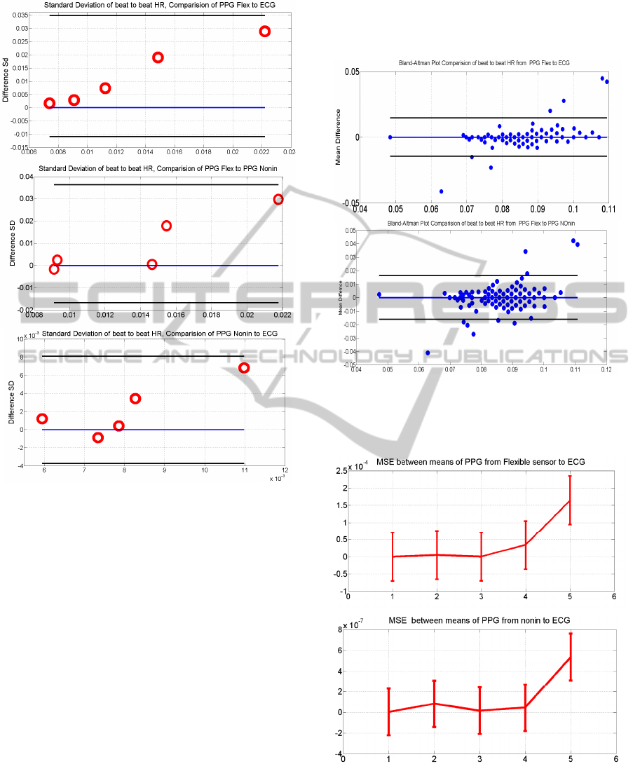
Figure 4: Bland and Altman analysis, comparison of SD of
beat-to-beat heart rate from PPG flexible to ECG, PPG
flexible to commercial PPG and PPG from commercial
device to ECG.
between the mean of the heart rate estimated by our
flexible PPG sensor, and the one of the Biopac ECG.
In this figure, the horizontal line indicates the MSE
for each subject, and the vertical lines reveal the
MSE standard deviation between all subjects.
Evidently, a consistency can be noticed between
the two error bars. That is, the MSE of the 5th
subject is greatest in two sets of measurements, and
the best estimation in both sets belongs to subject
number 1. This consistency validates the heart rate
variability measured by our fabricated flexible PPG
sensor. In addition, the very small MSE between the
heart rate means between the flexible PPG sensor
and the Biopac ECG establishes the high accuracy of
our fabricated sensor. Even though this MSE is
higher than the one between the commercial PPG
and the Biopac ECG, but our sensor benefits from a
lower cost and less complexity. Also, this higher
error can be inferred to the lower accurate laboratory
facilities available in academics compared the
enhanced high accuracy fabrication facilities in
industry.
Figure 5: Bland and Altman analysis between heart rate
obtained from PPG flexible to PPG commercial and ECG
as well as PPG from commercial device to ECG for
subject number 3.
Figure 6: MSE between means flexible PPG sensor of
heart rate from five subjects to ECG, in parallel to MSE
between means of heart rate from commercial PPG to
ECG.
MechanicallyFlexiableBiosensorforDetectionofPhotoplethysography
163

5 CONCLUSIONS
The proposed flexible PPG sensor has been studied
as an alternative solution to the commercial PPG
sensor and the ECG device for continuous
monitoring of heart rate. Standard deviation and
mean of intervals between cardiac cycles were
analysed. Accuracy of this sensor and the algorithm
were analysed by Bland and Altman analysis. The
MSE between the mean values of beat-to-beat heart
rates from flexible PPG sensor and those of Biopac
ECG was compared to those between the
commercial PPG and Biopac ECG. The results
confirm feasibility of obtaining heart rate from the
flexible PPG sensor for analysis of heart rate
variability.
ACKNOWLEDGEMENTS
The author wishes to thank Dr. Kouhyar Tavakolyan
for his valuable guidance.
REFERENCES
Chi, Y. M., Jung, T. P., & Cauwenberghs, G. (2010). Dry-
contact and noncontact biopotential electrodes:
methodological review. Biomedical Engineering,
IEEE Reviews in, 3, 106-119.Smith, J., 1998. The
book, The publishing company. London, 2
nd
edition.
Griffith, M. E., Portnoy, W. M., Stotts, L. J., & Day, J. L.
(1979). Improved capacitive electrocardiogram
electrodes for burn applications. Medical and
Biological Engineering and Computing, 17(5), 641-
646.
Bouwstra, S., Feijs, L., Chen, W., & Oetomo, S. B. (2009,
June). Smart jacket design for neonatal monitoring
with wearable sensors. In Wearable and Implantable
Body Sensor Networks, 2009. BSN 2009. Sixth
International Workshop on (pp. 162-167). IEEE.
Dresher, R. (2006). Wearable forehead pulse oximetry:
Minimization of motion and pressure artifacts
(Doctoral dissertation, Worcester Polytechnic
Institute).
Schäfer, A., & Vagedes, J. (2012). How accurate is pulse
rate variability as an estimate of heart rate variability?:
A review on studies comparing
photoplethysmographic technology with an
electrocardiogram. International Journal of
Cardiology.
Allen, J. (2007). Photoplethysmography and its
application in clinical physiological measurement.
Physiological measurement, 28(3), R1.
Karlen, W.; Raman, S.; Ansermino, J. M.; Dvvccont, G.
A. (July 2013), "Multiparameter Respiratory Rate
Estimation From the Photoplethysmogram,"
Biomedical Engineering, IEEE Transactions on,
vol.60, no.7, pp.1946, 1953.
Nilsson, L., Johansson, A., & Kalman, S. (2003).
Macrocirculation is not the sole determinant of
respiratory induced variations in the reflection mode
photoplethysmographic signal. Physiological
measurement, 24(4), 925.
BIODEVICES2014-InternationalConferenceonBiomedicalElectronicsandDevices
164
