
An Application to Interact with 3D Models Reconstructed from Medical
Images
F´elix Paulano, Juan J. Jim´enez and Rub´en Pulido
Computer Science Department, University of Ja´en, Ja´en, Spain
Keywords:
3D Interaction, Stereoscopy, CT Scans, Model Reconstruction.
Abstract:
Although the reconstruction of 3D models from medical images is not an easy task, there are many algorithms
to perform it. However, the reconstructed models are usually large, have a lot of outliers and have not a correct
topology. To interact with these models, the methods must be fast and robust. In this paper, we present an
application that enables the interaction with models reconstructed from medical images. The application uses
Marching Cubes to generate triangle soups from the medical scans. Then, the user can define models by
selecting sets of triangles. Once the models have been defined, the application allows to interact with them.
In addition, a detailed collision detection is calculated between the models in the scene not only to avoid that
models in the scene collide, but also to determine which triangles are overlapping. In addition, the calculation
of distances and nearest points provides visual aid when the user is interacting with the models. Finally, the
Leonar3Do system have been incorporated to improve the interaction and to provide stereo visualization. The
presented application can be used in the field of education since users can manipulate individual body parts
to examine them. Moreover, the application can be utilized in the preparation of an intervention or even as a
guide for it, since it enables the utilization of models reconstructed from real medical scans.
1 INTRODUCTION
Nowadays, it is very common to work with polygonal
models of the human body. In most cases, these mod-
els are generated from scans of real patients, since it
allows to customize the simulation. However, the re-
construction of 3D models from medical images is not
an easy task. Usually, the reconstructed geometry is
not topologically correct or even topological informa-
tion is not available. This geometry is huge and has
a lot of outliers in most cases. In addition, the differ-
ent models represented by the reconstructed geometry
are not isolated or labelled. For all these reasons, it is
necessary to develop tools that allow to interact with
the reconstructed geometry. These tools should in-
clude, among others, visualization, picking, area se-
lection and collision detection methods.(der Bergen,
2003)(Jim´enez et al., 2006).
Due to the fact that the different models repre-
sented by the reconstructed geometry are neither iso-
lated nor labelled, a tool that allows to select pieces
of geometry and define models from them is neces-
sary. This allows the user to interact with the models
independently. In addition, this type of tools helps
to clean from the scene the not interesting geome-
try. Once the models are defined, picking methods
are needed. Since this type of methods allows to se-
lect the model to interact with, they enable the inter-
action with a model independently from the others.
The integration of a detailed collision detection pro-
vides visual aid during the interaction and allows to
detect which models are colliding or even the collid-
ing zones. This information is useful to avoid that
two models collide and then generate a response to
the collision. Moreover, it also can be helpful to in-
form the user that the models collide, hence the user
can act accordingly during the interaction. If the col-
lision detection is not calculated, the user can find dif-
ficulties when placing the models is the scene. Since
time is a very important factor in medicine, stereo dis-
play and 3D interaction systems can be integrated to
improve the usability of the application and reduce
the usage time. Because of the complexity of the
reconstructed models, all these techniques and sys-
tems must be fast and robust. In recent years sev-
eral approaches to reconstruct 3D surfaces from med-
ical images have been presented. Some techniques
have been adapted from popular methods in compu-
tational geometry such as Delaunay triangulation and
Voronoi diagram (Lv et al., 2009). Other techniques
224
Paulano F., Jiménez J. and Pulido R..
An Application to Interact with 3D Models Reconstructed from Medical Images.
DOI: 10.5220/0004834902240229
In Proceedings of the 9th International Conference on Computer Vision Theory and Applications (VISAPP-2014), pages 224-229
ISBN: 978-989-758-009-3
Copyright
c
2014 SCITEPRESS (Science and Technology Publications, Lda.)
are based on Hierarchical Space Subdivision Schemes
(Boubekeur et al., 2006). In (Sharf et al., 2007) pre-
sented a method to interactively reconstruct surfaces
by using a prior distribution. On the other hand, the
extraction of contours, using a hierarchical spatial de-
composition, can be complemented with a table of
patterns to triangulate these contours (Pulido et al.,
2012). Applications that are usually used to work
with medical images are limited to 3D visualization
(Bhanirantka and Demange, 2002) and they usually
only allow to rotate the camera and to remove outliers
if it is necessary. In (Rautek et al., 2007), authors, in-
stead of using the traditional transfer function speci-
fication, use semantic layers to define the mapping of
volumetric attributes to a specific visual style. This
mapping is specified by rules which are evaluated
with fuzzy logic arithmetic. There are several toolk-
its which enable the manipulation of models recon-
structed from medical scans. ITK-SNAP (Yushkevich
et al., 2006) is a application that allows to interac-
tively segment structures from medical scans. MITK
(Wolf et al., 2004) combines VTK and ITK to provide
interaction with models reconstructed from medical
scans. Most of these methods and tools work with
volumetric models and do not allow to separate and
classify parts of the model and interact with them.
Thus, it could be useful to develop an application that
lets the user interact with a model individually. In
addition, 3D motion tracking devices could allow to
improve the user experience and could make the ap-
plication easy to use.
In this paper, we present an application that en-
ables the examination of 3D models, which are gen-
erated from medical images, in detail. This can be
used in the field of education since users can trans-
late and rotate individual body parts to examine them.
Moreover, proximity queries and collision detection
methods can help the user to place body parts in their
correct position. On the other hand, the application
can be used in the preparation of an intervention or
even as a guide for it, since it allows to work with
models reconstructed from real medical images. For
all the above reasons, the application can be used to
improve the visualization and thus the diagnosis.
2 OVERVIEW OF THE
APPLICATION
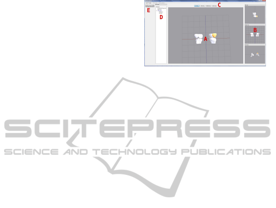
The graphic user interface is mainly composed of
four canvas and a hierarchy tree. The main canvas
(Figure1, A) allows to move the camera and the other
three show static views: side, front, and top view
(Figure1, B). All canvas can exchange their position
A
B
C
D
E
Figure 1: Screen capture of the application. A - Main can-
vas. B - Secondary canvas. C - Buttons to exchange canvas.
D - Hierarchical tree of the scene. E - Toolbars.
dynamically by clicking on any of the auxiliary can-
vas or using the buttons on the top of the window
(Figure1, C). This interface is similar to that used in
current medical visualization applications, hence it is
easy to use for professionals. On the left side of the
application window, an hierarchy tree shows the rela-
tion between the models and the model which is cur-
rently selected (Figure1, D). At the top of the window
there is a toolbar that allows to load medical images,
define models and their relationship, remove unneces-
sary parts, and enable the calculation of nearest points
and overlapping triangles (Figure1, E). The procedure
for using the application can be summarized as fol-
lows. First of all, the user must specify the medical
images to be loaded. Once the images are loaded,
the application generates a triangle soup from them.
Then, models must be identified and defined using an
area selector. Each time a model is defined, the user
can set the models to which it is related and the ap-
plication updates the hierarchy of models. Moreover,
the unnecessary parts and the outliers can be removed
by using the area selector. Defined models can be
selected, translated and rotated to place it correctly
in the scene. With the aim of providing assistance
during the interaction, the application calculates the
nearest points and the overlapping triangles. The def-
inition of the hierarchy of models allow to only calcu-
late collisions and distances between the models that
have some type of relationship. To improve the in-
teraction, the Leonar3Do input device can be used to
manipulate the models.
The generation of 3D geometric models from
medical images takes a few seconds. For instance,
it takes approximately 20 seconds to perform a recon-
struction from 200 medical images and 30 seconds
from 400. The resolution of the CT images utilized in
the test is 512x512 and the application is able to load
up to 400 medical images. Once the geometric mod-
els have been generated, the interaction is performed
in real time. To measure this, a Intel i7 2.80GHz
processor, 4GB RAM and a NVidia GeForce GTS
AnApplicationtoInteractwith3DModelsReconstructedfromMedicalImages
225

240 have been used. The application has been im-
plemented in C++ using the gcc compiler and fol-
lowing a MVC architecture. The user interface has
been developed using the Qt 4.7 library (Blanchette
and Summerfield, 2006). Therefore, the implemented
software can be compiled for various platforms. The
QtOpenGL module has been used to implement 3D
graphics. Furthermore, the application uses VTK
(Schroeder et al., 2006) to reconstruct the 3D models
and PQP (Larsen et al., 1999) to calculate a detailed
collision detection. To program the Leonar3Do, the
1.0 version of the LeoAPI has been utilized.
3 MODEL RECONSTRUCTION
Model reconstruction from medical data is a difficult
task due to the complexity of the human body struc-
tures. Once the medical images are obtained, the first
step consists in segmenting the images in order to iso-
late the areas of interest. After that, the result ob-
tained is utilized to reconstruct a 3D geometric model.
The results obtained depend on the techniques used in
each of the parts of the process.
Figure 2: Iso-surface rendering from 3D medical images by
using the Marching Cubes algorithm.
There are different sources such as Computed To-
mography (CT), Magnetic Resonance Imaging (MRI)
or Ultrasonic (US) techniques (Sachse, 2004) that
provides 3D datasets and values that represent physy-
cal quantities, e.g. proton densities in MRI, and at-
tenuation coefficients in CT. In the context of mod-
elling, these values can be used to extract features of
a patient, such as bone, muscle or fat. These medical
image sources can store the information in multiple
formats. The Digital Imaging and Communications
in Medicine (DICOM) standard is the most common
format. Our application allows to visualize 2D im-
ages and reconstruct models from 3D medical image
series and it has been tested with DICOM datasets and
images from The Visible Human Project repository
(NLM, 1986). To generate 3D data, the presented ap-
plication makes use of the Marching Cubes algorithm
[LC87] (see Figure 2). Marching Cubes is a fast and
simple method that allows to automatically generate
large polygonal datasets from volumetric data with
high resolution. The outcome of the algorithm is a
large soup of triangles without topology. However,
the presented application is able to deal with surfaces
that present holes by using the interaction techniques
described in the next section. Before the reconstruc-
tion process, a threshold based method is utilized to
segment the medical images in order to isolate the de-
sired tissue. The threshold depends on the type of tis-
sue and it is manually selected by the user. The seg-
mentation result is a volume which is used as a input
of the Marching Cubes algorithm.
Figure 3: Virtual representation of the Leonar3Do
(Leonar3Do, 2012).
4 INTERACTION
Once the 3D geometric model has been reconstructed,
our application enables the interaction with it. With
this aim, it has been integrated registration, collision
detection, picking and multi-view methods. These
methods allow the user to define 3D models from the
reconstructed geometry and interact with each of the
models independently. In addition, the detailed col-
lision detection and the multi-view implementation
provide visual aid to the user during the interaction.
In order to improve the interaction, the Leonar3Do
has been utilized. This is a virtual reality system that
enables a stereo interaction. The Leonar3Do system
mainly consist of a spatial input device, 3D glasses
and monitor-mountedsensors (figure 3). Both the bird
and the 3D glasses operate in six degrees of freedom
and sensors can track both the bird and the glasses.
To this end, the Leonar3Do system uses a technology
based on infrared sensors. The bird has two buttons,
one big and one small, which can be programmed. In
addition, the big button is sensitive to pressure. On
the other hand, 3D glasses enable stereoscopic vision.
For that, Leonar3Do can use either an active and a
passive system. While the passive system uses com-
monly polarizing lenses, the active system uses liquid
crystal shutters which are powered through an USB
port. In our case, the active system have been utilized.
VISAPP2014-InternationalConferenceonComputerVisionTheoryandApplications
226
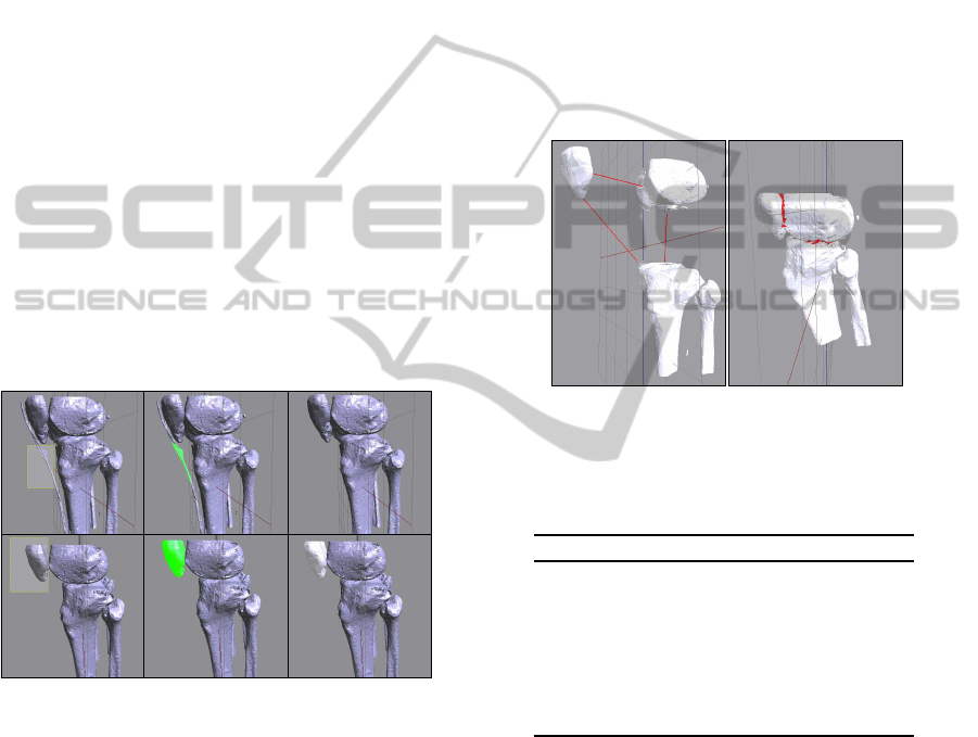
4.1 Registration of the Models
Due to the fact that the reconstructed geometry is a
triangle soup, there is no information about the model
to which each triangle belongs. To solve this defi-
ciency, an area selector has been implemented. This
tool allows to select a set of triangles and to define a
model from them (Figure 4, bottom). When a model
is defined by the area selector, the application allows
to relate it to another previously defined model. Thus,
it is possible to establish an hierarchy between the de-
fined models. Furthermore, the area selector can also
be used to remove triangles from the scene (Figure 4,
top). Each time the user makes a selection, the scene
has to be repainted to draw the selector. To solve this
problem, we avoid repainting the entire scene by us-
ing a frame buffer. In the rendering function, the con-
tent of the frame buffer is drawn in a 2D texture which
is located in the background of the scene. Moreover,
to determine which triangles have been selected they
are projected in the plane determined by the selec-
tor. Thus, it is only necessary to resolve a triangle-
rectangle test for each triangle in the scene. More-
over, as the intersection is calculated in 2D, it is easy
to implement new shapes for the selector.
Figure 4: The area selector is used to remove unnecessary
parts and to define models. Top, from left to right: selecting
the part to remove; the selected part is displayed in green;
Unnecessary parts have been removed from the scene. Bot-
tom, from left to right: selecting the triangles that represent
the patella; selected triangles are displayed in green; finally,
the defined model is shown in white.
4.2 Collision Detection
In order to implement the interaction between the
different models, we have not only to calculate the
collision but also to provide visual aid to the user.
Since the reconstructed models are triangle soups, it
is necessary to use algorithms that can work with that
type of models. These calculations are necessary to
avoid that two models overlap or even to inform the
user that the models are colliding (Figure 5, right).
Furthermore, the distances and the nearest points be-
tween two models can help the user to place models
that are not correctly positioned (Figure 5, left). The
leonar3Do system has been used to improve the col-
lision response. When a collision occurs, the spatial
input device emits a small vibration. Some algorithms
have been tested (Paulano et al., 2012) to choose the
best suited to this problem. In that work, algorithms
were tested with models with a complexity of up to
one million triangles. The results shown that the PQP
library (Larsen et al., 1999) is quite fast and robust.
This library uses swept sphere hierarchies to perform
collision detections. Moreover, PQP is able to calcu-
late different collision detection parameters, such us
distances, overlapping triangles, or closest points.
(a) (b)
Figure 5: a) Calculation of the nearest points between some
models. b) Overlapping triangles are displayed in red.
Table 1: Picking time using the ray picking method based
on the PQP library. Run in a computer with Intel i7
2,80GHz, 4GB RAM, NVidia GeForce GTS 240.
Triangles in the scene Picking time (s)
58206 0.0671
253696 0.2868
428411 0.3845
478462 0.4231
513658 0.4608
546308 0.4949
577827 0.5206
4.3 3D Picking
After the models are defined, the application allows
to select them by picking.To this end, the spatial in-
put device is used. By moving this device, a 3D
cursor in the scene is manipulated. This cursor en-
ables the selection of previously defined models by
using the small programmable button. If a model is
selected, the spatial input device manipulates the se-
lected model instead of the 3D cursor. Apart from
this, each selected model can be rotated and trans-
lated by using the mouse. These transformations are
performed independently from the rest of the mod-
els in the scene. In order to enable the selection of
AnApplicationtoInteractwith3DModelsReconstructedfromMedicalImages
227
models, we decided to implement a method based on
ray picking. The method consist of throwing a ray
from the observer that pass through the cursor posi-
tion and calculating the collision between the ray and
all the objects in the scene. In order to check the col-
lisions, we have used the PQP library. In this way, the
data structures previously constructed are reused. To
measure the efficiency of the method, some tests have
been performed. As shown in table 1, the method
calculates the ray picking in a reasonable good time,
even when there are several hundred of thousand of
triangles in the scene. Moreover, since it is based on
PQP, the method is also robust.
4.4 Multi-canvas
Although the 3D view provides extra information to
the user, doctors and radiologists usually work with
a 2D view of the area of the patient. For this reason,
the application includes four canvas: a canvas with a
free camera and three canvas with static cameras. The
first one is the main canvas and the other three can-
vas represent a top, side, and front view respectively.
However, the four canvas can exchange their posi-
tions dynamically by clicking them or by pushing the
buttons located at the top of the window. Instead of
implementing multi-canvas using several glViewport
(Shreiner, 2010), we decided to use four QGLWidget
to implement the four canvas. In this way, we can
take advantage of all the functionality that is already
implemented in the QtOpenGL module.
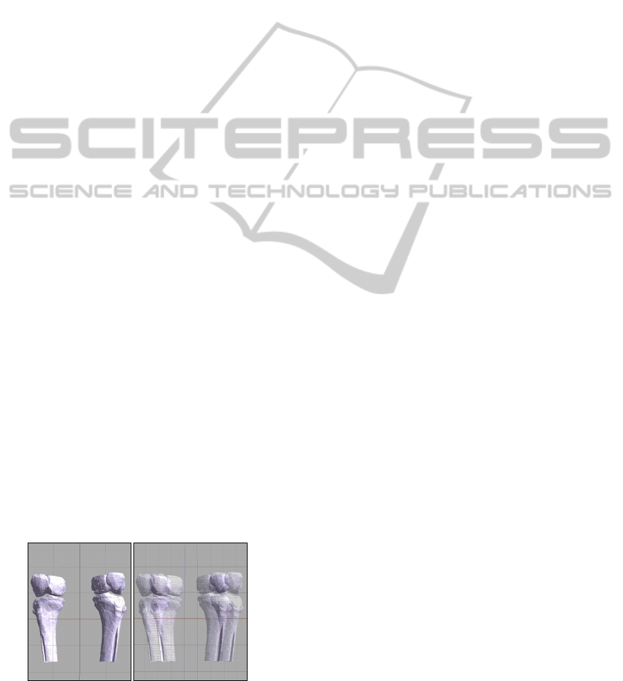
4.5 Stereo Visualization and Immersion
The Leonar3Do system have been used to provide
stereo visualization in the main canvas by using active
glasses (see figure 6). This visualization system al-
lows the user to better understand the scene and makes
easy to select objects by using the spatial input device.
In addition, the stereo visualization increases the feel-
ing of immersion when using the application.
The Leonar3Do API (LeoAPI) is divided into two
main parts: the tracking API and the stereoscopic
Figure 6: Screenshot of the main canvas of the application.
Left, non-stereo mode. Right, stereo mode.
API. The first one allows to know where both the spa-
tial input device and the glasses are situated. As said
in previous sections, the spatial input device has been
used to manage a 3D cursor that enables the selec-
tion of models in the scene and to manipulate selected
models. On the other hand, the position of the glasses
is used to manage the camera, hence when the user
move the head, the camera moves accordingly. To
do this, both the translation and the rotation of the
user head are considered. This increases the feeling
of immersion and makes the application easy to use.
It is important to consider that the camera position is
modified by the LeoAPI, hence that it is placed at the
focal point. The stereoscopic API enables the imple-
mentation of the stereo view and it is responsible for
generating the final frame of the application, whether
or not the stereo mode is activated. In our case, the
stereo API is responsible for drawing the main canvas
of the application. Because of this responsibility, it is
necessary to disable the auto-bufferswapping because
the LeoAPI already does it. Moreover, the LeoAPI
fill a projection matrix which have to be provided to
OpenGL before rendering the main canvas. However,
this projection matrix can be modified before passing
it to the OpenGL pipeline.
5 CONCLUSIONS
In this work, an application to interact with 3D mod-
els reconstructed from medical data has been devel-
oped. Unlike other existing applications that enable
3D visualization of medical images, the presented ap-
plication can interact with the 3D models in terms
of geometry because it previously performs a recon-
struction. Moreover, the application enables a real-
time interaction, although the reconstruction gener-
ates large models with no topology. Finally, the fol-
lowing tasks are proposed as future work:
• The presented application can be applied to pre-
pare various types of surgical procedures. The
surgeon could generate a 3D model from real pa-
tient data and simulate the interaction with the
damaged area before the intervention.
• Measure the usability of the application. Experts
will test the application and propose improve-
ments to make the interaction more realistic.
• Obtain topologically correct meshes from trian-
gle soups. These meshes would enable the use
of more efficient algorithms to perform the inter-
action. In addition, different body parts could be
labelled during the reconstruction step, avoiding
the selection and registration of the models.
VISAPP2014-InternationalConferenceonComputerVisionTheoryandApplications
228
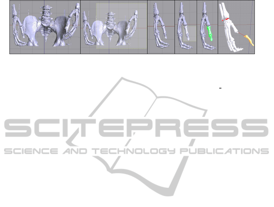
1 2 3 4 5 6
Figure 7: Example of using the application. 1 - Triangle soup generated from CT scans. 2 - Selection of some unnecessary
triangles. 3 - Cleaned scene. 4 - Selection of a model. 5 - Selected model. 6 - Interaction with defined models.
• Incorporate a method to remove outliers automat-
ically that avoids the user having to delete them
manually. This would allow to obtain less com-
plex models and improve the interaction.
ACKNOWLEDGEMENTS
This work has been partially supported by the Min-
isterio de Econom´ıa y Competitividad and the Euro-
pean Union (via ERDF funds) through the research
project TIN2011-25259.
REFERENCES
Bhanirantka, P. and Demange, Y. (2002). OpenGL volu-
mizer: a toolkit for high quality volume rendering of
large data sets. In Symposium on Volume Visualiza-
tion and Graphics, 2002. Proceedings. IEEE / ACM
SIGGRAPH, pages 45–53. IEEE.
Blanchette, J. and Summerfield, M. (2006). C++ GUI Pro-
gramming with Qt 4. Prentice Hall Open Source Soft-
ware Development Series. Prentice Hall PTR.
Boubekeur, T., Heidrich, W., Granier, X., and Schlick, C.
(2006). Volume-Surface Trees. Computer Graphics
Forum, 25(3):399–406.
der Bergen, G. V. (2003). Collision Detection in Interactive
3D Environments. Elsevier.
Jim´enez, J. J., Og´ayar, C. J., Segura, R. J., and Feito, F. R.
(2006). Collision Detection between a Complex Solid
and a Particle Cloud assisted by Programmable GPU.
In Vriphys: 3rd Workshop in Virtual Realitiy, Interac-
tions, and Physical Simulation, pages 43–52.
Larsen, E., Gottschalk, S., Lin, M., and Manocha, D.
(1999). Fast proximity queries with swept sphere vol-
umes. Technical report, Department of Computer Sci-
ence, UNC Chapel Hill.
Leonar3Do (2012). Leonar3do. http://www.leonar3do.com.
[Online; accessed 2-October-2013].
Lv, S., Yang, X., Gu, L., Xing, X., and Pan, L. (2009). De-
launay mesh reconstruction from 3D medical images
based on centroidal voronoi tessellations. In Compu-
tational Intelligence and Software Engineering, 2009.
CiSE 2009. International Conference on, pages 1–4.
NLM (1986). The visible human project. http://www.nlm.
nih.gov/research/visible/visible
human.html.
Paulano, F., Jim´enez, J. J., Pulido, R., and Ogayar, C. J.
(2012). A comparative study of implemented collision
detection strategies. In Proceedings of the Interna-
tional Conference on Computer Graphics Theory and
Applications (GRAPP 2012), pages 485–490.
Pulido, R., Jim´enez, J. J., and Paulano, F. (2012). Surface
reconstruction from 3d medical images based on tri-
tree contouring. In Proceedings of the International
Conference on Computer Graphics Theory and Appli-
cations (GRAPP 2012), pages 175–181.
Rautek, P., Bruckner, S., and Gr¨oller, E. (2007). Seman-
tic layers for illustrative volume rendering. IEEE
transactions on visualization and computer graphics,
13(6):1336–1343.
Sachse, F. (2004). 5. digital image processing. In Compu-
tational Cardiology, volume 2966 of Lecture Notes in
Computer Science, pages 91–118. Springer Berlin.
Schroeder, W., Martin, K. M., and Lorensen, W. E. (2006).
The Visualization Toolkit: An Object-Oriented Ap-
proach to 3D Graphics. Kitware, Inc.
Sharf, A., Lewiner, T., Shklarski, G., Toledo, S., and Cohen-
Or, D. (2007). Interactive topology-aware surface re-
construction. ACM Trans. on Graphics, 26(3):43.
Shreiner, D. (2010). OpenGL Programming Guide: The
Official Guide to Learning OpenGL, Versions 3.0 and
3.1. OpenGL Series. Addison-Wesley.
Wolf, I., Vetter, M., Wegner, I., Nolden, M., Bottger, T.,
Hastenteufel, M., Schobinger, M., Kunert, T., and
Meinzer, H.-P. (2004). The Medical Imaging Interac-
tion Toolkit (MITK) a toolkit facilitating the creation
of interactive software by extending VTK and ITK.
In Medical Imaging 2004, pages 16–27. International
Society for Optics and Photonics.
Yushkevich, P. A., Piven, J., Hazlett, H. C., Smith, R. G.,
Ho, S., Gee, J. C., and Gerig, G. (2006). User-guided
3D active contour segmentation of anatomical struc-
tures: Significantly improved efficiency and reliabil-
ity. NeuroImage, 31(3):1116–1128.
AnApplicationtoInteractwith3DModelsReconstructedfromMedicalImages
229
