
Automatic Creation of an Efficient Image Filter
based on the Genetic Algorithm for Evaluation of Veins
Koji Kashihara
Institute of Techonology and Science, The University of Tokushima, 2-1 Minamijyousanjima, Tokushima, Japan
Keywords: Genetic Algorithm, Expectation Maximization Algorithm, near Infrared Camera, Image Filtering.
Abstract: Instead of expensive and complicated diagnostic equipment, low-cost infrared cameras can record vein
images noninvasively and simply. However, the recorded image may induce low contrast and a worse
signal-to-noise (S/N) ratio. To solve this problem, an effective image filtering method to catch vein shapes
will enable the early detection of disease. Therefore, a new filtering method based on the genetic algorithm
(GA) with the expectation maximization (EM) algorithm was proposed for the analysis of vein images
acquired from a near-infrared (780 nm) CCD camera. The new filter was automatically designed by the GA
to modify the worse S/N ratio of vein images, with an unknown correct image answer. If the proposed
filtering method is incorporated into the e-healthcare application, it could be widely distributed through
smart phones or tablets.
1 INTRODUCTION
Sitting on a narrow seat during long air travel
increases the risk of dyspnea and acute myocardial
infarction triggered by leg deep venous thrombosis
(Feltracco, Barbieri, Bertamini, Michieletto, and Ori,
2007). Venous insufficiency may also cause varicose
veins owing to incompetent valves (Callam, 1994).
Medical doctors and experts must operate large,
expensive, and complicated medical equipment such
as ultrasonic diagnostic equipment and X-ray
systems (Bergqvist and Jaroszewski, 1986) to
diagnose patients with circulatory diseases.
Instead of such equipment, a near-infrared
camera (Kashihara, Ito, and Fukumi, 2012; Zharov,
Ferguson, Eidt, Howard, Fink, and Waner, 2004)
can easily and noninvasively visualize venous
shapes. However, vein images taken by the near-
infrared camera may result in low contrast and a
worse signal to noise (S/N) ratio. The auto-tuning
function or manual camera settings of photographic
parameters may also induce low image quality.
Accordingly, creating an effective image filter will
address this issue and lead to the accurate detection
of venous states.
Standard image processing such as equalization
and binarization (Soni, Gupta, Rao, and Gupta,
2010; Yakno, Saleh, and Rosdi, 2011) can enhance a
low-contrast image; on the other hand, important
information on complicated and thin lines may be
lost. If the filter kernel parameters determining the
feature of each image are explored in detail, optimal
image processing would be obtained instead of the
customary methods.
The purpose of this study was to create an
effective filtering method to detect vein shapes in
the low-contrast image from a near-infrared camera.
The image filter was automatically designed by the
genetic algorithm (GA) with the expectation
maximization (EM) algorithm (GA-EM algorithm).
The GA-EM algorithm could find the best
combination of convolution kernel values for a new
filter.
2 DESIGN OF A NEW FILTER
A new filter for vein images was designed by real
coded GA with the EM algorithm. The GA approach
can search for the convolution filter kernel at the
maximum fitness. The EM algorithm search for the
optimal parameter in Gaussian mixture models
(Bilmes, 1998). The EM algorithm could separate
the area of target veins from background, estimating
parameters for two components of the Gaussian
mixture model.
506
Kashihara K..
Automatic Creation of an Efficient Image Filter based on the Genetic Algorithm for Evaluation of Veins.
DOI: 10.5220/0004913505060510
In Proceedings of the International Conference on Health Informatics (HEALTHINF-2014), pages 506-510
ISBN: 978-989-758-010-9
Copyright
c
2014 SCITEPRESS (Science and Technology Publications, Lda.)

2.1 Ga Process
An individual is represented by an array of real
values indicating the convolution kernel values. The
GA consists of the following three operators:
selection, crossover, and mutation.
The initial population of individuals is randomly
generated within a set range. Each individual is
represented by real numbers; its score is calculated
from a fitness function. The fitness function for the
GA is based on a convolution filter kernel values to
an input image. The convolution kernel updated by
the GA could highlight a region of interest (ROI),
reducing external noise in the image.
A selection operation determines the individuals
for the generation of offspring. Next, a crossover
operation combines two individuals to generate an
offspring. A blend crossover (BLX-α) operator
(Eshelman and Schaffer, 1993) was selected for this
study. Furthermore, a mutation operator randomly
changes some individuals, altering the variables of a
selected individual to facilitate the diversity in the
population. The mutation can avoid falling into a
local solution.
The above GA operators were repeated to update
the population and created the next generations,
improving the fitness of the population. The GA
program stopped after some generations.
2.2 EM Algorithm
The fitness function values for the GA were
computed from the log likelihood of the Gaussian
mixture model. The Gaussian mixture model is
described as:
)()(
1
k
K
k
k
xGxp
,
(1)
where
T
d
xxx ],...,[
1
is the d-dimensional data
vector.
k
is the mixture ratio of a distribution and
the ratio must be
0
k
and
1
1
K
k
k
. G means the
normal distribution with the parameters of
},{
kkk
corresponding to the mean vector and
the covariance matrix. K shows the number of the
models (K = 2, the vein and background for this
study).
The EM algorithm consists of the expectation (E-
step) and maximization (M-step) steps, which are
alternately applied until the log-likelihood value
converges to an optimal value. The observed data x
mean the brightness values after applying a proposed
filter (d = 1), and x
},...,{
1 N
xx
(N data samples,
the total number of pixels in a target image). The
parameter values (
0
,
0
, and
0
) were initialized,
and the E-M steps were repeated.
2.2.1 E-step
The posterior probability of the latent variables (z)
for the Gaussian mixture model can be calculated as
follows.
,
(2)
2.2.2 M-step
The new parameters [
)1( t
] are identified by
estimating the latent variables (z
ik
) in order to
maximize the log likelihood of the complete data.
This process is consistent with calculating the
parameters
)1( t
k
,
)1( t
k
, and
)1(
t
k
for the Gaussian
mixture model:
,
,
(3)
where
N
i
ikk
zN
1
)(
.
3 EVALUATION METHODS
The novel image filter was evaluated for the
detection of actual venous changes with an unknown
image answer.
3.1 Measurement Environment
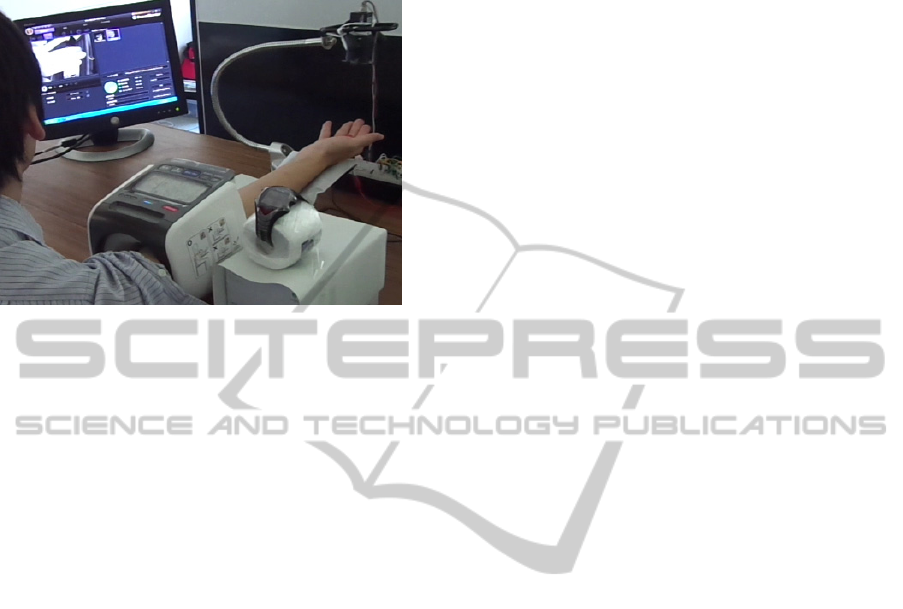
Figure 1 shows the measurement environment for
vein shapes. Finger veins as a target for image
analysis were recorded by a CCD camera (Toshiba
Teli Co., CS8620Hi) with near-infrared light-
emitting diodes (wavelength of 780 nm). A filter to
cut visible light (Fuji Film Co., IR760) was set in the
front of the camera. Photographic parameters in the
camera (contrast, focus, gain, exposure, etc.) were
manually set to optimal values before the actual
),(
),(
)(
1
kk
K
j
j
kkk
ik
xG
xG
z
N
N
k
t
k
)1(
k
N
i
iik
t
k
N
xz
1
)1(
)(
k
N
i
T
kikiik
t
k
N
xxz
1
)1(
))()((
AutomaticCreationofanEfficientImageFilterbasedontheGeneticAlgorithmforEvaluationofVeins
507
measurement. The recorded images were modified
by the filters (Microsoft Visual C++ 2010 with Open
CV Ver. 2.1).
Figure 1: Measurement environment for vein shapes and
states during the measurement of blood pressure.
The subjects were healthy male volunteers (n = 4;
age ± S.D. = 26.8 ± 8.2 years). The experiment was
conducted in accordance with the Declaration of
Helsinki. A signed informed consent form was
obtained from each participant. The participants
refrained from eating, drinking, or smoking at least
two hours prior to the experiment.
3.2 Procedures
To induce a great change in the venous states, the
forearm was pressed by a sphygmomanometer and
finger veins were recorded by a CCD camera. The
vein images were extracted at the prestimulus
(before 10 s of starting the blood pressure
measurement), maximum pressure, and recovery
(after 90 s of stopping the measurement) period.
3.3 Applied Filters
After a typical Gaussian filter was applied to a target
image, the GA process was performed. The fitness
function for the GA was a convolution filter kernel.
The kernel values were updated by the GA and it
was applied to vein images within the ROI (60 × 120
pixels).
The initial population of individuals (kernel
values) in the GA parameters is randomly generated
within a set range between -10 and 10. The
population size was set at 40; the tournament size for
selection was 20; the crossover probability was 0.6;
the mutation probability was 0.1. The parameter for
a blend crossover (BLX-α) operator was set at α =
0.4. The procedure for the GA was repeated until
reaching 100 generations. The designed filter kernel
had a fixed size array (3 × 3) of numerical
coefficients. Once the novel image filter was created
under the prestimulus condition in each subject, the
same filter kernel was applied to the target images in
all experimental conditions.
The EM algorithm for the GA was iterated five
times, considering the computing time. The repeat
count was determined by considering the calculation
time for the GA. The pixel values in a target image
were initially normalized. The initial parameters for
the Gaussian mixture model were set at
0
= (0.5,
0.5),
0
= (-0.5, 0.5), and
0
= (0.1, 0.1) in the two
distributions. In the case of division by zero or an
infinite value, the fitness function value received a
large penalty score. To avoid the singular value
problem, white or black (0 or 255) areas of an image
also had a penalty if over 1%.
3.4 Analysis
To confirm the strict accuracy in the novel filter, the
venous changes during a blood pressure stimulus
were evaluated by the Gaussian fitting of a venous
line. So that the target venous line and X axis would
cross at a right angle, the image was rotated at an
angle of some degrees. The ROI (20 × 10 pixels)
was set for the part of a venous line, and 10 pixel
values of the inverted Y axis were averaged at every
point on the X axis. The Gaussian approximation
based on a modified Levenberg-Marquardt method
was applied to the target distribution:
y = a exp[(-(x-b)
2
) / (2c
2
)] + d (4)
The x and y are the values of the X axis and the
estimated brightness levels, respectively. The
parameters a and c indicate the amplitude and width
of the distribution, b is the peak location, and d is the
offset value.
4 EXPERIMENTAL RESULTS
Figure 2 shows the vein images with and without
image filtering under the prestimulus, maximum
pressure, and recovery period (90 s later). The ROI
was set for the area of the middle finger, referencing
a fixed marker. The average values (n = 4) of
systolic and diastolic pressures were 116.5 ± 14.7
and 68.3 ± 11.7 mmHg, respectively. The pulse
wave was 71.3 ± 6.2 beats/min.
HEALTHINF2014-InternationalConferenceonHealthInformatics
508
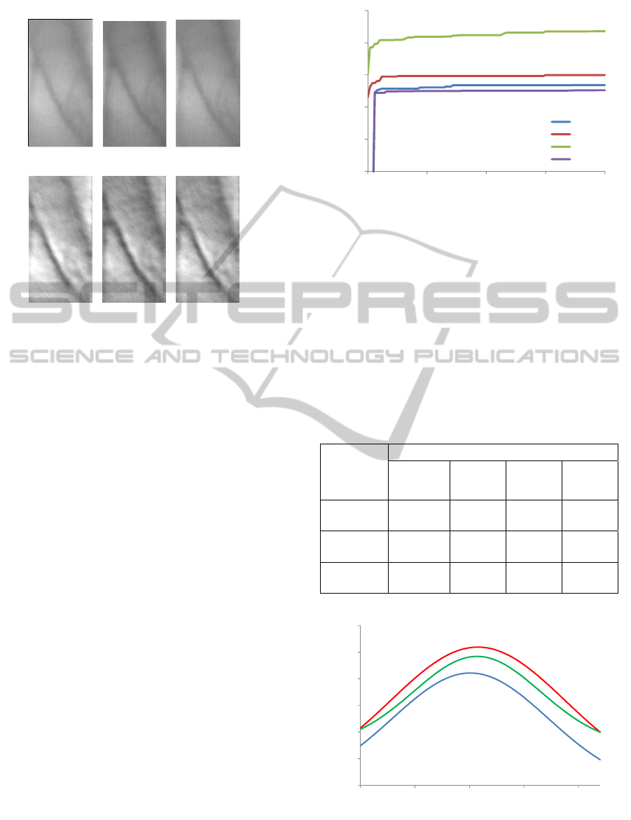
(a) No filter
(b) New filter
Figure 2: A typical example of vein images before (a) and
after (b) the image filtering process within the region of
interest (ROI) at the prestimulus (left), maximum pressure
(middle), and recovery period (right).
The recorded images showed low-contrast resolution
and contained external noise. The venous lines
appeared to be thicker at the maximum pressure than
at the prestimulus and the recovery period. The new
filter was especially effective for highlighting the
vein shapes.
Figure 3 represents the change in the maximum
values of the fitness function during the GA process
(n = 4). These values remarkably increased after five
generations; they gradually converged to a constant
value at around 50 generations.
The intensity histogram of vein images with no
filter and the new filter included bimodal
distribution, indicating the area of veins and
background. Although venous changes were
quantified by using the EM algorithm, the left
distribution (i.e., low intensity) showing venous
areas was not able to be modified by the fitting
function without an image filter. On the other hand,
the features of venous areas were sufficiently
extracted by using the EM algorithm under the
proposed filtering process. The distance of the two
distributions was longer with the proposed filter than
with no filter, showing the separation of the veins
and background area.
As shown in Figure 4, Gaussian fitting was
applicable to the intensity distribution in the ROI
vertically across a venous line in order to estimate
the accuracy of the proposed filter during a blood
pressure stimulus. Table 1 summarizes the estimated
Figure 3: Change in the maximum value of the fitness
function during the GA process (n = 4).
parameters of a Gaussian function. Sensitivity of
venous changes was increased by applying the new
filter, and the difference in the image characteristics
was enlarged. A greater change of the parameters
was observed at the maximum pressure period. In
special, the parameter c would have reflected the
increased amount of deoxyhemoglobin and the
enlarged venous areas. This result suggests the
detection of venous changes with high accuracy.
Table 1: Fitting parameters for experimental conditions.
Conditions
Gaussian Fitting Parameters
Amplitude
(a)
Peak
location
(b)
Width
(c)
Offset
(d)
Prestimulus
86.7
(55.5)
10.1
(1.5)
7.2
(7.3)
-2.3
(63.2)
Max.
pressure
102.4
(67.0)
10.8
(1.4)
8.0
(7.9)
1.4
(71.7)
Recovery
67.1
(24.3)
10.7
(2.5)
5.8
(4.1)
29.8
(18.5)
a to d, parameters in Eq. (4); ( ) means S.D.
Figure 4: Gaussian fitting functions (average responses; n
= 4) at the prestimulus (blue), maximum pressure (red),
and recovery period (green).
1000
1500
2000
2500
3000
3500
0 25 50 75 100
Fitness function [-]
Generation [-]
No.1
No.2
No.3
No.4
Subjects
0
20
40
60
80
100
120
0 5 10 15 20
255 - Brightness value [-]
X axis
pre
max
post
AutomaticCreationofanEfficientImageFilterbasedontheGeneticAlgorithmforEvaluationofVeins
509

5 DISCUSSION
Vein images from a near-infrared camera may result
in the low quality because of the worse S/N ratio.
Therefore, the efficient filtering process will be
necessary for quantifying venous changes. The
proposed filtering method based on the GA-EM
algorithm sufficiently modified the low-contrast vein
images during a blood pressure stimulus, under an
unknown correct image answer. However, the ability
of the EM algorithm is influenced by the method of
selection of the initial parameters. Furthermore, the
design of a novel filter with the EM algorithm must
avoid singularity or note identifiability (Casella and
Berger, 2002).
The same filtering procedure is not optimal for
all situations because the accuracy of image
processing depends on inter- or intra- individual
variability of vein shapes. The typical image
processing will need the adjustment of the camera or
filtering parameter settings every measurement
environment or an acquired image, by trial and error.
The proposed filtering method will sufficiently
improve the images obtained from such situations.
6 CONCLUSIONS
The image filter made by the GA-EM algorithm was
able to efficiently detect vein shapes recorded by an
infrared camera. The filtered images were quantified
with the EM algorithm to assess venous changes
during the blood pressure measurement. The
proposed imaging filter was able to modify its kernel
array, adapting the feature of target images. In future
studies, the optimal range of parameters for the GA-
EM algorithm should be investigated to catch
abnormal veins at an early stage. If the proposed
filtering method is incorporated into the e-healthcare
application, it could be widely distributed through
smart phones or tablets.
ACKNOWLEDGEMENTS
This study was partially funded by a Grant-in-Aid
for Scientific Research (C) from Japan Society for
the Promotion of Science (KAKENHI, 25330171).
REFERENCES
Feltracco, P., Barbieri, S., Bertamini, F., Michieletto, E.,
Ori, C., 2007. Economy class syndrome: still a
recurrent complication of long journeys. Eur. J.
Emerg. Med., vol. 14, pp. 100–103.
Callam, M. J., 1994. Epidemiology of varicose veins. Br.
J. Surg., vol. 81, pp. 167–173.
Bergqvist, D., Jaroszewski, H., 1986. Deep vein
thrombosis in patients with superficial
thrombophlebitis of the leg. Br. Med. J. (Clin. Res.
Ed.), vol. 292, pp. 658–659.
Kashihara, K., Ito, M., Fukumi, M., 2012. Estimation of
venous shapes acquired from CMOS camera images.
Proc. of the Eighteenth Korea-Japan Joint Workshop
on Frontiers of Computer Vision (FCV2012), pp. 47–
52.
Zharov, V. P., Ferguson, S., Eidt, J. F., Howard, P. C.,
Fink, L. M., Waner, M., 2004. Infrared imaging of
subcutaneous veins. Lasers Surg. Med., vol. 34, pp.
56–61.
Soni, M., Gupta, S., Rao, M. S., Gupta, P., 2010. A new
vein pattern-based verification system. International
Journal of Computer Science and Information Security,
vol. 8, pp. 99–105.
Yakno, M., Saleh, J. M., Rosdi, B. A., 2011. Low contrast
hand vein image enhancement. Proc. of 2011 IEEE
International Conference on Signal and Image
Processing Applications (ICSIPA), pp. 390–392.
Bilmes, J. A., 1998. A gentle tutorial of the EM algorithm
and its application to parameter estimation for
Gaussian mixed and hidden Markov models. U.C.
Berkely, TR–97–021.
Eshelman, L. J., Schaffer, J. D., 1993. Real-coded genetic
algorithms and interval-schemata. Foundations of
genetic algorithms, vol. 2, pp. 187–202.
Casella, G., Berger, R. L., 2002. Statistical Inference, 2nd
ed., Pacific Grove, CA: Duxbury.
HEALTHINF2014-InternationalConferenceonHealthInformatics
510
