
Brown Adipose Tissue Participate in Lactate Utilization
during Muscular Work
VD. Son’kin, EB. Akimov, RS. Andreev, AV. Yakushkin, AV. Kozlov
Department of Physiology, Russian State University of Physical Education, Sports, Youth and Tourism, Moscow, Russia
Keywords: Skin Temperature, Ramp Test, Brown Adipose Tissue, Lactate, Glucose, Anaerobic Threshold.
Abstract: In an experiment involving five healthy volunteers studied the dynamics of the skin temperature of the back
and neck, combined with the dynamics of blood glucose and lactate during treadmill ramp test and 10
minutes of the recovery period. Skin temperature decreases in all cases at the beginning of the ramp test, but
after reaching the anaerobic threshold temperature increases rapidly and reaches a maximum at the time of
the refusal of work or shortly thereafter. Since the moment of reaching the anaerobic threshold and to the
end of the observation period strong positive correlation between the maximum temperature of the selected
area of the body surface and lactate content in the peripheral blood is observed. Blood glucose levels do not
correlate with the skin temperature. The data obtained can be used as some evidence in favor of the
hypothesis of the participation of brown adipose tissue in lactate utilization.
1 INTRODUCTION
Brown adipose tissue (BAT) today is one of the
most thoroughly studied objects in the human body.
As it has recently been shown, it is widely
distributed in adults (Virtanen et al., 2013) and at the
same time this tissue is associated with the
possibility of normalization of carbohydrate and fat
metabolism and the ability to prevent the
development of obesity and the metabolic syndrome
effects (Cypess et al., 2009). The studies of
molecular, cellular and physiological mechanisms of
BAT especially intensified after discovering muscle
peptide “irisin” (Boström et al., 2012), which is
produced during exercise and has hormonal effects
on fat cells, contributing to their transformation into
mitochondria-rich structures similar to BAT cells
(Spiegelman, 2013). Soon after that an experiment
with the rat showed that physical exercise
significantly activates specific membrane transporter
lactate (De Matteis et al., 2013), not long before
detected in mice BAT cells (Iwanaga et al., 2009).
Thus, new evidence has been obtained for
previously expressed hypothesis that BAT is
involved in the homeostatic reactions not only in the
case of exposure to cold, or high-calorie foods, but
also in the case of strenuous exercise, contributing to
the rapid utilization of lactate (Son’kin et al., 2010).
Meanwhile, to obtain direct evidence for the
involvement of human BAT in lactate utilization
during physical work is not easy, because the main
method of in vivo study of BAT activity is positron
emission tomography (PET), unsafe with repeated
use over a short time. Infrared thermography is a
good alternative to this method. It is totally harmless
method that gives reliable results in the case of
registration of the dynamic changes in temperature.
This method allows to register the projection zone of
thermal radiation thermogenic subcutaneous
structures on the skin (Lee et al., 2011).
The principle of BAT cells operation is that they
contain a large amount of mitochondria - specific
uncoupling protein UCP1, which is embedded in the
mitochondrial membrane. Its activity leads to
termination of the ATP synthesis together with high
intensity of mitochondrial oxidation (Cypess et al.,
2009). Free energy formed in this reaction is
released as heat. It is the base for "warming"
(thermoregulatory) BAT effect (so-called "non-
shivering thermogenesis"). This type of metabolism
is useful not only to maintain temperature
homeostasis, but also for "burning" excess amounts
of certain substrates, in particular - the nutrient that
allows the body possessing BAT, maintain
homeostasis substrate and prevent excessive fat
accumulation (Harms and Seale, 2013). The removal
of heat from the source - BAT - occurs in all
possible ways, including infrared radiation, which is
97
Son’kin V., Akimov E., Andreev R., Yakushkin A. and Kozlov A..
Brown Adipose Tissue Participate in Lactate Utilization during Muscular Work.
DOI: 10.5220/0005080100970102
In Proceedings of the 2nd International Congress on Sports Sciences Research and Technology Support (icSPORTS-2014), pages 97-102
ISBN: 978-989-758-057-4
Copyright
c
2014 SCITEPRESS (Science and Technology Publications, Lda.)

projected onto the surface of the skin. Modern
thermal imaging matrix technology allows
completely harmless and non-invasive study of the
distribution of thermal fields on the surface of the
human body, and this kind of dynamic thermogram
can be used successfully to identify the active brown
fat in humans (Lee et al., 2011; Sacks and Symonds,
2013), which, as shown by PET studies in
combination with histochemical study biopsies, is
most often located in adults’ neck and
supraclavicular depots (Sacks and Symonds, 2013;
Cypess et al., 2009; Virtanen, 2009).
The purpose of this study was to investigate the
dynamic changes of maximum surface temperature
in the upper half of the back and dorsal surface of
the neck in conjunction with the content of blood
lactate during exhaustive physical work and
recovery hoping to find signs of lactate utilization
by BAT.
2 ORGANIZATION AND
METHODS
The investigation was conducted at the Moscow
State Centre for athletes testing. Treadmill ramp test
was used as a model of strenuous exercise. The
standard initial belt speed was 7 km / h, it was
increased every 10 s at 0.1 km / h. 5 healthy
physically active men volunteers aged 20-35
participated in the experiment. Before the load test,
all the participants were granted access - conclusion
of a cardiologist, and gave written informed consent
to participate in the research. The research program
was approved by the Ethics Committee RSUPE.
Morphological and functional characteristics of
the subjects are shown in table 1.
At rest, during the test, and within 10 minutes of
recovery some physiological parameters of the
subjects were recorded: heart rate (HR), ventilation,
and gas exchange. At rest, before the beginning of
the test and then every 2-3 minutes during work and
recovery blood samples were collected from the
distal phalanx of the finger, for the determination of
glucose and lactate. The anaerobic threshold value
was determined individually by the dynamics of
blood lactate level under the control of pulmonary
ventilation (PV) and СО2 emission.
Used equipment: treadmill HP Cosmos, gas
analyzer Metamax 3B, heart rate monitor Polar RX
800, glucose and lactate analyzer Biosen C-line,
infra-red video camera NEC TH 9100SL.
Dynamic registration of thermogram was
produced in video mode with a frequency of 4
frames / s, while the imager was located at a height
of 1.4 m above ground level at a distance of 3 m
from the subject, being on a treadmill. While
processing the thermogram with the help of the
specialized software Image Processor ® current
maximum temperature at selected area of the skin
(Fig. 1) reflecting the thermal radiation projection of
most heated subcutaneous structures was fixed.
Room temperature was maintained at 21-22 °C.
Thermogram registration started after 10-15 minutes
of adaptation to the test room temperature.
Statistical analysis of the results was performed
by means of MS Excel.
3 RESULTS
Fig. 1 shows examples of infrared thermal images,
on the basis of which the maximum temperature on
the selected area of the skin surface of the back and
neck was calculated. Dotted line at the
thermogramms allocates surface area of the back,
including the back of the neck, where the maximum
temperature was automatically recorded throughout
the experiment in the video at 4 frames per second.
As seen from the thermograms the hottest areas of
the skin at all stages of the experiment are found at
the back of the neck.
Before performing the test, most of the selected
surface of the back and neck has a temperature in the
range 32,5-33,0°C. During work at speeds below the
anaerobic (lactic) threshold skin surface cools back
through perspiration, and only in the neck keeps the
temperature above 32°C. When the work is
completed, the back surface thermogram represents
a mosaic picture, which contains some parts of a
fairly high temperature, interspersed with the areas
that remain cold. The hottest areas in this case are on
the skin of the neck, under which, as is known, loci
BAT depots are located.
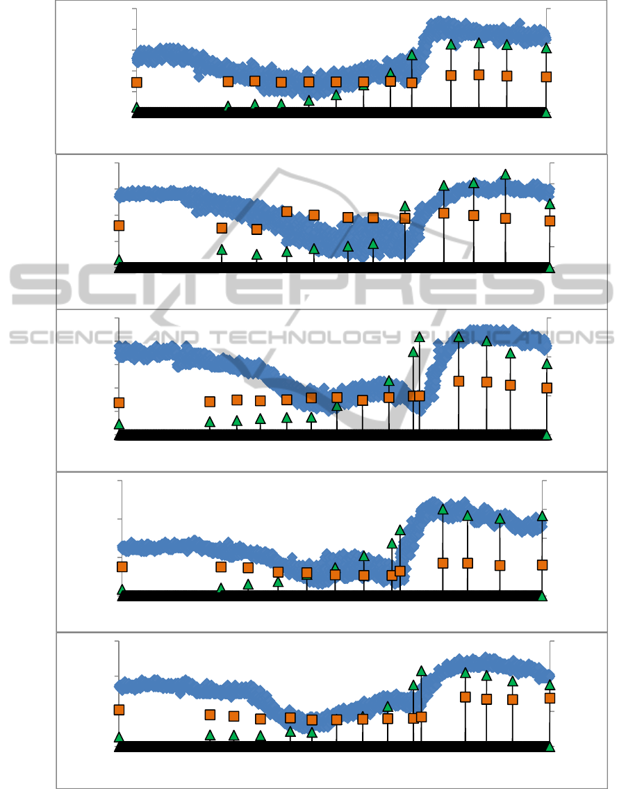
Dynamic changes in temperature of each of our
subjects in conjunction with the dynamics of lactate
and glucose in the peripheral blood are shown in
Fig. 2. It is clearly seen that all five subjects show
the same pattern: the temperature of the skin during
the period of adaptation to the experimental
conditions is either declining slightly or does not
change, then it is gradually decreasing during the
execution of ramp test, reaching a minimum at a
speed of 11-13 km / hour, and then begins to
increase rapidly and reaches a maximum at the time
of the refusal of work or a bit later. During the
icSPORTS2014-InternationalCongressonSportSciencesResearchandTechnologySupport
98
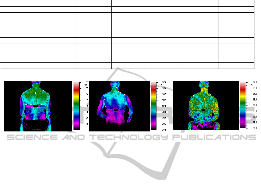
Table 1: Morpho-functional characteristics of subjects.
Subject A.I. G.A. Sh.D. Ya.A. Z.A.
Age, years 20 35 23 28 24
Body mass, kg 78 95,5 62 70 71,5
Body height, sm 175 192 170 176 174
Body mass index 25,5 25,9 21,5 22,6 23,6
VO2max, l/min 4,9 5,8 4,5 3,9 5,5
Anaerobic threshold, km/h 14,0 14,6 14,8 12,6 14,6
Duration of test, min:s 15:25 14:40 18:30 14:05 18:30
Heart rate max., 1/min. 197 186 197 193 192
Lactate max., mM/l 10,06 8,92 12,58 15,03 10,78
Glucose min/max, mM/l 4,34/5,44 3,65/5,20 4,11/6,88 3,53/5,69 3,78/7,04
Rest before ramp test During execution of ramp test Recovery after ramp test
Figure 1: Examples of infrared thermal images obtained at different stages of testing. Subject – Ya.A.
recovery period, the temperature is gradually going
down, but by the end of 10 minutes still does not
reach the level recorded before the ramp test.
Noteworthy is the fact that the curve of
temperature variation is very unstable and the graph
looks like a broad band due to the large scatter of the
data. This is due, firstly, to a rather complex picture
of the functional manifestations of skin temperature
- in fact plural functions are involved in the process:
nervous control, and skin blood flow, and sweating,
all of these factors interact in a complex manner,
which leads to a large scatter in the data.
Also in this case the temperature profile was
made in the course of movement, and though the test
body was relatively motionless in a horizontal plane,
it nevertheless constantly fluctuated in the vertical
plane, thus creating an additional disturbance for
measuring temperature in dynamic observations.
However, the general trend of the temperature
dynamics is not only evident for each subject , but
practically identical for all 5 participants.
Superimposed on the line of temperature
dynamics markers, reflecting changes of lactate and
glucose in the peripheral blood, allow to notice some
interesting facts.
Firstly, there is a large apparent difference in the
dynamics of changes of lactate and glucose during
the ramp test: Lactate increases exponentially
throughout the test time, while the glucose in most
cases varies little during work, and rises to a
relatively high level only with the start of recovery.
The range of variation of lactate is much broader
than the relatively narrow range of variation of
glucose.
Secondly, no connection between the dynamics
of glucose and skin temperature is visible. But there
is a demonstrative interaction between temperature
and lactate: while the load is below the anaerobic
threshold, skin temperature depends on the lactate
level in moderately negative manner, and if the load
goes above the anaerobic threshold we see strongly
positive correlation (Fig. 3).
We obtained a very high coefficient of correlation
between the two indicators (R2> 0.94). This can
testify in favor of the assumption of a causal
relationship between lactate levels and the level of
skin temperature after reaching the
anaerobic threshold. Careful consideration of
individual curves ensures that in all 5 cases, increase
in lactate level precedes raise of the temperature.
The question of what is a direct trigger for the
activation of structures, thermal radiation of which is
projected onto the skin and is fixed by infrared
device, requires further detailed study.
4 DISCUSSION
The fact that skin temperature initially decreases
during intense muscular work, and then may rise in
some of the subjects, and the beginning of this
increase is linked to the achievement of anaerobic
BrownAdiposeTissueParticipateinLactateUtilizationduringMuscularWork
99
Subject
Rest Ramp test Recovery
A.I.
G.A.
Sh.D.
Ya.A.
Z.A.
Figure 2: Individual dynamics of maximal skin temperature, blood lactate (triangles) and glucose (squares) during ramp test
and recovery.
0
5
10
15
32
33
34
35
36
37
0
0
0
0
0
7,7
8,4
9,2
10
10,7
11,5
12,2
13
13,8
14,5
15,3
16,1
0
0
0
0
0
0
0
Lactate,Glucose,
mM/l
Temperature,°С
Velosity,km/h
0
2
4
6
8
10
29
31
33
35
37
0
0
0
0
0
7,5
8,2
8,9
9,6
10,4
11,1
11,8
12,5
13,2
14
14,7
15,4
16,2
16,8
17,6
1,9
0
0
0
0
Lactate,Glucose,
mM/l
Temperature,°С
Velosity,km/h
0
5
10
15
31
32
33
34
35
36
0
0
0
0
6,9
7,9
8,7
9,6
10,4
11,1
12
12,8
13,6
14,4
15,2
16,1
16,8
17,7
0
0
0
0
0
0
0
Lactate,Glucose,
mM/l
Temperature,°С
Velosity,km/h
0
5
10
15
20
31
33
35
37
0
0
0
0
0
7,6
8,3
9
9,8
10,6
11,3
12
12,7
13,5
14,2
15
15,8
16,5
17,2
17,9
0
0
0
0
Lactate,Glucose,
mM/l
Temperature,°С
Velosity,km/h
0
5
10
15
30
32
34
36
0
0
0
0
6,9
7,9
8,7
9,6
10,4
11,1
12
12,8
13,6
14,4
15,2
16,1
16,8
17,7
0
0
0
0
0
0
0
Lactate,Glucose,
mM/l
Temperature,°С
Velosity,km/h
icSPORTS2014-InternationalCongressonSportSciencesResearchandTechnologySupport
100
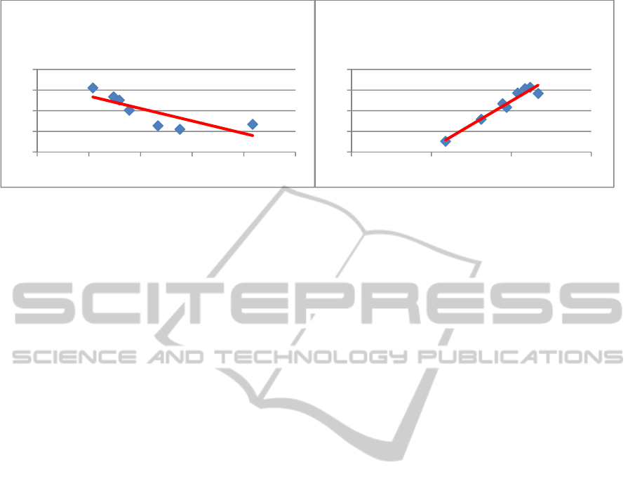
Figure 3: The relationship between the maximum surface temperature of the skin of the back and neck and lactate content in
peripheral blood before the anaerobic threshold (left) and after it (right).
threshold, was previously reported through forehead
thermographing (Akimov, Son’kin, 2011). In this
paper it was shown that such a reaction is typical for
2/3 of the subjects, whereas 1/3 shows no increase in
temperature at loads above the forehead anaerobic
threshold. However, it is impossible to
unambiguously correlate these events with the
activity of BAT, as this tissue under the skin of the
forehead is missing. We should rather speak about
the change in the overall thermal state of the body.
An entirely different matter is the maximum
surface temperature of the back, and especially the
neck, where according to PET and biopsy studies the
most significant fragments of BAT or analogues
thereof are located (Sacks and Symonds, 2013;
Virtanen et al., 2009), having a powerful
metabolism. Maximum temperature in these areas of
the skin increases very fast right before the refusal of
work. High level of the temperature is also observed
during the recovery (at least 10 minutes), and this
level closely correlates with the dynamics of blood
lactate. In this case we have to emphasize that there
is no correlation of the level of blood glucose with
this temperature curve. These results are difficult to
interpret differently than to associate BAT obviously
increased activity with targeted utilization of lactate.
Brown adipose tissue in the last 2-3 years has
become well known among physiologists as an
active participant of the metabolic processes in the
human body (Harms M., Seale P., 2013;
Spiegelman, B., 2013; Virtanen, K.A. et al., 2013).
Due to its uncoupled mitochondria, brown adipose
tissue is involved in maintaining temperature
homeostasis and glucose homeostasis (Cypess A.M.
et al., 2009; Lee Y.-H. et al., 2014; Sacks H. and
Symonds M., 2013). The latest research suggests
BAT also participate in maintaining lactate
homeostasis (De Matteis et al., 2013; Son’kin V. et
al., 2010).
First to describe the thermal effect of BAT under
cyclic physical work were Japanese authors (Shibata
and Nagasaka, 1987), who used a thermocouple to
measure the temperature in BAT in rats while
running on a treadmill, and it was about 0.5 degrees
higher than the rectal one. However, this does not
imply that the activation of BAT is somehow related
to the metabolism of lactate. Relatively recently, it
was shown that in mice BAT cells have specific
transmembrane lactate transporter MST1. Through
the activity of this molecule lactate penetrates inside
the mitochondria and becomes available for
oxidation (Iwanaga et al., 2009). And finally, a
group of Italian researchers recently showed that
running training leads to a twofold increase in the
content and activity of MST1 in rat BAT (De
Matteis et al., 2013).
5 CONCLUSIONS
Given the fact that the muscles can produce
hormone irisin during contractile function, and this
hormone stimulates the conversion of white fat cells
into "beige" cells, which are similar by the metabolic
activity with BAT (Boström et al., 2012; Harms and
Seale, 2013; Lee at el., 2014; Spiegelman, 2013),
begs the question of the biological sense of the
phenomenon. If BAT or its metabolic analogs are
able to utilize lactate produced during muscular
work, and thereby fulfill another kind of homeostatic
activity - the question of the biological sense gets a
clear and unequivocal answer. But in this case there
rise a plurality of application issues related to the
ability to use the newly opened physiological
phenomena in the labor process and the sport, not
just a series of measures in prophylactics of obesity
pandemic.
y=34.32‐ 0.60X
R²=0.64
31
32
33
34
35
012345
TemperaturevsLactate
beforeanaerobicthreshold
y=29.88+0,46X
R²=0.94
32
33
34
35
36
0 5 10 15
TemperaturevsLactate
afteranaerobicthreshold
BrownAdiposeTissueParticipateinLactateUtilizationduringMuscularWork
101

ACKNOWLEDGEMENTS
The authors thank Professor A.Tonevitskii for active
participation in the planning of these studies on the
origin of the idea stage, as well as Professor A.
Meygal and Professor S. Levushkin for productive
discussions on the results of our experiments.
REFERENCES
Akimov EB, Son’kin VD. (2011) Skin Temperature and
Lactate Threshold during Muscle Work in Athletes.
Human Physiology, 37(5): 621-628.
Boström P, Wu J, Jedrychowski MP, et al. (2012) A
PGC1-a-dependent myokine that drives brown-fat-like
development of white fat and thermogenesis. Nature,
481: 463–468
Cypess AM, Lehman S, Williams G, et al. (2009)
Identification and importance of brown adipose tissue
in adult humans. N Engl J Med; 360: 1509–1517
De Matteis, R., Lucertini, F., Guescini, M., Polidori, E.,
Zeppa, S., Stocchi, V., Cinti, S., et al. (2013) Exercise
as a new physiological stimulus for brown adipose
tissue activity. Nutr Metab Cardiovasc Dis. 23(6):582-
590.
Harms M., Seale P. (2013) Brown and beige fat:
development, function and therapeutic potential.
Nature Medicine 19 (10) :1252
Iwanaga T, Kuchiiwa T, Saito M. (2009) Histochemical
demonstration of monocarboxylate transporters in
mouse brown adipose tissue. Biomed Res. 30(4): 217-
225.
Lee P., Greenfield J. R. and Ho K. K. Y. (2011) Hot fat in
a cool man: infrared thermography and brown adipose
tissue. Letter to the editor. Diabetes, Obesity and
Metabolism 13: 92–93.
Lee, Y.-H., Mottillo, E.P., and Granneman, J.G. (2014)
Adipose tissue plasticity from WAT to BAT and in
between. Biochim Biophys Acta, 1842(3):358-69.
Sacks H. and Symonds M.E. (2013) Anatomical Locations
of Human Brown Adipose Tissue Functional
Relevance and Implications in Obesity and Type 2
Diabetes. Diabetes, 62: 1783–1790
Shibata H, Nagasaka T. (1987) The effect of forced
running on heat production in brown adipose tissue in
rats. Physiol Behav.; 39 (3): 377-80.
Son’kin V, Kirdin A, Andreev R, Akimov E. (2010)
Homeostatic Non-Shivering Thermogenesis in
Humans: Facts and Hypotheses. Human Physiology,
36 (5): 599-614
Spiegelman, B. M. (2013). Regulation of Adipogenesis:
Toward New Therapeutics for Metabolic Disease.
Diabetes, 62(6): 1774–1782.
Virtanen, KA, Lidell ME, Orava J, Heglind M, et al.
(2009) Functional brown adipose tissue in healthy
adults. N Engl J Med.; 360(15): 1518-25.
Virtanen, K.A., Van Marken Lihtenbelt, W.D., and
Nuutila, P. (2013). Brown adipose tissue functions in
humans. Biochim Biophys Acta, 1831(5):1004-1008.
icSPORTS2014-InternationalCongressonSportSciencesResearchandTechnologySupport
102
