
Design of a Myelin Basic Protein Biosensor based on EnFET
Technology
Jorge Guerrero, Roberto Ambrosio and Amanda Carrillo
Departamento de Ingenieria Electrica y Computacion, Instituto de Ingenieria y Tecnologia,
Universidad Autonoma de Ciudad Juarez, Avenida del Charro 450N, Ciudad Juarez, Mexico
Keywords: Biosensor, EnFET, Transducer, Myelin Basic Protein, Multiple Sclerosis.
Abstract: In this work, the design of a biosensor based on FET technology have been proposed, simulating the
modification to the gate of an Ion Sensitive Field-Effect Transistor (ISFET) with a synthetic polymer to
entrap the desired analyte which contains Myelin Basic Protein (MBP). This analyte is generally used in test
to find out if someone is suffering a demyelinating disease, and is commonly detected by Enzyme Linked
Immunosorbent Assays (ELISA). Based on this principle, we propose a simpler method, fundamented on
Enzyme Field-Effect Transistor (EnFET) technology in order to develop a new device applied to the
diagnosis of demyelinating diseases. Simulation examples are used in order to demonstrate the functionality
for this type of biosensor to its exposure to MBP at concentrations of 10
-4
to 10
-1
mol/L, where the amount
of analyte in the receptor located at the top of the gate will determine the level of voltage applied to create a
channel and activate the device.
1 INTRODUCTION
The diagnosis of diseases is one of the principal
concerns related to all aspects of life. One of the
most alarming diseases is from the degenerative
kind. These diseases are related to the motor and
sensory function of the body, which are in charge of
the nervous system, where the communication from
cell to cell is conducted by neurons. If someone
suffers from a demyelinating disease, the myelin
sheath of the neuron loses myelin causing a bad
conduction of nerve pulses.
Recent statistics carried out in 2013 on behalf of
the Multiple Sclerosis International Federation
(MSIF) reveal that 2.3 million people suffer from
multiple sclerosis (Thompson et al., 2013). This has
increased the interest from physicians and
researchers in search of new methods of analysis.
Currently, demyelinating diseases are diagnosed
by methods that get results after several tests.
Performing a lumbar puncture to remove
cerebrospinal fluid from the central nervous system
and subsequently perform an Enzyme Linked
Immunosorbent Assay (ELISA) test to obtain the
levels of myelin is one of the most common tests
because of its economy in comparison with other
methods such as Magnetic Resonance Imaging
(MRI) (Holland et al., 2007).
Recent investigations have developed
immunosensors based on different receptors to
detect myelin basic protein in order to have
alternatives to the existing methods of diagnosis,
reducing the consumption of time and costs (La
Belle et al., 2007). Encouraging the development of
new devices to be implemented both at the area of
research and medical application of demyelinating
diseases. However, the complexity of certain
immunosensors makes them not suitable for mass
production, but it is one of the main advantages of
FET technology.
Although FETs can be aseptically manufactured
and hermetically sealed, the biocompatibility of the
materials with which they are made is the key so that
they can be implemented for biomedical purposes;
these devices are called biologically sensitive field-
effect transistors (BioFETs). Some applications of
BioFETs have been already studied, like the
detection of DNA (Ozsoz, 2007), proteins (Park et
al., 2005) and enzymes (Zayats et al., 2000). In
principle, every charged molecule located in the
solute that can be bound to the surface can be
detected by a BioFET. In this work the objective
was to design a biosensor that can detect myelin in
78
Guerrero J., Ambrosio R. and Carrillo A..
Design of a Myelin Basic Protein Biosensor based on EnFET Technology.
DOI: 10.5220/0005180700780082
In Proceedings of the International Conference on Biomedical Electronics and Devices (BIODEVICES-2015), pages 78-82
ISBN: 978-989-758-071-0
Copyright
c
2015 SCITEPRESS (Science and Technology Publications, Lda.)

order to help the research of demyelinating diseases,
based on BioFET technology.
2 METHOD
A BioFET can be constructed from an ISFET,
modifying the gate by a coupling of different
biological recognition elements and processing the
output signal. Basically, an EnFET is composed of
an analyte, a receptor, a transducer and a signal
acquisition system (Figure 1).
Figure 1: Schematic diagram of the EnFET operating
principle.
The selected analyte was MBP (Myelin Basic
Protein) which properties and interactions have been
already studied (Boggs 2006). This analyte can be
immobilized by polymer entrapment or covalent
attachment. Based on the chemical structure of MBP
(Figure 2), the chosen method for the analyte
immobilization was polymer entrapment; making a
hybrid gel conformed by polyvinyl alcohol (PVA),
Tetraethyl orthosilicate (TEOS) and glutaraldehyde
(GA) the receptor. Where the implementation of GA
as a selective immobilizer to MBP has been used in
related works (Burak et al., 2013).
Figure 2: Schematic diagram of the EnFET operating
principle.
The most important point for the device is the
transferring of data from the part of biologic
recognition to the transduction of the signal. To
achieve this task, an ISFET is used as a transducer.
Basically, ISFETs are implemented to measure the
concentration of ions in an electrolytic solution, and
are used in biosensor development due to their
favourable characteristics such as sensitivity,
miniaturization, fast response and low cost
(Bergveld, 2003a). An ISFET threshold voltage can
be calculated by the following equation (Bergveld,
2003b):
Ф
2∅
(1)
Where
represents the reference electrode
potential,
the electrochemical potential at the
dielectric-electrolyte interface,
the surface
potential of the solution, Ф
the work function of
the semiconductor,
the density of accumulated
charge in the oxide-semiconductor interface,
the
density of accumulated charge in the oxide,
the
density of accumulated charge in the region of the
interface close to the semiconductor,
the
capacitance of the oxide layer and 2∅
the
difference between half of the band gap and the
Fermi level.
From the expression given in (1) all potentials
are constant, except the electrochemical potential
(
) that depends on the ionic concentration of the
solution, which can be calculated as follows:
(2)
Substituting (2) on (1) the expression can be reduced
to
Ф
2∅
(3)
Since the threshold voltage is a function of
, the
drain current will be influenced by the changes in
the electrochemical potential
. Where the drain to
source current of an ISFET in the linear region is
given by the following expression (Lee et al., 2009):
2
(4)
Where
represents the charge-carrier effective
mobility in the channel,
the capacitance of the
oxide layer, the channel width, the channel
length,
the gate to source voltage,
the
threshold voltage for zero substrate bias and
the
drain to source voltage.
Once
and
are theoretically calculated, all
the parameters applied to the simulation of the
ISFET can be defined.
DesignofaMyelinBasicProteinBiosensorbasedonEnFETTechnology
79
3 SIMULATION
The transducer device was simulated using
SILVACO a Technology Computer Aided Design
(TCAD) tool, its software models semiconductor
fabrication and device operation. In this work the
ATHENA and ATLAS modules were used. The
proposal device was designed to have a dense mesh
in the regions of drain, source and channel, selecting
a substrate material of Silicon (Si) and a dielectric
compound layer of SiO
2
/Si
3
N
4
deposited over the
channel region. This layer is commonly used in
ISFET sensors as a sensitive membrane to H
+
and
OH
-
ions and it is implemented to approach the
sensibility of the device to a Nernstian value of 59
mV/pH (Kühnhold and Ryssel, 2000). Finally the
source, drain and gate regions were defined with its
electrodes to be used as contacts, the final structure
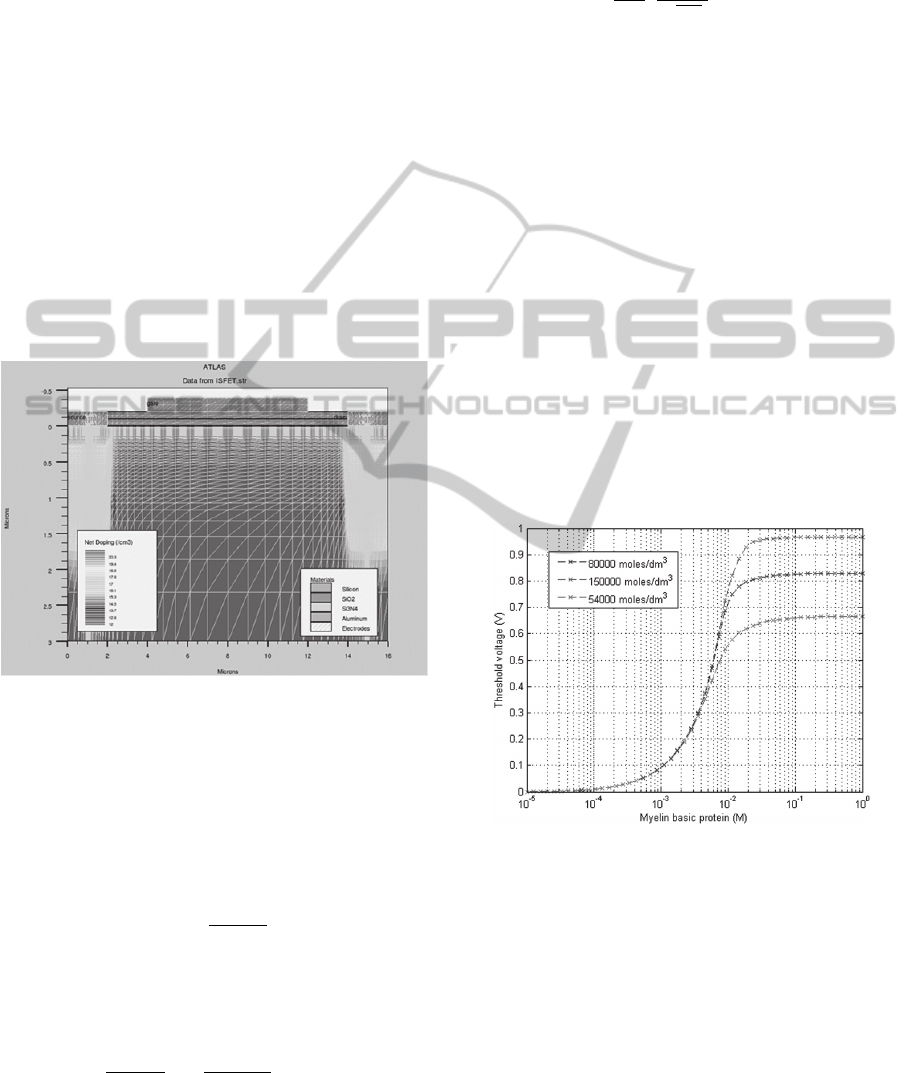
is shown in Figure 3.
Figure 3: Resulting structure made on the ATHENA
module and used as an input for ATLAS.
As a part of the design, an analytical model was
developed based in the mechanism of diffusion in
the interface between the hybrid gel
(PVA/TEOS/GA) and Si
3
N
4
layers located over the
gate of the ISFET based on the Fick law of
diffusion.
∗
,
(5)
Where D is the diffusion coefficient of the studied
biochemical species in cm
2
/s and c(x, t) is the
concentration level of that species represented as
follows:
,
,
(6)
The diffusion coefficients (D) in the solution of
different biochemical species are calculated from the
relation of Einstein-Stokes.
1
6
4
√
3
(7)
A MATLAB program was developed to solve the
Fick law of diffusion, and also to simulate the
generation of biochemical species in the electrolyte-
dielectric interface based on the Michaelis-Menten
kinetics.
4 RESULTS
Figure 4 and 5 represents the simulation results of
the MATLAB program for three different enzyme
concentrations of the layer in moles/dm
3
. Figure 4
shows an increase in the output voltage depending
on the analyte concentration in the surface of the
device in M. As can be seen, the concentration of the
sample containing MBP located in the surface
between the values of 10
-4
to 10
-1
M have a voltage
response from 0 to 1 V, which shows an increase
from 10
-3
to 10
-2
M and reaching a constant value at
10
-1
M.
Figure 4: Output curves representing the changes in
threshold voltage of the EnFET for different
concentrations of MBP depending of concentration of
enzyme.
Figure 5 represents the increase of reaction
velocity, depending on the solution concentration
based on the Michaelis Menten enzyme kinetics,
where the solution concentration is gradually
increased from 0 to 0.5 mol/L and the reaction
velocity increases until it reaches a maximum where
it keeps a steady value.
BIODEVICES2015-InternationalConferenceonBiomedicalElectronicsandDevices
80

Figure 5: Output curves representing the velocity of
reaction rate in the EnFET for different values of solution
concentration depending of concentration of enzyme.
Based on the results obtained at the MATLAB
simulation and the structure made in SILVACO, a
voltage level was applied as gate bias in the module
of ATLAS in order to check if the threshold voltage
of the device is in range of the output voltage
generated as EnFET response (Figure 6).
Figure 6: Characteristic curve obtained from ATLAS.
Figure 6 shows that the device turns on at a gate
voltage between 0.5 and 0.7 V, matching the
threshold voltage level generated in Figure 4, which
corroborates that the EnFET biosensor switches on
at those levels.
5 CONCLUSIONS
The main objective of this work was to design a
biosensor based on the operation of an EnFET in
order to detect MBP, using the output electrical
characteristics due to the changes in concentration of
the analyte. The threshold voltage was calculated
and related to the electrical characteristics and the
fabrication process of the transducer. Based on
simulations, the device haves a voltage range from 0
to 1 Volt, having a concentration level between the
values of 10
-4
to 10
-1
M of MBP, which is
compatible with the FET technology for this
application.
In comparison to other EnFET designs, the
structure is also based on silicon technology,
modifying the gate material by other ion sensitive
membranes depending on the implementation. They
also express results as the ones described in Figures
4 and 5. But the comparison of threshold voltage or
output voltage of EnFET sensors to the design is
always based on experimental basis. In this work,
the use of a TCAD tool was used to simulate the
response of the transducer, to ensure it activates in
the range of the voltage generated from the diffusion
of MBP in the (PVA/TEOS/GA) membrane and the
concentration of substrate generated.
Although the presented work is only based in
theoretical grounds, it provides a scheme of how to
elaborate a biosensor based on techniques applied to
microelectronics, also taking its advantages. For
future investigations the characterization of the
materials and the development of the EnFET based
on flexible electronics can be applied. Also the
results obtained should be verified by practical
experiments in the future, in order to be applied on
in vitro tests.
REFERENCES
Thompson J., Uitdehaag B., Taylor B., Holloway E.,
Tremlett H., Pandit L., Bettaglia M., 2013. ATLAS of
MS 2013, Summers Editorial & Design, London, 6
th
edition.
Holland, N., Murray T., Reingold, S., 2007. Multiple
Sclerosis: A Guide for the Newly Diagnosed. Demos
Medical Publishing, New York, 3
rd
edition.
La Belle, J., Bhavsar, K., Fairchild, A., Das, A., Sweeney,
J., Alford, T. L., Wang J., Bhavanandan, V., Joshi, L.,
2007. A cytokine immunosensor for multiple sclerosis
detection based upon a label-free electrochemical
impedance spectroscopy, Biosensors and
Bioelectronics, 23, 428–431.
Ozsoz M., 2007. Electrochemical DNA Biosensors. Pan
Stanford Publishing, Singapore, 1
st
edition.
Park, K., Kim, M., Choi, S., 2005. Fabrication and
characteristics of MOSFET protein chip for detection
of ribosomal protein, Biosens, Bioelectron, 20, 2111-
2115.
Zayats, M., Kharitonov, A., Katz, E., Bückmann, A.,
Willner, I., 2000. An integrated NAD+- dependent
enzyme-functionalized field-effect transistor (ENFET)
DesignofaMyelinBasicProteinBiosensorbasedonEnFETTechnology
81

system: development of a lactate biosensor. Biosens,
Bioelectron, 15, 671-680.
Boggs, J. M., 2006. Myelin basic protein: a
multifunctional protein. Cellular and Molecular Life
Sciences CMLS, 63, 1945-1961.
Burak, D., Emergul E., Canan Y., Kaan C., 2013. Myelin
basic protein immunosensor for multiple sclerosis
detection based upon label-free electrochemical
impedance spectroscopy, Biosensors and
Bioelectronics, 46, 53-60.
Bergveld, P., 2003a. Thirty years of ISFETOLOGY, What
happened in the past 30 years and what may happen in
the next 30 years. Sensors and Actuators B, 88, 1-20.
Bergveld, P., 2003b. ISFET, Theory and Practice. In
Proceedings of the IEEE Sensor Conference in
Toronto, IEEE.
Lee, C., Kim, S., Kim, M., 2009. Ion-Sensitive Field-
Effect Transistor for Biological Sensing. Sensors, 9,
7111-7131.
Kühnhold, R., Ryssel, H. 2000. Modeling the pH response
of silicon nitride ISFET devices. Sensors and
Actuators B, 68, 307-312.
BIODEVICES2015-InternationalConferenceonBiomedicalElectronicsandDevices
82
