
Wound Area Assessment using Mobile Application
Ivan Miguel Pires
1,2
and Nuno M. Garcia
1,3
1
Instituto de Telecomunicações, University of Beira Interior, Covilhã, Portugal
2
Altranportugal, Lisbon, Portugal
3
ECATI, Universidade Lusófona de Humanidades e Tecnologias, Lisbon, Portugal
Keywords: Wound, Detection, Contours, Area, Mobile Device, Mobile Application, Wound Area Assessment,
OpenCV, Java, Threshold, Image Processing, Color Histograms, Gauss Filter, Mobile Platforms, Android,
iOS.
Abstract: This research aims to discover methods for the detection of the area of a wound using mobile devices. These
devices have low memory and low processing capacity and they need the use of low complexity operations
to identify a wound. The calculation of the wounded area consists of three phases, there are: image
acquisition, image processing, surface reconstruction and calculations. This research is related to the use a
mobile device to identify wound contours and area in a captured image. This image can be captured with a
camera in a smartphone and the wound area is calculated based on the distance of the surface area and the
resolution of the image captured. The main study in this research is the image processing in a mobile device,
due to the limitations of these devices. However, the application developed during this research was
developed for desktop, using the OpenCV library that is compatible with the Android platform and Java
desktop technologies. During this research, the developed code written in Java will be easily adapted to the
Android platform. The desktop application developed is available in a free repository for testing.
1 INTRODUCTION
The wound area assessment is an important research
to estimate the evolution of wound healing and the
healthcare professional can change the treatment of
the patients (Ousey and Cook, 2011; Russell, 1999).
Currently this topic is under a lot of research, but a
method to estimate the wound area hasn’t been
discovered. This is a challenging task due to the
complexity of the wound, variable lighting
conditions and time constraints in clinical
laboratories (Loizou et al., 2013). Wounds have
some characteristics related to color and texture, but
these characteristics aren’t the same in all wounds
images and people in the world. Therefore, research
in this area hasn´t finished and future research can
improve the wounds characterization. The research
studies in this area have many purposes, such as
verify the existence of a chronic wound, the
existence of an infected wound, the origin of the
wound and other aspects that classify and
characterize a wound.
This analysis is related to the image capture and
processing of a wound (Lazarus et al., 1994). The
image processing and consequent wound area
estimation has several phases, such as: image
acquisition, image processing, surface reconstruction
and calculations.
The image acquisition includes the process by
witch the user makes use of a camera to capture a
wound image. This process has some problems, such
as ambient lighting, digital camera quality and
resolution, as well as other problems. To minimize
this problem, the next phase is image processing.
This phase includes, the application of filters
(blurring the images to minimize the imperfections
of the images) and the thresholding of the images
(converting the images to black and white, related to
the pixel color intensity). In the next phase, some
authors are doing the surface reconstruction, after
the minimization of possible errors in calculations in
the image-processing phase. In the calculations
phase, the contours of a wound are identified and the
wound area is the area within the contours.
This study is very important (Ousey and Cook,
2011; Russell, 1999), because it allows healthcare
professionals to control their patient’s state of
wound healing and it can improve the treatment of
271
Miguel Pires I. and M. Garcia N..
Wound Area Assessment using Mobile Application.
DOI: 10.5220/0005236502710282
In Proceedings of the International Conference on Biomedical Electronics and Devices (SmartMedDev-2015), pages 271-282
ISBN: 978-989-758-071-0
Copyright
c
2015 SCITEPRESS (Science and Technology Publications, Lda.)

wounds, which this occurs with a correct method
and within a short period of time. In the 22nd annual
meeting of the Wound Healing Society (WHS) held
in 2012, the standards for wound healing procedures
and proposed recommendations for evaluating
optimal wound treatments were set (Loizou et al.,
2013).
The challenge of wound area assessment is very
complex, because the captured images contains
various sources of environmental noise, which are
difficult to control, such as lightning, contrast,
distance to the objects and quality of images. Many
people have done some research, but this research
hasn’t established a consensus in external factor
correction in images. The research studies have a
purpose to create an automatic algorithm,
programmatically implemented and mathematically
validated to calculate the wound area, but this
research depends on the approach of the researcher
to consider some variables or not. Manually, this
research is complex too, because the wound area
hasn’t a defined geometric shape. Thus, in some
cases of research without images, people preferred
to measure the wound perimeter instead of the
wound area.
Mobile technology is currently the most used and
the mobile devices in current days have a camera
with a good quality of image capture. Some devices
have a proximity sensor. The presence of the
proximity sensor allows the estimation of the wound
area with better quality. Using a mobile device
equipped with sensors the value of wound area
calculated is more approximate to the real value.
This purpose can be attained based on various
programming languages and frameworks. To
estimate the wound area there exist various methods
for image processing. Initially, in this paper, it is
analysed the wound area measurement using
MATLAB with Image Processing library. Next, in
this research is analysed the wound area
measurement with Java language and OpenCV
library compatible with the most used mobile
platforms (i.e., Android and iOS operating systems).
This paper was organized in five sections. In
section 2, other researches done by other authors are
presented. In section 3, an overview of the methods
for wound area estimation using images is described.
Section 4 relates the wound images analysis and
wound area measurement to mobile devices,
presenting new methods or sensors utilization with
these devices. Section 5 shows the conclusions of
this research. At the end, the literature references
used for this paper will be presented.
2 RELATED WORK
During the last years, various research authors
addressed the wound area measurement and they
have different perspectives about this topic. In
general, research on this topic consists in the use of
images to estimate the wound area measurement
using a device (e.g., computer, laptop, smartphone
or tablet) for image processing (Krouskop et al.,
2002). Some research focuses in the use of other
sensors, but it wasn’t very investigated and
validated. In relation to the use of images, the
approach to this topic can be divided in some parts,
there are: verification of a existence of a wound in
the image obtained for image processing,
identification of the contours of the wound and
calculate the area between the contours (this
corresponds to the wound area). This process isn’t
linear and objective and depends on the way the
research authors view the problems. Some authors
attempt to identify a wound with a relation to
standard colors of wounds, other authors attempt to
identify by textures and other authors use the join of
standard features of texture and color of wound.
Some research presents the identification of the
existence of infection in a wound region and the type
of wound.
This research area is important for the healthcare
professionals to control, with an easy method, the
wound-healing rate in a patient during the time of
healing (Ousey and Cook, 2011; Russell, 1999).
Thus, it allows a healthcare professional to verify the
wound state and change the treatment used to the
correct treatment related to the evolution of the
wound healing. This is really important in chronic
wounds (Casas et al., 2011; Papazoglou et al., 2010),
because the area of this type of wounds doesn’t
decrease constantly during the wound healing time
and it can increase and decrease inconstantly during
the time of wound healing. The chronic wounds are
more frequently in elderly people, diabetic people or
people with chronic diseases. The health defences of
these people are weakened and their wounds take
longer to heal.
Thus, some research authors have done various
studies related to this topic. The wound-healing rate
is primarily quantified by the change of wound area
and the authors attempt to define a standardized and
objective technique to assess the progress of wound
healing, by means of texture image analysis (Loizou
et al., 2013). The texture of wounds depends of the
location of wounds in people’s body. In the studies
done by the authors of (Loizou et al., 2013) some
tasks where done related to the image processing,
BIODEVICES2015-InternationalConferenceonBiomedicalElectronicsandDevices
272

these were image pre-processing, segmentation,
texture and geometrical analysis together with visual
expert’s to assess the wound healing evaluation.
These authors use a total of 77 digital images
collected in 11 different subjects with foot wounds.
These images were taken every third day, for 21
days, by an inexpensive digital camera under
different lighting conditions (Loizou et al., 2013).
The images collected were intensity normalized, and
wounds were automatically segmented using a
segmentation system based on snakes. With these
experiments, the authors of (Loizou et al., 2013), in
order to identify features that quantify the rate of
wound healing, had extracted 56 different texture
features and 4 different geometrical measures. Thus,
the authors of (Loizou et al., 2013) discovered that
the texture features indicate the progression of
wound healing and some texture features increase
(mean, contrast, roughness and radial sum), while
some other texture features decrease (sum of squares
variance, sum variance, sum average, entropy,
coarseness, EE-laws texture energy measures and
the Hurst coefficients for fractal dimension one and
two analysis) with the progression of wound healing
process. When they compare the different features at
two different time points during wound healing
process, they access the rate of wound healing, but
the comparison of all geometrical measures
extracted from wounds at two different points
doesn’t present important information about the
wound healing. So, these authors create a simple
method that uses some texture features to monitor
the wound healing process, to reduce costs, provide
standardization and improve the treatment quality
for patients and provide a valuable tool in clinical
wound evaluation (Loizou et al., 2013).
Related to the importance of wound healing
control in chronic wounds (Russell, 1999), the
authors of (Papazoglou et al., 2010) present a new
algorithm implemented in MATLAB software
validated in, approximately, 50 animal images and
100 human images. The images for the tests were
captured with a common inexpensive digital camera
and in various lighting conditions. These authors
make a comparison of results in animal images and
human images and compare the manual wound
boundary (obtained in Adobe Photoshop) and the
automatic wound boundary (obtained in MATLAB
software), obtaining very small errors in wound area
measurement. This research depends of the
resolution of the processed wound images, but the
authors of (Papazoglou et al., 2010) proposed and
evaluated a highly accurate algorithm for wound
segmentation which requires a minimal manual
input by using a combination of both red-green-blue
(RGB) and L*a*b color spaces, as well as a
combination of threshold and pixel-based color
comparing segmentation methods.
Other authors have developed systems with
automatic algorithms to measure various parameters
of wounds, such as area, perimeter, width and height
of wounds using images. A example of a system
developed by authors of (Plassmann and Jones,
1998) is the MAVIS system used to automatically
measure the dimensions of skin wounds. In this
system, the method of measurements is based on
color segmentation algorithms and this method is
able to segment images related to healthy skin,
wound tissue and epithelialisation tissue. The
method considers the RGB color planes, hue,
saturation and grey-level intensity. The RGB color
planes were only examined in isolation, showing
that straightforward thresholding of color planes
cannot produce a good segmentation, which
distinguishes between wound and skin tissues. The
wound segmentation with this method is only
partially successful if only the one-dimensional
color histograms were taken into consideration,
while using a 3-dimesional (3D) RGB histogram
space, the color volume clusters may be more widely
separated and a better segmentation result can be
achieved. Some authors, such as (Mekkes and
Westerhof, 1995; Nayak et al., 2009; Wannous et al.,
2008; Wannous et al., 2007), consider they made
some progress using 3D RGB color histogram
clustering technique to assess the wounds healing.
The research of (Mekkes and Westerhof, 1995)
shows that clusters in RGB space for a given tissue
type formed an irregularly shaped 3D cloud, and
therefore simple thresholding along the R, G and B
axis wouldn’t help to segment the image into some
tissue types. The segmentation of wounds in color
images based on the use of the black-yellow-red
classification scheme to evaluate the debridement
activity of wounds have some techniques presented
by other authors (Gammal and Popp, 1995;
Gallagher, 2012). The segmentation of wound
images consists in the image-processing phase.
The method of pre-processing images
corresponds to the first phase of wound area
measurement. The second phase consists in the
identification of contours of wounds, but some
research join the first and second phases and in the
first phase (corresponding to the segmentation,
threshold and other tasks for pre-processing images)
identifies the contours of the wound. The Support
Vector Machine classifiers (SVM) can be used to
perform region segmentation of the wound tissue
WoundAreaAssessmentusingMobileApplication
273

followed by extraction of the contours of the wound
(Kolesnik and Fexa, 2004). In the study presented in
(Kolesnik and Fexa, 2004), the authors used 50 RGB
images, which were manually delineated by experts
as training data, and then tested their method using
23 new RGB wound images. The SVM algorithm
used by (Kolesnik and Fexa, 2004) was able to
correctly classify roughly 94% of the pixels as either
wound or non-wound, compared with the expert’s
manual tracings.
Many research used SVM algorithms (Kolesnik
and Fexa, 2006) with various purposes on this topic.
Authors of (Giger et al., 2008) start using a 3D
model for wound measurements using uncalibrated
vision techniques and a color classification wound
tissues, combining shape and color analysis in a
single tool for real tissue surface measurements. A
database with images of different tissue types in
uncontrolled lighting environments was created
(Giger et al., 2008), applying a correction method to
reduce color shifts. The problem unsolved by all
authors researching in this topic is the difficulty to
control environmental conditions in the experiments
and in the use of the system by other people. Then,
color and texture tissue descriptors are extracted
from tissue regions of the images database, for the
learning stage of an SVM region classifier, and
apply unsupervised color region segmentation on
wound images and classify the tissue regions. The
SVM algorithm used by (Giger et al., 2008) obtains
an overlap score in the result of automatic
segmentation driven classification, (66 % to 88%) of
tissue regions higher than that obtained by
clinicians.
Multidimensional color histograms in SVM
classifiers for automatic extraction of wound region
from an image are used in some research (Kolesnik
and Fexa, 2005). The authors of (Kolesnik and Fexa,
2005) compare the performance of the multi-
dimensional histogram sampling with several
existing techniques for quantization of 3D color
space and this increased the performance of wound
segmentation by about 25%. Many research authors
researched about the creation of systems to measure
size and tissue type of wounds, using images taken
by a digital camera and complex systems were
created. For example, the system constructed by
(Wild et al., 2008) takes about 90 seconds per lesion
and, if the user needs a report with suggestions for
therapy, the system needs 4 minutes.
Instead of the use of SVM classifiers, some
research uses Artificial Neural Networks (ANNs)
algorithms (Acha et al., 2005; Navas et al., 2013;
Song, 2012; Song and Sacan, 2012), such as the
Multi-Layer Perceptron (MLP) and the Radial Basis
Function (RBF) with parameters determined by a
cross-validation approach. There are then applied
with supervised learning in the prediction procedure
for the wound identification, and their results are
compared. The results obtained with ANNs are
satisfactory and this reveals that this method can, in
the future, improve the techniques of wound area
measurement and identification, making it a
promising tool to assist in the field of clinical wound
evaluation.
The automatic systems for wound area
measurements are very useful for telemedicine
systems, because the patient can send a digital
photography to the system and the healthcare
professional is able to check the patient’s wound
state and change the treatments at distance
(Wannous et al., 2010). These systems need to allow
for practical image acquisition conditions, such as
digital camera type, lighting, and viewpoint of
wounds. In general, the telemedicine system consists
in a website or platform for users/patients and
professionals interaction. The systems presented in
(Wannous et al., 2010) were obtaining results of
79.3% between classified tissues and the medical
reference, which compares favorably with the
average score of 69.1% obtained by a single
clinician during the validation tests. The research
authors conclude that these systems are very
important for improving the health treatment in a
world with more people. These systems are very
useful for e-learning systems, because the use of
Web platforms allows to explaining this topic to the
students. The e-learning systems apply the
techniques present in some research and consider the
phases of image processing already present. These
systems improve the teaching-learning relations and
provide a better assessment among students than
traditional methods (Prodan et al., 2010b). All
systems implemented are Web-based (Kim et al.,
2003; Prodan et al., 2009) or software developed in
MATLAB software or in other programming
languages, such as the Java programming language
(Cuautle, 2007; Prodan et al., 2009; Prodan et al.,
2006; Prodan et al., 2010a) and others, using various
frameworks. These systems for classifying the
wounds, in general, comprise four phases, and these
are (Kumar et al., 2013): pre-processing images,
image segmentation, feature extraction and
classification. At the end of this, the system will be
able to measure the wound area and other features of
wounds. The web-based systems (Kim et al., 2003,
Prodan et al., 2009) consist in three-tier layer system
for the user to send a digital image and the image is
BIODEVICES2015-InternationalConferenceonBiomedicalElectronicsandDevices
274

processed in a server, showing the results of wounds
area during the time of wound healing.
Other authors (D, 2006) developed a desktop
application in the VB.NET language for Microsoft
systems. This algorithm consists in various steps,
these are: the digitalization of the outline (perimeter)
of an image from right to left, the digitalization of
the outline (perimeter) of an image from left to right,
the digitalization of the outline (perimeter) of an
image from top to bottom, the digitalization of the
outline (perimeter) of an image from bottom to top
and, finally, the calculation of the area enclosed by
that outline. This system has an objective to use a
database and it consists in a non-invasive, accurate,
consistent, efficient and easy wound measurement
system.
The differential evolution method for estimating
the wound healing is used in some research and
obtains best results. This method uses the K-Nearest
Neighbor (KNN) algorithm to classify the wound
healing. This method includes many phases, there
are (Aslantas and Tunckanat, 2007): read image,
detect entire wound, fill gaps, dilate the image, fill
interior gaps, remove connected objects on border
and smooth the object. This method obtains good
results during the validation tests and it obtains very
low errors.
The planimetric techniques for assessing the
wound area and perimeter with reliability and low
errors are used by authors of (Mayrovitz and
Soontupe, 2009). The automated systems are very
important for a correct wound area measurement,
because, in some cases, the manual measurement is
very difficult.
Nowadays, the use of smartphones equipped
with camera and various sensors is very common the
people and these equipments are practical for wound
measurement (Wannous et al., 2011; Cuautle, 2007;
Foltynski et al., 2013; Hettiarachchi et al., 2013;
Perera and Chakrabarti, 2013; Sikka et al., 2012).
These devices can improve the telemedicine
techniques and treatment at distance (Vivanco et al.,
2011). The mobile applications structure is the same
of desktop applications using frameworks designed
for the wound area measurement. In general, the
captured images are sent to an images database to
improve the reliability of the method and send the
processing tasks to the server that has more capacity
to do complex tasks of image processing. These
applications can apply all research studies already
presented, but the user can access to the wound area
over time in various places. These systems have the
same problem of digital images, because all
variables in the environment are difficult to control.
In fact, the use of these practical devices is low-cost
and it will be able to adapt in hospitals for correct
and practical measurements, because, in recent days,
the research in this topic has improved. Recently,
many authors developed various frameworks to
estimate the wound area using the Java
programming language (Cuautle, 2007), and XML
(eXtended Markup Language) descriptors (Prodan et
al., 2009; Prodan et al., 2006) as reference, for
Android platform or other mobile platforms. The
mobile health improves the treatments in various
areas, such as control of a healing wound and the
patient’s health state in various parameters (Friesen
et al., 2013, Perera and Chakrabarti, 2013). For
desktop applications, mobile applications or web-
based applications, this needs a study and
development of a framework for the programming
language used and an image descriptor (in general, it
is defined in XML) for classifying the images. This
work is very difficult, because images have many
features, such as granularity, texture, color and the
wounds depends of various factors.
In current days, many applications, frameworks
and methods have been developed and are in
advanced research state. The tracings of the Visitrak
method were quick, easy, and inexpensive to
perform and noninvasive for the patient (H et al.,
2009). The Visitrak method considers the foot
curvature and removed the subjectivity associated
with manual square counting. The method was both
valid and repeatable in the measurement of wounds
> 25mm
2
in size. The Pressure Ulcer Scale for
Healing tool was designed to track pressure ulcer
healing by monitoring wound parameters of length
times width, exudate amount and tissue type (H et
al., 2009). The PSST system was designed to
describe wound healing in pressure ulcers,
consisting of 15 scored (used to assess variables of
wound size and depth, tissue characteristics and
wound exudate, whereas the non-scored items
examined wound location and shape) and two non-
scored items (H et al., 2009). The Sessing Scale is a
seven-stage scale designed to measure progress in
wound healing over time, with each stage describing
wound tissue attributes throughout the wound
healing process (H et al., 2009). The Sussman
Wound Healing Tool is based on an acute model of
wound healing, which describes tissue status and
size throughout the wound healing process (H et al.,
2009). Other mobile application for mobile devices
is MOWA (Mobile Wound Analyzer) (Healthpath,
2011). MOWA differentiates types of tissues found
in pressure ulcer, analyzes photos taken with the on
board camera or uploaded pictures from other
WoundAreaAssessmentusingMobileApplication
275
sources, identifies three types of tissues in the bed of
the lesion (necrotic, fibrinous and granulation) and
calculates the area of the lesion and indicates the
treatment. WoundRight (Technologies, 2013) is an
other mobile wound care application that offers
advanced wound, ostomy, and continence
documentation with the ability to add individual
treatments, and perform detailed assessments.
WoundRight (Technologies, 2013) performs a
powerful accurate and consistent assessments for
wounds and generates progress, area and dimension
charts of wounds and calculates and analyzes wound
data to drive better care and better results. These
applications improve ulcer therapy, telemedicine,
assistance quality, follow up of the cure,
communication, collaboration, home care assistance
and medical/nurse training and education, reducing
use of ineffective products, care times and
hospitalization time.
The next sections present some methods of
wound area estimation using images and the
inclusion of the use of mobile equipments for
executing this analysis of wound area.
3 METHODS OF ESTIMATION
OF WOUND AREA USING
IMAGES
The estimation of wound area has already a lot of
research focus on the use of images for creating
automatic algorithms to identify the wound and
estimate the wound area. For this process, various
research identify a lot of methods to estimate the
wound area with more or less accuracy and
reliability, depending of the research authors. In this
section, a lot of methods of estimation of wound
area using images are described.
The methods of estimation of wound area are
part of software implemented in desktop and mobile
applications and exists various types of methods.
The process of the methods researched can do
automatic or manual procedures to identify the
wound area and measure the wound area.
The basic method of wound area measurement
consists in the phases focused by other authors in
other research studies presented in the section 2. So,
after segmenting images by color or texture or a
mixed segmentation by color and texture and
consequent wound contours detection, the results of
wounds area are in an enclosed contour over the area
of the plain image (Hettiarachchi et al., 2013). In
order to calculate the enclosed pixel area, a flood fill
is used to separate internal and external pixels
(Hettiarachchi et al., 2013). After this process,
histogram method, that consists in separate the
colors by intensity, is used to calculate the number
of pixels within the wound, which is then scaled to
the actual size using the initial calibration triangle
(Hettiarachchi et al., 2013).
A free hand (FH) drawing (Van Poucke et al.,
2010) method is based on the simply holding down
of the mouse button and dragging to draw the
margin of the wound bed. In fact of the difficulty to
calculate the wound area, this method was compared
with other method in (Van Poucke et al., 2010) and
the mean of two wound area values obtained by two
methods is considered the approximated value of the
wound area. The method used in the comparison is a
method based on a closed polygon (CP) (Van
Poucke et al., 2010) graph algorithm, which is a
technique where the margin of the wound bed is
drawn with multiple lines that eventually meet. For
this comparison, the authors of (Van Poucke et al.,
2010) used a set of 2285 images of wounds and any
method that is considered clinically accepted,
because the values are very different, as it is possible
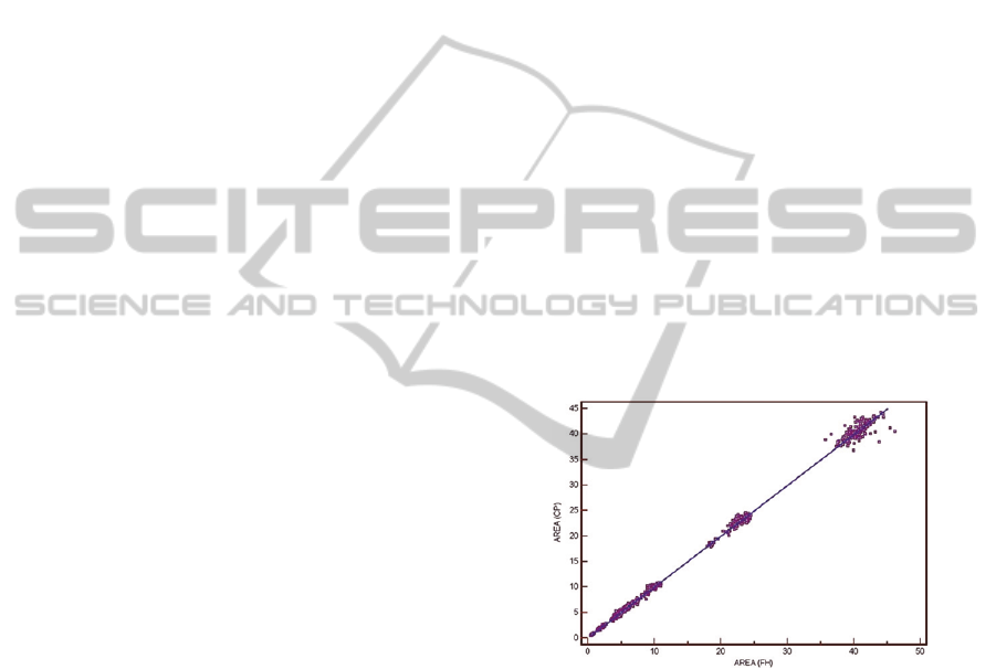
see in figure 1.
Figure 1: Correlation of area measured with free hand and
closed polygon with line of equality (Van Poucke et al.,
2010).
In figure 1 is possible to view that the wound
areas identified differs in the use of free hand
drawing method and a closed polygon graph
algorithm. So, these methods are acceptable to some
healthcare professionals, but not accepted for others,
because these methods obtain large errors in wound
area measurement.
The other computer-aided method for wound
area measurement consists in the use of tools related
to the image processing, such as Adobe Photoshop,
to open a JPG image, defining manually the border
of wound in image and calculate the pixel value of
the region selected as wound and record this to a
Microsoft Excel sheet (Li et al., 2012). This process
BIODEVICES2015-InternationalConferenceonBiomedicalElectronicsandDevices
276

is repeated and some independent research authors
statistically measure the values of wound area based
on the values in the sheet. The traditional
transparency-based wound healing assessment is an
efficient method and clinically accepted by
healthcare professionals (Li et al., 2012). This
method uses a transparency film for marking out the
margins of individual wounds. The outlines regions
as well as the suitable standard area control(s) were
cut off along the margin with an electronic cutter
and weighed by an analytical balance and the
weights of transparency pieces were converted to the
areas one by one by dividing the weight of the
checked region (Y) with the weight of standard unit
(X) and then times the known area of the standard
unit (Z), applying, in the end, statistical corrections
for the obtain a better wound area measurement (Li
et al., 2012).
The chronic wounds have very irregular
dimensions and various methods created aren’t very
accurate. But, for chronic wounds, it is important to
control the wound healing. Thus, a reasonable
approach to determining wound size during a brief
patient encounter would be to document the wound’s
linear measurement – that is, perpendicular linear
dimensions (D, 2006). Normally, in the wound’s
linear measurement, the researchers measure a
wound as a shape, such as a rectangle or an ellipse.
The area of an ellipse is calculated by measuring two
perpendicular diameters, such as maximum diameter
(major diameter) and maximum diameter
perpendicular to the first diameter (minor diameter)
(D, 2006) and this area have an error between 16%
and 40% of the real area (Casas et al., 2011). This
method is simple and relatively cheap, but the
method isn’t precise, because it assumes the wound
area as a simple shape and the wound can have an
irregular form (D, 2006). By other research authors,
this method is called ruler based method (Nemeth et
al., 2010). If the area selected is a rectangle, the area
may be overestimated by 10% to 45% with less
accuracy for smaller wounds (Casas et al., 2011).
An other method consists in the placing of a
transparent film over the wound and tracing the
outline with a permanent marker (Casas et al., 2011,
Nemeth et al., 2010). After this process, the
transparent film is placed on a metric grid and the
area is calculated by counting the number of squared
millimeters contained within the outline (Casas et
al., 2011). This method has a large probability to
have a human error in tracing and the trace is
subjective, depending of the person that does the
trace (Casas et al., 2011). After this, the area can be
measured with a digital photography of the
transparent film with the trace to measure the value
of wound area (Casas et al., 2011). The wound area
can be estimated with a planimeter (Nemeth et al.,
2010).
Other vision-based techniques use either
stereophotogrammetry (SPG) or structured lighting
to obtain wound images (Nemeth et al., 2010). For
stereophotogrammetry, two or more photographs of
the same wound are taken from slightly different
angles and the photographs are used to produce a 3D
model of the wound in a computer (Nemeth et al.,
2010). Then, a computer traces the wound border
and the wound area can be calculated.
Various research authors attempt to create an
algorithm to estimate the wound area using images,
but all methods have influence of external factors,
such as lightning, shadow, and others, in the image
and these cause errors in wound area estimation.
Despite errors, some algorithms are clinical accepted
to help the measurement of wound area.
In the next section, this document will explain
how to use mobile devices, such as smartphones,
tablets and other hand-held devices, for the
estimation of the wound area and the methods,
languages and frameworks, which is possible to use.
4 WOUND AREA ESTIMATION
USING A MOBILE DEVICE
In the last decade, the use of mobile devices has
been increasing, because these equipments
experienced a reduction in price and an increase of
memory and processing capacities (Heggestuen,
2013). Now, one in every 5 people in the world own
a smartphone and one in every 17 people own a
tablet (Heggestuen, 2013). The two platforms
responsible for the largest market share are Android
operating system (owned by Google) and iOS
operating system (owned by Apple) (Bosomworth,
2013). Smartphones usually integrate various
sensors to perform tasks that are related to the use of
the phone in a telecommunications or multimedia-
browsing context, such as camera, accelerometer,
proximity sensor and others. These equipments
allow the user to do complex tasks in movement
without dependency of a desktop computer, because
these equipments can connect to the Internet to send
data for processing tasks that needs a server for
remote processing, storing into a remote database
and visualization of remote data processed.
These devices have a lot of applications available
in the online application stores, to manage and
WoundAreaAssessmentusingMobileApplication
277
processing data and do other tasks. Applications
related to the wound area measurement are low,
because it is very difficult to measure the wound
area and various research don’t have a consensus
about the wound area measurement techniques.
Generally, these equipments have a capacity to take
photographic images of a wound with the embedded
camera in the smartphone. Recently, various
research authors saw the benefits of the use of
smartphones for wound area measurement and a lot
of research was done in this topic. The two mobile
applications available in application online stores
about wound area measurement are MOWA (Mobile
Wound Analyzer) (Healthpath, 2011) and
WoundRight (Technologies, 2013).
MOWA is a non-invasive software that makes
use of a camera of a smartphone to allow the user to
take photos or upload photos of the wound to
analyze, differentiating the types of tissues (necrotic
tissue, fibrinous tissue and granulation tissue) and
calculating the wound area and indicating the
treatment. This application does some tasks
automatically and other tasks are manual (needs
human interaction). The manual tasks are taking a
photo, designing the mask, setting parameters and
sending a JPG and PDF file via e-mail. The
automatic tasks are analyzing tissues, calculating the
wound area, defining directions/suggestions in
treatment and creating an analysis report in PDF.
MOWA is registered as a medical device, is fast
(analysis process takes less than 3 minutes), is easy
to use, improves ulcer therapy, improves the quality
of assistance, helps to identify and measures the
ulcer tissues, defines the priorities in treatment,
suggests the therapeutic treatment, improves the
follow up of the cure, automates the clinical
documentation, improves the communication and
collaboration, helps medical/nurse training and
education, facilitates sector study investigations,
eases effectiveness monitoring of new products,
reduces the use of ineffective products, reduces care
times, reduces hospitalization time, supports home
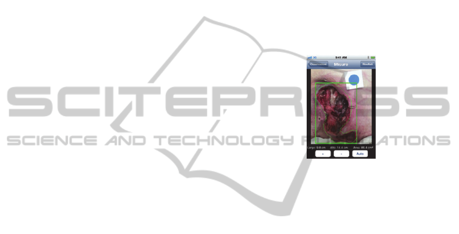
care assistance and supports telemedicine. Figure 2
shows the screen of application, calculating the
wound area of a digital image uploaded to the
application and using the automatic method of
measurement. MOWA application is only for iPhone
and it is paid.
WoundRight is a software application that offers
advanced wound, ostomy, and continence
documentation with the ability to add individual
treatments, performs detailed assessments and brings
convenient and immediate wound care to the patient.
This application takes the tablet anywhere and
collects your data with or without the Internet,
performs powerful, accurate and consistent
assessments for wounds, generated progress, area
and dimension charts, shares patients data securely
with their affiliated accounts, tracks vitals, medical
conditions, and report on open or closed wounds,
calculates and analyzes wound data to drive better
care and better results, reduces redundancy, keeps
compliant, improves revenues and decrease wound
care costs by lowering re-hospitalization rates and
increasing referral rates and decreases maintenance
costs. WoundRight application is only for tablets, is
supported for Android and iOS operating systems
and it is free.
Figure 2: Screen of MOWA application calculating the
wound area (Healthpath, 2011).
Many other research studies exist about the
wound area measurement related to mobile devices.
These authors define algorithms implemented in
various programming languages and use various
frameworks. The Android development should be
done in Java programming language and in this
programming language exists a lot of studies,
frameworks and libraries available for wound image
processing to measure the wound area. The iOS
development should be done in Objective-C
programming language and in this programming
language exists a minor number of studies,
frameworks and libraries available for wound image
processing to measure the wound area.
The study presented in (Hettiarachchi et al.,
2013) consists in an application, implemented for
the Android operating system, to provide a practical
fast and non-invasive technique to monitor the
wound healing process. The process starts with the
pre-processing of an image captured with camera of
the mobile device. In pre-processing phase, the
active contour is predefined and centered on the
wound, cropping is utilized for centering the wound.
In this phase, the unnecessary artifacts such as
clothing, limb borders and backgrounds are removed
and users demarcate the crop section by marking the
diagonal points of a rectangle on the device’s touch
BIODEVICES2015-InternationalConferenceonBiomedicalElectronicsandDevices
278

screen. Next, all images are resized to 500 pixels,
but maintain the original ratio. Finally, in this phase,
the saturation plane of the HSV color model is
extracted as the base for the active contour algorithm
since it displayed the best contrast between infected
and normal skin and the image is further smoothed
using a Gaussian filter of 31x31 dimensions with
σ=5 in order to remove artifacts that may have
otherwise attracted the active contour erroneously.
The next phase corresponds to the segmentation of
the image. The segmentation is based on active
contour models, which identifies the wound border
irrespective of coloration and shape, and the user,
providing higher control and accuracy, can modify
the segmentation. Finally, the wound area
measurement is further normalized to remove effects
of camera distance and angle and the area
corresponds to the area between the contours. The
results obtained in this application have an accuracy
level of 90%.
The mobile applications for estimating the
wound area have a lot of research that explain the
benefits of these applications (Perera and
Chakrabarti, 2013; Friesen et al., 2013; Casas et al.,
2011). About the method analyzed in the section 3,
ruler measures and transparency tracings were used
by authors of (Nemeth et al., 2010) for measuring
wound size often have low accuracy and reliability
and they created a new method used in a wound
measurement device (WMD) designed by them. The
device designed in (Nemeth et al., 2010) allows the
wound area measurement with to assess for accuracy
over distance from the wound surface, as well as
camera angle skew, using the inter-rater and a intra-
rater methods. Both intra-rater and inter-rater
reliability proved to be significantly higher than
conventional methods, such as ruler measures and
transparency tracings and these methods have an
average 2.65% error rate in the accuracy
measurement based on two black and white shapes
with known areas. The two methods that used a
rectangular approximation of area had positive bias
meaning that they typically over-estimated the area,
whereas the two techniques that traced the borders
under-estimated the areas (Nemeth et al., 2010).
Usually, the process of wound area measurement
using a mobile device consists in three main phases,
these are (Andrade et al., 1999, Gonzalez and
Woods, 1992; Zaffari, 2006): image pre-processing,
image segmentation and wound area measurement.
In the image pre-processing, to obtain better results
of the contours, a filter for remove the noise is
applied, such as the Gaussian Blur. After applying
the filter, the adaptive threshold should be done to
convert the image to black and white colors. Next,
the dilate operation is applied to fill the cracks. After
these three tasks, closed contours can be achieved.
For wound area measurement exists a framework,
developed for all platforms in the market to do the
image processing with easy methods. This
framework is named OpenCV (Bradski and Kaehler,
2008; Marengoni and Stringhini, 2009) and it allows
to do the tasks referring to pre-processing image
phase. In the image segmentation phase, this
framework has methods to identify the contours of a
wound, using various algorithms, such as Canny
Edge Detection or others. And for the end phase, this
framework has a method to calculate the area
between the contours. So, the use of OpenCV
framework is easy and facilitates the tasks for
wound area measurement. This is compatible with
Android applications (developed in Java) and iOS
applications (developed in Objective-C). Other
frameworks are developed in the Java programming
language (Cuautle, 2007; Prodan et al., 2006) and
can be adapted to mobile applications for Android
operating system.
Most of all methods researched for desktop
applications can be adapted to mobile device
applications (Friesen et al., 2013) and have
possibility to estimate the wound area, processing
the image on the device, after capturing the image
with the embedded camera, or sending to a server,
via Internet, and the image processing will be
performed remotely (Foltynski et al., 2013; Sikka et
al., 2012; Vivanco et al., 2011). The user can see the
results on the device, such as AreaMe (Foltynski et
al., 2013), that the results was compared with the
results of Visitrak and SilhouetteMobile (Wannous
et al., 2011) systems.
The use of mobile technology increases the ease
and accuracy in monitoring a wound treatment. This
is especially important in chronic wounds, because
they require greater control and adaptation of
treatment. This is a topic that is in constant
investigation and evolution to improve the existent
methods.
At the end of this research, a Java Desktop
application was available at a free repository and the
source code is available for future research studies.
5 CONCLUSIONS
In conclusion, the wound area assessment is a good
topic for research, because in the last years there
have been some improvements, but the algorithms
developed in some cases have big errors. The
WoundAreaAssessmentusingMobileApplication
279

existence of a lot of research indicates that this
research in this topic isn’t ended and is a very
interesting research.
The research of the wound area assessment can
be done manually (low precision) or automatically
(in the last years has a evolution in this methods). To
manually process for wound area measure, the
healthcare processional use metrics to measure the
wound area, which commonly is measured as a
geometric form, such as a rectangle. For automatic
process, the healthcare professional uses software
for image processing that identifies a wound based
on color and/or texture of wound images. The
wound image characteristics are stored in a database
for future comparisons. This process is done in three
phases, these are: image pre-processing,
segmentation and wound area measurement.
In the image pre-processing phase is identified
the existence of a wound in the image, by color
histograms or texture comparisons and the image
needs to be applied a low-pass filter, threshold and
dilatation of image to obtain a closed contour. To
verify the existence of a wound in the image a K-NN
or a SVM algorithm is applied for the verification.
In the segmentation phase, the contours of the
wound image are identified. For identify the
contours various methods exist, for example using a
geometric shape, discarding some parts out of the
wound or identify the pixels by the color.
For the wound area measurement phase, the
result obtained is the area between the contours. This
research is very important especially for the area of
chronic wounds, because these wounds need to be
monitored during the healing time, because this area
doesn’t decrease constantly during the healing time.
The use of mobile devices allows the wound area
measurement with better precision, because is good
to identify the camera distance of the wound, so it
allows to estimate the real wound area anywhere in
movement, using a proximity sensor of the mobile
device. For the process to estimate the wound area in
a mobile device, various frameworks exist in various
programming languages for help to the development
of the applications, such as OpenCV framework and
others.
This research is very difficult, because is not
possible to control some environmental variables,
such as lightning, noise and quality of camera, but
some algorithms implemented are clinically
accepted to help the healthcare professionals in
telemedicine. The wound image processing in
mobile device can be done with processing image in
the application or send the image by Internet to a
server and receive the results data in the smartphone.
As future work, this research topic needs to be
continuing improve for these applications can be
used commonly in a hospital to improve the
treatments of the patients. The use of automatic
systems has advantages and disadvantages, but
normally the use of automatic systems is more
precise than manual measurements.
ACKNOWLEDGMENTS
This work was supported by FCT projectPEst-
OE/EEI/L A0008/2013 (Este trabalho foi suportado
pelo projecto FCT PEst-OE/EEI/LA0008/2013).
The authors would also like to acknowledge the
contribution of the COST Action IC1303 –
AAPELE – Architectures, Algorithms and Protocols
for Enhanced Living Environments.
REFERENCES
Acha, B., Serrano, C., Acha, J. I. & Roa, L. M. 2005.
Segmentation And Classification Of Burn Images By
Color And Texture Information. J Biomed Opt, 10,
034014.
Andrade, E. Processamento Digital De Imagens - Pdi.
Centro De Estudos – FundaçÃo São Lucas.
Aslantas, V. & Tunckanat, M. 2007. Differential
Evolution Algorithm For Segmentation Of Wound
Images. 1-5.
Bosomworth, D. 2013. Mobile Marketing Statistics 2013
[Online]. Available:
Http://Www.Smartinsights.Com/Mobile-
Marketing/Mobile-Marketing-Analytics/Mobile-
Marketing-Statistics/ [Accessed January 12th 2014].
Bradski, G. & Kaehler, A. 2008. Learning Opencv, United
States Of America, O'reilly.
Casas, L., Castaneda, B. & Treuillet, S. 2011. Imaging
Technologies Applied To Chronic Wounds: A Survey.
4th International Symposium On Applied Sciences In
Biomedical And Communication Technologies.
Barcelona, Spain.
Cuautle, N. F. 2007. System For Face Detection On A
Mobile Phone Using Java Technology. Master Thesis,
University Of Oslo.
D, L. 2006. Design And Implementation For Wound
Measurement Application. Primary Intention 2006,
14, 56-58, 60-63, 66.
Filho, O. M. & Neto, H. V. 1999. Processamento Digital
De Imagens, Rio De Janeiro, Brasport.
Foltynski, P., Ladyzynski, P. & Wojcicki, J. M. 2013. A
New Smartphone-Based Method For Wound Area
Measurement. Artif Organs.
Friesen, M. R., Hamel, C. & Mcleod, R. D. 2013. A
Mhealth Application For Chronic Wound Care:
Findings Of A User Trial. Int J Environ Res Public
BIODEVICES2015-InternationalConferenceonBiomedicalElectronicsandDevices
280

Health, 10, 6199-214.
Gallagher, B. A. 2012. Wound Bed Assessment Using
Calibrated Images And Representation In Openehr.
Master Of Science In Health Informatics, University
Of Dublin.
Gammal, S. & Popp, R. 1995. A Color Image Analysis
System (Cd-Cwa) To Quantify Wound Healing Of
Ulcers. Skin Resear Techn, 1.
Giger, M. L., Wannous, H., Lucas, Y., Treuillet, S. &
Karssemeijer, N. 2008. Efficient Svm Classifier Based
On Color And Texture Region Features For Wound
Tissue Images. 6915, 69152t-69152t-10.
Gonzalez, R. C. & Woods, R. E. 1992. Processamento De
Imagens Digitais, Brasil, Editora Edgar Bucher Ldta.
H, P., M, M., J, T., P, P., S, S. & Ji, S. 2009. Assessment
Of Wound Healing: Validity, Reliability And
Sensitivity Of Available Instruments. Wound Practice
And Research, 17, 208-217.
Healthpath. 2011. Mowa - Mobile Wound Analyzer For
Iphone And Ipad - Healthpath [Online]. Available:
Http://Www.Healthpath.It/Imowa.Html [Accessed
January 12th 2014].
Heggestuen, J. 2013. One In Every 5 People In The World
Own A Smartphone, One In Every 17 Own A Tablet
[Chart] [Online]. Available:
Http://Www.Businessinsider.Com/Smartphone-And-
Tablet-Penetration-2013-10 [Accessed January 12th
2014].
Hettiarachchi, N. D. J., Mahindaratne, R. B. H., Mendis,
G. D. C., Nanayakkara, H. T. & Nanayakkara, N. D.
2013. Mobile Based Wound Measurement. 2013 Ieee
Point-Of-Care Healthcare Technologies (Pht), 298-
301.
Kim, H. M., Lowery, J. C., Hamill, J. B. & Wilkins, E. G.
2003. Accuracy Of A Web-Based System For
Monitoring Chronic Wounds. Telemed J E Health, 9,
129-40.
Kolesnik, M. & Fexa, A. 2004. Segmentation Of Wounds
In The Combined Color-Texture Feature Space.
Medical Imaging 2004: Image Processing, Pts 1-3,
5370, 549-556.
Kolesnik, M. & Fexa, A. 2005. Multi-Dimensional Color
Histograms For Segmentation Of Wounds In Images.
Image Analysis And Recognition, 3656, 1014-1022.
Kolesnik, M. & Fexa, A. How Robust Is The Svm Wound
Segmentation? In: Ieee, Ed. Signal Processing
Symposium, 2006 Rejkjavik. Ieee, 50-53.
Krouskop, T. A., Baker, R. & Wilson, M. S. 2002. A
Noncontact Wound Measurement System. J Rehabil
Res Dev, 39, 337-45.
Kumar, K. S., Asokan, L., M.P, P. & Reddy, B. E. 2013.
Assessment Of The Wound-Healing Process By
Accurate Single View Issue Classification And Depth
Estimation For Telemedicine. International Journal
Of Engineering And Advanced Technology (Ijeat), 2,
467-475.
Lazarus, G. S., Cooper, D. M., Knighton, D. R., Margolis,
D. J., Pecoraro, R. E., Rodeheaver, G. & Robson, M.
C. 1994. Definitions And Guidelines For Assessment
Of Wounds And Evaluation Of Healing. Archives Of
Dermatology, 130, 489-493.
Li, P. N., Li, H., Wu, M. L., Wang, S. Y., Kong, Q. Y.,
Zhang, Z., Sun, Y., Liu, J. & Lv, D. C. 2012. A Cost-
Effective Transparency-Based Digital Imaging For
Efficient And Accurate Wound Area Measurement.
Plos One, 7, E38069.
Loizou, C. P., Kasparis, T. & Polyviou, M. 2013.
Evaluation Of Wound Healing Process Based On
Texture Image Analysis. Journal Of Biomedical
Graphics And Computing, 3.
Marengoni, M. C. & Stringhini, D. 2009. Tutorial:
IntroduçÃo À Visão Computacional Usando Opencv.
Rita, Xvi.
Mayrovitz, H. N. & Soontupe, L. B. 2009. Wound Areas
By Computerized Planimetry Of Digital Images:
Accuracy And Reliability. Adv Skin Wound Care, 22,
222-9.
Mekkes, J. R. & Westerhof, W. 1995. Image Processing In
The Study Of Wound Healing. Clinics In
Dermatology, 13, 401-407.
Navas, M., Luque-Baena, R. M., Morente, L., Coronado,
D., Rodriguez, R. & Veredas, F. J. 2013. Computer-
Aided Diagnosis In Wound Images With Neural
Networks. Advances In Computational Intelligence, Pt
Ii, 7903, 439-448.
Nayak, R., Kumar, P. & Galigekere, R. R. 2009. Towards
A Comprehensive Assessment Of Wound-
Composition Using Color-Image Processing. 2009
16th Ieee International Conference On Image
Processing, Vols 1-6, 4133-4136.
Nemeth, M., Sprigle, S. & Gajjala, A. 2010. Clinical
Usability Of A Wound Measurement Device. 36th
Annual American Spinal Injury Conference. Nashville.
Ousey, K. & Cook, L. 2011. Understanding The
Importance Of Holistic Wound Assessment. Practice
Nursing, 22, 308-314.
Papazoglou, E. S., Zubkov, L., Mao, X., Neidrauer, M.,
Rannou, N. & Weingarten, M. S. 2010. Image
Analysis Of Chronic Wounds For Determining The
Surface Area. Wound Repair Regen,
18, 349-58.
Perera, C. & Chakrabarti, R. 2013. The Utility Of Mhealth
In Medical Imaging. Journal Of Mobile Technology In
Medicine, 2, 4-6.
Plassmann, P. & Jones, T. D. 1998. Mavis: A Non-
Invasive Instrument To Measure Area And Volume Of
Wounds. Measurement Of Area And Volume
Instrument System. Med Eng Phys, 20, 332-8.
Prodan, A., Campean, R., Revnic, C. & Prodan, R. 2009.
Strategies For Wound Image Understanding. 2009
Ieee/Acs International Conference On Computer
Systems And Applications, Vols 1 And 2, 1018-1024.
Prodan, A., Campean, R., Rusu, M., Revnic, C., Mitrea, P.
& Prodan, R. 2010a. Toward A Model For Wound
Healing Simulation. 115-120.
Prodan, A., Rusu, M., Campean, R. & Prodan, R. 2006. A
Java Framework For Analysing And Processing
Wound Images For Medical Education. 20th European
Conference On Modelling And Simulation Ecms 2006,
421-426.
Prodan, A., Rusu, M., Revnic, C., Campean, R., Mitrea, P.
WoundAreaAssessmentusingMobileApplication
281

& Prodan, R. 2010b. Intelligent E-Tools For Wound
Image Understanding And Evaluation. 8-13.
Russell, L. 1999. The Importance Of Wound
Documentation And Classification. Br J Nurs, 8,
1342-3, 1346, 1348 Passim.
Sikka, N., Carlin, K. N., Pines, J., Pirri, M., Strauss, R. &
Rahimi, F. 2012. The Use Of Mobile Phones For
Acute Wound Care: Attitudes And Opinions Of
Emergency Department Patients. J Health Commun,
17 Suppl 1, 37-42; Quiz 42-3.
Song, B. 2012. An Automated Wound Identification
System Based On Image Segmentation And Artificial
Neural Networks. Master Of Science In Biomedical
Engineering, Faculty Of Drexel University.
Song, B. & Sacan, A. 2012. Automated Wound
Identification System Based On Image Segmentation
And Artificial Neural Networks. 1-4.
Technologies, W. 2013. Woundright - Mobile Wound Care
Software [Online]. Woundright Technologies.
Available:
Http://Www.Woundrightapp.Com/Woundright.Html
[Accessed January 12th 2014].
Van Poucke, S., Nelissen, R., Jorens, P. & Vander
Haeghen, Y. 2010. Comparative Analysis Of Two
Methods For Wound Bed Area Measurement. Int
Wound J, 7, 366-77.
Vivanco, J., Neighbour, R., Hamel, C., Mukhi, S., Friesen,
M. R. & Mcleod, R. D. 2011. A Smartphone
Application For Remote Wound Treatment And
Documentation Compliance. In: Ieee (Ed.) Resna.
Toronto, Canada: Ieee.
Wannous, H., Lucas, Y. & Treuillet, S. 2011. Enhanced
Assessment Of The Wound-Healing Process By
Accurate Multiview Tissue Classification. Ieee Trans
Med Imaging, 30, 315-26.
Wannous, H., Lucas, Y., Treuillet, S. & Albouy, B. 2008.
Mapping Classification Results On 3d Model: A
Solution For Measuring The Real Areas Covered By
Skin Wound Tissues. 2008 3rd International
Conference On Information And Communication
Technologies: From Theory To Applications, Vols 1-5,
930-935.
Wannous, H., Treuillet, S. & Lucas, Y. 2007. Supervised
Tissue Classification From Color Images For A
Complete Wound Assessment Tool. Conf Proc Ieee
Eng Med Biol Soc, 2007, 6032-5.
Wannous, H., Treuillet, S. & Lucas, Y. 2010. Robust
Tissue Classification For Reproducible Wound
Assessment In Telemedicine Environments. Journal
Of Electronic Imaging, 19.
Wild, T., Prinz, M., Fortner, N., Krois, W., Sahora, K.,
Stremitzer, S. & Hoelzenbein, T. 2008. Digital
Measurement And Analysis Of Wounds Based On
Colour Segmentation. European Surgery, 40, 5-10.
Zaffari, C. A. 2006. Visualização E Processamento
Digital De Imagens Médicas. Master Of Science,
Universidade Católica Do Rio Grande Do Sul.
BIODEVICES2015-InternationalConferenceonBiomedicalElectronicsandDevices
282
