
Vertebral Metrics
Automation of a Non-invasive Instrument to Analyse the Spine
A. Gabriel
1,2
, C. Quaresma
2,3
and P. Vieira
1,2
1
Center of Atomic Physics, Faculty of Sciences and Technology New University of Lisbon, Caparica, Portugal
2
Department of Physics, Faculty of Sciences and Technology New University of Lisbon, Caparica, Portugal
3
CEFITEC, Faculty of Sciences and Technology New University of Lisbon, Caparica, Portugal
Keywords: Spinal Column, Biomechanics, Non-invasive, Medical Device, Stereo Vision.
Abstract: Back pain is a major health problem in modern society. It is known that the main responsible for this
symptom are the biomechanical changes in the spinal column. Thus, it is important to develop an instrument
that evaluates in a global way the spinal column in a standing position. For that purpose, a complete
innovative and non-invasive system – the Vertebral Metrics – has been built. The Vertebral Metrics is a
semi-automatic equipment designed to identify the spatial position of each spinal process from the first
cervical to the first sacral vertebra. Using a camera and a laser diode the recognition is achieved with
software capable of distinguishing prominent blue marks that identifies each spinous process. After the
validation process it was concluded that the Vertebral Metrics is a reliable and valid instrument. However,
the time required for a completely scan is still too long for practical use. Thus, we are presently working on
the automation of this medical device by developing a new prototype. The process will comprise several
modifications in the equipment as the introduction of fluorescent markers, UV lights and two video
cameras. The identification of the vertebras will be performed by the stereo vision method. The software
must be adapted to the new assembly.
1 INTRODUCTION
Back pain has affected humans throughout out
recorded history and it is well documented to be a
major health problem in modern society (Alexandre,
2001, Dankaerts, 2006). It is the leading cause of
activity limitation and work absence (Dionne, 2006)
so it has a significant impact on individuals,
families, communities, governments and business
(Hoy, 2010). In fact, the incidence of rachialgia is so
high that it must be studied as if it were an epidemic
and social disease (Knoplich, 2003; Galukande,
2005). Most researchers suggest that about 80% of
the individuals experience back pain at some point
of their life and, from those, 80 to 90% of the pain is
caused by mechanical changes in the spine (Najm,
2003; Quaresma, 2010).
Currently, many options are available to evaluate the
spine. There are several types of diagnostic imaging
technologies to assist in identifying the anatomical
changes, responsible by back pain. However, very
few of them are ionising radiation free and do not
allow the analysis of the spinal column in a vertical
standing position. Furthermore they tend to evaluate
only parts of this osseous structure (Secca, 2008).
The radiological studies, particularly X-rays, are the
most widely used methods for assessing the spinal
column curvatures in a global way, however, since it
uses ionizing radiation, it is not possible to use them
in general population (Harlick, 2007; Pinel-Giroux,
2006). X-rays are associated with 0.6 – 3.2% of
cumulative risk of cancer to age 75 years old
(Gonzales, 2004) so its use must be reduced to a
minimum.
Non-invasive equipment for evaluation of the spine
is commercially available, but often only perform
partial scans of the spine or are extremely expensive
(Hinman, 2004; Quaresma, 2013). Thus, the
development of non-invasive methods to evaluate
the spine in a standing position in a global way is
needed to attain a better insight into back disorders
(Quaresma, 2009).
For that purpose, the Vertebral Metrics was built. It
is a non-invasive system which is able to identify the
X, Y and Z position of each vertebra, from the first
cervical to the first sacral vertebra. The mechanical
equipment was originally planned and built to be
applied to pregnant women (Quaresma, 2009). After
150
Gabriel A., Quaresma C. and Vieira P..
Vertebral Metrics - Automation of a Non-invasive Instrument to Analyse the Spine.
DOI: 10.5220/0005276101500155
In Proceedings of the International Conference on Biomedical Electronics and Devices (BIODEVICES-2015), pages 150-155
ISBN: 978-989-758-071-0
Copyright
c
2015 SCITEPRESS (Science and Technology Publications, Lda.)
an automation process, a semi-automatic prototype
was developed. This prototype can be applied in
general population. Its setup uses a camera and a
laser diode to measure the spatial coordinates of
each spinal process. Using an adequate blue marker
to identify the spinal processes, the recognition is
achieved with software capable of distinguish the
prominent blue marks in the skin. However, the time
required for data acquisition is about three minutes.
Artefacts in measurements are affecting the
performance of the system because individuals can
not stand still for such a long period of time. This
constraint limits the practical use of the equipment.
For that reason, the further development of the
Vertebral Metrics has become necessary. The aim of
this paper is to present a third improved prototype
that is being developed. This system will have
several differences comparing to the previous
prototypes. With the new equipment tests will be
faster with an improved resolution.
2 AUTOMATION OF THE
VERTEBRAL METRICS
The main purposes of the automation of the
Vertebral Metrics are the improvement of the
resolution and time required for data acquisition.
For a better understanding of the instrument that
is under study the axis system considered will be
defined as following: the transversal distance as X
coordinate, the antero-posterior distance as the Y
coordinate and the height as Z coordinate.
Several modifications will be performed
comparing to the existing prototype. The equipment
that is being projected will move along the Z
direction only. It has been designed to have two
video cameras and two UV lights that will be fixed
in specific positions. A fluorescent dye will be used
to identify the projection of the spinal processes.
This dye emits a specific wavelength when exposed
to the ultra-violet radiation. Also the triangulation
method, that uses a RGB camera and a laser diode,
will be replaced by the stereo vision method. The
software has to be adapted to perform scans with the
new model.
The characteristics of the automated system will
be described in the following topics.
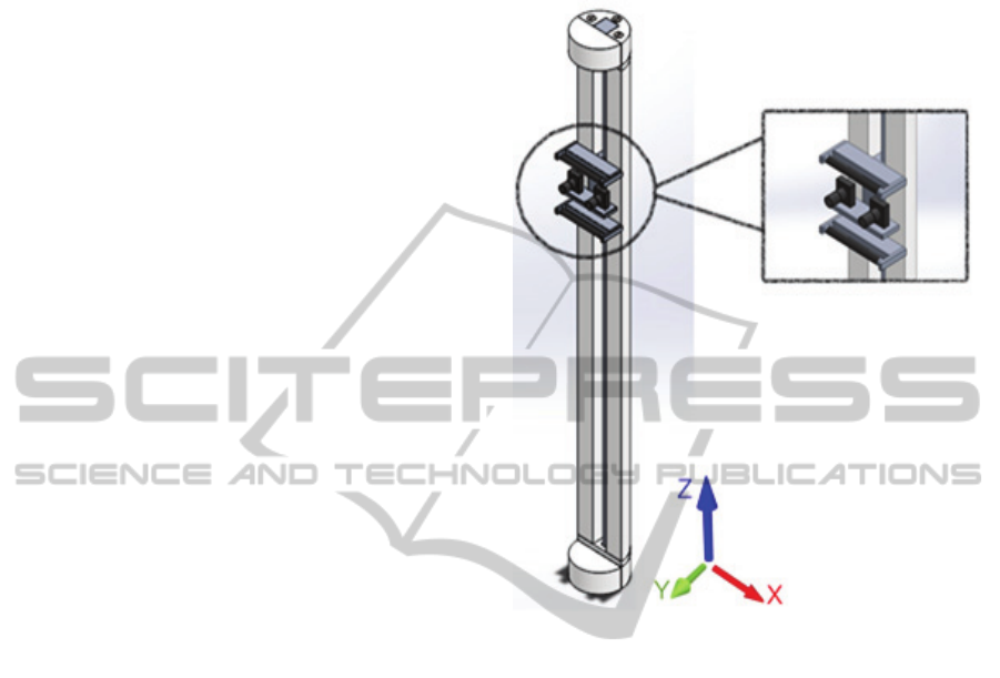
2.1 Mechanical System
The design of the mechanical system has been
already projected using SolidWorks™ (Figure 1). It
is a vertical structure which has a bracket coupled.
This bracket consists of two identical video cameras
positioned between two ultraviolet lights.
Figure 1: Scheme of the mechanical system.
There are two main goals for the mechanical
structure: resolution and speed. The instrument is
being constructed based on the following pre-
requisites:
- The mechanical apparatus should move all the
hardware necessary for the image acquisition in the
Z direction (up and down).
- Must move 1000 mm in 30 seconds (33.3
mm/s).
- Must have a communication protocol
controlled by software.
For the vertical positioner a linear stage will be
used. It has a high helix pitch lead screw providing
high-speed positioner. The motor will be step-by-
step type for precision positioning. As in the
previous prototype, the motion and control of the
motor will be performed by a microcontroller
PIC16F877 from Microchip™. In addition, the
microcontroller will also monitor limit switches,
control serial RS232 communications and control
lights relays.
In terms of RS232 communication a protocol
will be developed based on instructions and replies.
VertebralMetrics-AutomationofaNon-invasiveInstrumenttoAnalysetheSpine
151
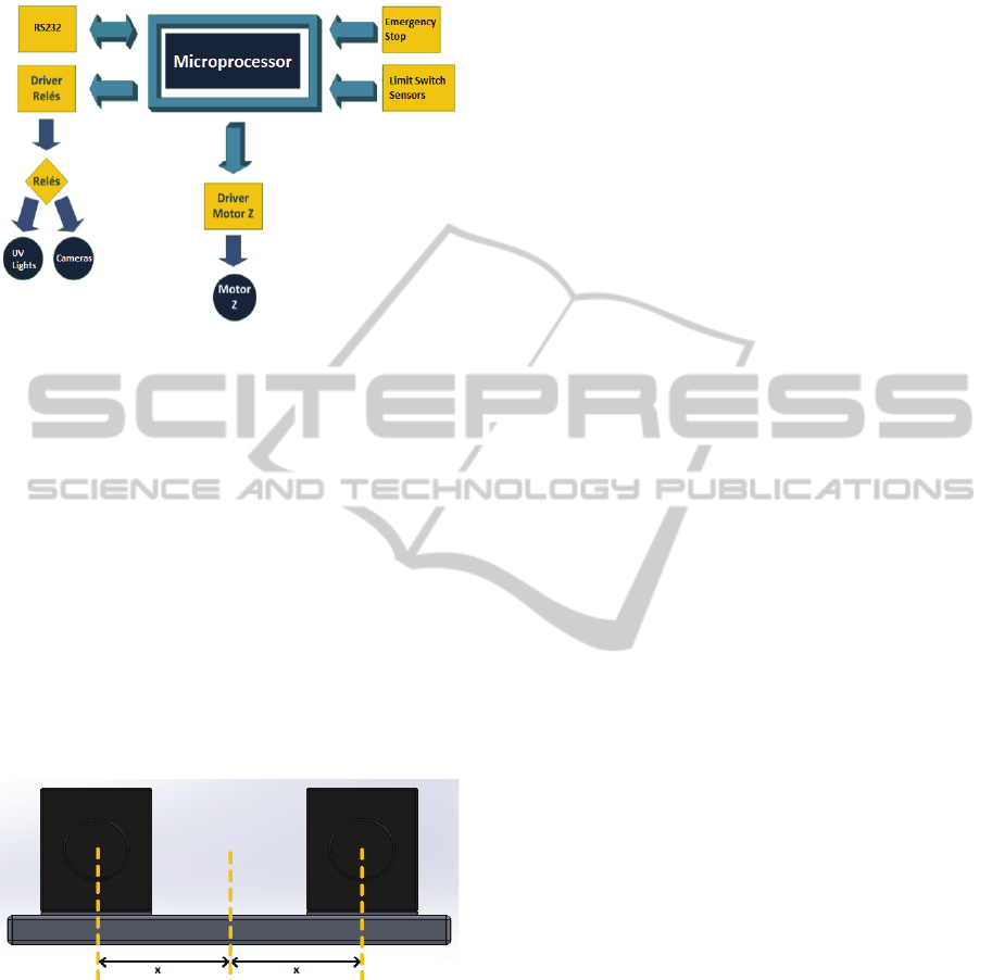
Therefore, the microprocessor will only answer and
execute commands from the computer.
Figure 2: Scheme of the communication system.
2.2 Determination of Distances Using a
Stereo Vision System
Stereo vision is a method that provides 3D
perception by means of two different images of the
same scene. To develop a stereo vision system some
specific elements are required. The geometry of the
system is also a relevant concern.
In order to engineer a stereo vision system, two
video cameras will be fixed side by side in a
horizontal support with parallel optical axes. Both
cameras will be equidistant from the middle of the
support (Figure 3). This distance has to be studied in
order to maximize the performance of the
equipment.
Figure 3: Scheme of the stereo vision system (x0z plane).
The system must have at least 1 mm of resolution
in all directions (X, Y and Z). To achieve the desired
resolution several calculations that involve
trigonometric equations have been studied in detail.
It was concluded that any camera available in the
market would be sufficient to identify the X and Z
coordinates. However, powerful cameras are
required to assess the Y coordinate. The selected
cameras are from IDS™, model UI-3480ML. They
are monochromatic cameras with 2560x1920
resolution. A lens with 12 mm of focal distance
(Optica Goyo from IDS™) will be coupled to each
camera.
Software must be developed in order to determine
the spatial position of each spinal process through
the stereo vision method. Different approaches of
the image processing algorithms are still being
studied at this stage of the project.
2.3 Detection of the Spinal Processes
Two programming softwares - Matlab™ and Visual
Studio™ - will be used to develop all software
including the algorithms to perform the detection of
the spinal processes. The image processing
algorithms will be implemented in Matlab™
(MATrix LABoratory, a numeric computer
environment for programming). Visual Studio™ will
be used to define functions for communication with
the mechanical equipment as well as the user
interface.
Selecting an appropriate marker to identify the
patients’ skin above the vertex of each the spinal
process is the first step required to the development
of the image processing algorithms. The marker will
be a fluorescent dye. When excited by ultraviolet
light this dye emits a well-known wavelength that
must be different from the wavelength of the
ultraviolet light. Presently, several spectroscopic
tests are being performed with multiple dyes in order
to choose the most adequate (Figure 4). It is
noteworthy that the dyes under study are suitable for
skin.
Only wavelengths emitted by the marker will be
detected by the cameras. The emission spectrum of
the ultraviolet lights (Figure 4c) has a clear peak at
400 nm so the wavelength of the emission peak of
the adequate markers must be quite different. By
observing all the emission spectrums it was
concluded that the dyes iv and v might be adequate
markers (Figure 4g and Figure 4h, respectively)
because they emit between 625 nm and 675 nm.
Spectral tests are still being performed with those
two dyes in order to achieve the most appropriate.
Optical filters must be positioned in front of each
camera in order to prevent other wavelengths. As the
emission wavelength of the iv and v dyes comprises
the red region of the visible spectrum, the red
component of the RGB image was used to simulate
the filter (Figure 4b). Both dyes are completely
distinguishable from the skin, therefore, the
positioning of the filters in front of the cameras may
simplify dramatically the definition of the
algorithms.
BIODEVICES2015-InternationalConferenceonBiomedicalElectronicsandDevices
152
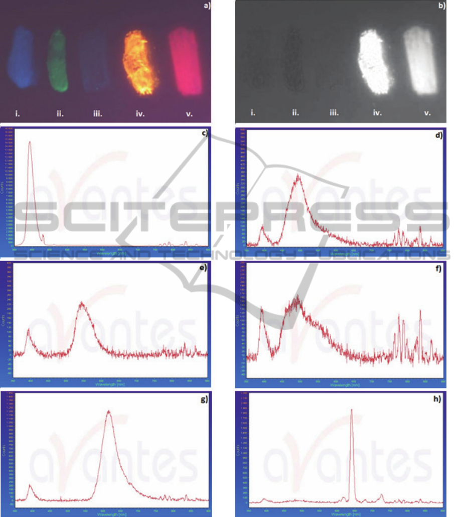
Figure 4: Study of fluorescent dyes. a) RGB picture of the fluorescent dyes in skin; b) Red component of the previous
image; c) Emission spectrum of the UV lights; d) Emission spectrum of the first dye (i); e) Emission spectrum of the second
dye (ii); f) Emission spectrum of the third dye (iii); g) Emission spectrum of the fourth dye (iv); h) Emission spectrum of
the fifth dye (v).
While the vertebral metrics equipment is operating
the data acquisition process will be simple. In the
beginning of each scan the physician has to move
the mechanical system until the first sacral vertebra
is seen by the cameras. An intuitive interface is
being developed to assist the positioning. After the
physician orders the mechanical system begins its
upward movement. The software has a cyclical
VertebralMetrics-AutomationofaNon-invasiveInstrumenttoAnalysetheSpine
153

behaviour wherein the cameras acquire a pair of
images simultaneously every 20 ms. Each pair of
images is saved in memory and analysed by the
image processing algorithms when the scan is
completed. It is noteworthy that data acquisition will
be achieved in such a short period of time – about 30
seconds - because the image processing will be
performed after a full scan of the spine.
The start position of the mechanical system, its
vertical speed and the time between the acquisition
of each pair of images as well as the stereo vision
method are essential to determine the spatial position
of the vertebras. The physician has the possibility of
save data acquired in a single file. By running this
file in a specific software the 3D reconstruction of
the spinal column can be observed. An example that
uses data acquired with the second prototype is
presented in Figure 5.
Figure 5: 3D reconstruction of the spinal column.
3 CONCLUSIONS
The present study will contribute to the automation
of a non-invasive equipment that is a complete
innovation in health – the Vertebral Metrics.
Throughout this article, the characteristics of the
new prototype of the Vertebral Metrics were
described. At this moment a few steps were already
taken: the scheme of the new prototype was drawn,
the characteristics of the cameras were determined
through trigonometric equations and studies with
fluorescent dyes are being performed. The future of
the project will comprise the implementation of
software, image processing algorithms and
instrumentation. Furthermore, the equipment must
be validated. Tests in people with biomechanical
changes of the spine will be performed with
Vertebral Metrics and compared with X-ray (the
gold standard) in order to prove that it is reliable
system.
The differences between the previous prototypes
and the system that is being built will be particularly
relevant regarding the acquisition time required to
collect the individuals data. The stereo vision
method will improve the resolution and the accuracy
of the equipment. As data acquisition will be
performed faster it will also allow the analysis of the
postural adjustments. However, the patient’s skin
above the vertex of the spinal processes still needs to
be marked by the physician.
Due to its non-invasive characteristics the
Vertebral Metrics will be a powerful tool to evaluate
the spinal column. In a few seconds the new
equipment will provide a comprehensive overview
of the spine as it will perform a complete scan in
about 30 seconds. Furthermore, it will became
available a three dimensional analysis of the vertex
of the spinal processes.
This unique and already patented device will
avoid unnecessary invasive methodologies as well as
qualified staff for screening and prevention of spinal
disorders because it will allow repeated scans
without causing damage to the individuals. It may be
used in different contexts such as ambulatory public
and private (healthcare centre / doctor’s office),
hospital and as a potential screening device for back
pain in the general population. Using the Vertebral
Metrics will also became available the definition of
the most appropriate intervention methodologies to
each particular clinical case.
ACKNOWLEDGEMENTS
The authors would like to acknowledge NGNS-
Ingenious Solutions for the support provided.
REFERENCES
Alexandre, N., Moraes, M., 2001. Modelo de avaliação
fisio-funcional da coluna vertebral. In Rev. Latino-am
Enfermagem Março, 9(2): 67-75. RESEARCH GATE.
Dankaerts, W., O’Sullivan, P. B., Burnett, A. F., Straker,
L. M., 2007. The use of a mechanism-based
classification system to evaluate and management of a
patient with non-specific chronic low back pain and
motor control impairment - A case report. In Man Ther
12: 181-19. PUBMED.
Dionne, C. E., Dunn, K. M., Croft, P. R., 2006. Does back
pain prevalence really decrease with increasing age? A
systematic review. In Age Ageing35(3):229-34.
OXFORD JOURNALS.
Galukande, M., Muwazi, S., Mugisa, D. B., 2005.
Aetiology of low back pain in Mulago Hospital,
Uganda. In Afr Health Sci5:164-167. PUBMED.
BIODEVICES2015-InternationalConferenceonBiomedicalElectronicsandDevices
154

Gonzales, A., Darby, S., 2004. Risk of cancer from
diagnostic X-rays: estimates for the UK and 14 other
countries. In Lancet 345-341. PUBMED.
Harlick, J., Milosavlsejevic, S., 2007. Palpation
identification of spinous processes in the lumbar spine.
Man Ther. 12: 56-62. PUBMED.
Hinman, M., 2004. Comparison of Thoracic Kiphosis and
Postural Stiffness in Younger and Older Women. In
Spine Journal, 4: 413-417. PUBMED.
Hoy, D., Brooks, P., Blyth, F., Buchbinder, R., 2010. The
epidemiology of low back pain. In Best Practice &
Research Clinical Rheumatology 24:769-781.
SCIENCE DIRECT.
Jordão, A., Duque, P., Quaresma, C., Vieira, P., 2011.
Development of Vertebral Metrics – An instrument to
study the vertebral column. In Biodevices 2011 –
Proceedings of the International Conference on
Biomedical Electronics and Device: 224-229.
SCITEPRESS.
Knoplich, J., 2003. Enfermidades da coluna vertebral,
Robe Editorial. São Paulo. 3
rd
edition.
Najm, W., Seffinger, M., Mishra, S., Dickerson, V.,
Adams, A., Reinsch, S., Murphy, L., Goodman, A.,
2003.Content validity of manual spinal palpatory
exams – A systematic review. In BMC
Complementary Altern Med 3:1. SPRINGER LINK.
Pinel-Giroux, F., Mac-Thiong, J., Guise, J., Labelle, H.,
2006.Computorized assessment of sagittal curvatures
of spine – Comparison between Cobb and tangent
circles tehniques.In Spinal Disord Tech vol. 19(7):
507-512. PUBMED.
Quaresma, C., João, F., Fonseca, M., Secca, M. F.,
Veloso, A., O’Neil, J. G., Branco, J., 2009. Validation
of Vertebral Metrics: A Mechanical Instrument to
Evaluate Posture of the Spinal Column. In World
Congress on Medical Physics and Biomedical
Engineering 25:711-713. SPRINGER LINK.
Quaresma, C., João, F., Fonseca, M.,Secca, M. F., Veloso,
A., O’Neill, J. G., Branco, J., 2010. Comparative
evaluation of the tridimensional spine position
measured with a new instrument (Vertebral Metrics)
and an optoelectronic system of stereophoto-
grammetry. In Med Biol Eng Comput 48: 1161-1164.
PUBMED.
Quaresma, C., Dias, I., Secca, M. F., O’Neill, J. G.,
Branco, J., 2013. Métrica Vertebral: a Aplicação de
uma Nova Tecnologia na Análise da Coluna Vertebral.
In Acta Med Port 2013 May-Jun 26(3): 226-230.
RESEARCH GATE.
Secca, M. F., Quaresma, C., Santos, F., 2008. A
Mechanical Instrument to Evaluate Posture of the
Spinal Column in Pregnant Women. In IFMBE
Proceedings 22: 1781-1784. SPRINGER LINK.
VertebralMetrics-AutomationofaNon-invasiveInstrumenttoAnalysetheSpine
155
