Machine Learning Techniques and the Existence of Variant Processes
in Humans Declarative Memory
Alex Frid
1
, Hananel Hazan
2
, Ester Koilis
3
, Larry M. Manevitz
3,5
, Maayan Merhav
4
and Gal Star
3
1
Edmond J. Safra Brain Research Center, University of Haifa, Haifa, Israel
2
Network Biology Research Laboratory, Technion, Haifa, Israel
3
Computer Science Department, University of Haifa, Haifa, Israel
4
German Center for Neurodegenerative Diseases (DZNE), Magdeburg, Germany
5
Center of Information and Neural Networks, National Institute of Information and Communications Technology,
Suita, Osaka, Japan
Keywords: Machine Learning, Classification, functional Magnetic Resonance Imaging (fMRI), Feature Selection,
Support Vector Machines, Radial Basis Function Kernel, Declarative Memory, Information Biomarkers.
Abstract: This work uses supervised machine learning methods over fMRI brain scans to establish the existence of
two different encoding procedures for human declarative memory. Declarative knowledge refers to the
memory for facts and events and initially depends on the hippocampus. Recent studies which used patients
with hippocampal lesions and neuroimaging data, suggested the existence of an alternative process to form
declarative memories. This process is triggered by learning mechanism called "Fast Mapping (FM)", as
opposed to the 'standard' "Explicit Encoding (EE)" learning procedure. The present work gives a clear
biomarker on the existence of two distinct encoding procedures as we can accurately predict which of the
processes is being used directly from voxel activity in fMRI scans. The scans are taken during retrieval of
information wherein the tasks are identical regardless of which procedure was used for acquisition and by
that reflect conclusive prediction. This is an identification of a more subtle cognitive task than direct
perceptual cognitive tasks as it requires some encoding and processing in the brain.
1 INTRODUCTION
Human declarative memory is defined as the
conscious information recollection of facts and
events (Squire, 1992). Under the "standard model"
theory for adult declarative memory systems, novel
information is encoded explicitly into the memory
using, amongst other brain parts, the hippocampus
(McClelland et al., 1995). This standard,
hippocampal dependant memory is acquired through
intentional "Explicit Encoding (EE)" procedure. The
encoded information is then slowly transferred from
the hippocampus to the neo-cortex where it becomes
permanently stored (Squire and Alvarez, 1995;
Frankland and Bontempi, 2005). Overtime, the
initially hippocampal dependant memories become
independent of the hippocampus. It has been
suggested that this re-organization process is done
during sleep (Gais et al., 2007).
Amongst toddlers, the process of rapid language
acquisition occurs prior to the full development of
the hippocampus (Bauer, 2008; Uematsu et al.,
2012). Moreover, some evidence from hippocampal
injured subjects demonstrated an ability to acquire
information which seems to have declarative-like
characteristics despite severe damages in the
hippocampus (Sharon et al., 2011; Merhav et al.,
2014) and so must involve a different brain network
than the one engaged by "EE". This alternative
learning mechanism is called "Fast Mapping (FM)".
It is unknown if the memory representations
following FM undergo consolidation processes,
similar to memories gained through EE. However,
since it was shown that patients with hippocampal
damages as well as healthy controls could learn and
store information acquired via FM for a week
(Sharon et al., 2011; Merhav et al., 2014), the
scheme used to explain memory consolidation of
other declarative memories cannot be applied for
FM in a straightforward manner.
It remains somewhat controversial as to whether
the FM is available for acquisition of words among
amnesic patients (Warren and Duff, 2014). Proving
that FM methods are mostly based on brain
114
Frid, A., Hazan, H., Koilis, E., Manevitz, L., Merhav, M. and Star, G..
Machine Learning Techniques and the Existence of Variant Processes in Humans Declarative Memory.
In Proceedings of the 7th International Joint Conference on Computational Intelligence (IJCCI 2015) - Volume 3: NCTA, pages 114-121
ISBN: 978-989-758-157-1
Copyright
c
2015 by SCITEPRESS – Science and Technology Publications, Lda. All rights reserved
structures outside of the hippocampus area opens
possibility for therapeutic approach for people with
damages in these areas.
In this work we aim to demonstrate the
distinctiveness of brain systems, which support EE
and FM memory process, by extracting activity
patterns directly from brain data. Functional
magnetic resonance imaging (fMRI) captures
information from thousands of different localities
(voxels) of the brain simultaneously. Multivariate
pattern analysis approach (MVPA) (Norman et al.,
2006) utilizes these activities by looking for changes
in BOLD signal across different voxels. Different
methods can be used for analysis on such complex
data depending on the question of study (retrieval or
decoding stimuli, mental states, behaviours and
other variables of interest). A growing number of
studies (Mitchell et al. 2008; Kriegeskorte et al.,
2006; Nawa and Ando, 2014; Atir-Sharon et al.,
2015) shows ability in using machine learning
methods for analysis of neuroimaging data.
Nevertheless, the feasibility to achieve successful
results using machine learning on fMRI multivariate
data is not trivial and relies on the sensitive choice
of features to be considered in the analysis.
2 RELATED WORK
The mechanism of FM was examined among healthy
individuals (Gilboa et al., 2011; Atir-Sharon et al.,
2015). It was shown that two learning mechanisms,
EE and FM, can be discriminated from fMRI data
during memory acquisition using machine learning
based classifier. In addition, memories acquired
during scanning were tested for recollection success
later, outside the fMRI machine. Successful
accuracy results were achieved when identifying
scans corresponding to successful and unsuccessful
recollection within EE group and within FM group,
for each participant separately and cross-participant.
However, the different nature of the procedures
used for acquisition of information (EE and FM),
does not allow for complete control over the task
with regard to the behavioural experience.
Therefore, the possibility remained that the
successful classification obtained in the experiment
is a result of differences in the acquisition
procedures and not in the learning mechanisms.
To overcome this limitation, in another study
(Merhav et al., 2015), the neural correlates of FM
and EE were explored during a retrieval procedure,
designed to be identical for both mechanisms. In
addition, the study was focused on overnight re-
organization of memory representations, following
both EE and FM. Findings suggested that, despite
the identical retrieval tasks, memories that were
gained through FM induced distinct neural
substrates from those involved EE (Merhav et al.,
2015). While retrieval of data learned through EE
engaged the expected hippocampal and vmPFC
related network, retrieval of information acquired
through FM immediately engaged an ATL related
network, typically supporting well-established
semantic knowledge. In addition, analysis of
neuroimaging data associated with EE showed the
expected overnight changes in network connectivity
where for FM minimal overnight changes were
presented. The analysis was performed by a
multivariate technique of Spatiotemporal Partial
Least Squares (PLS), helping to identify assemblies
of brain regions that co-vary together.
3 CURRENT STUDY
In this study, fMRI brain data was captured during
retrieval of memories, acquired through either EE or
FM. The goal is to provide a biomarker directly
from these fMRI scans using machine learning
methods. Such classification ability based on the
neural activity data gives strong evidence for the
existence of distinct neural processes associated with
EE and FM.
Multivariate classification is performed on fMRI
features obtained during memory recollection, where
tasks performed by the participants are identical for
EE and FM. We also perform classification to
explore re-organization processes following both
learning mechanisms. Classification was performed
over brain scans which were acquired either 30
minutes before scanning (recent memory) or a day
before scanning (remote memory).
Regarding the distinction between the two
memory processes during recollection, we address
two questions:
1. Is it possible to distinguish between the two
learning modes (i.e. EE and FM) based on
neural activity information collected during
the recollection of memories?
2. Is it possible to distinguish between items
learned recently and remotely?
Machine Learning Techniques and the Existence of Variant Processes in Humans Declarative Memory
115
4 EXPERIMENT PROCEDURE
4.1 Participants
The experiment, full details of which can be seen in
Merhav et al. (Merhav et al., 2015), was conducted
in Rotman Research Institute at Baycrest, Canada.
Here we mention the salient points.
32 participants (20 females) were recruited and
randomly assigned to one of the two groups (EE or
FM). All participants were English native speakers,
right-handed and had no history of neurological or
psychiatric disorders and no learning disabilities. A
written informed consent was obtained according to
Baycrest’s Research Ethics Board’s guidelines.
Gender and age distributions (10 females in each
group) were similar in the FM and in the EE groups,
respectively. The two groups also did not differ on
the number of years of education, I.Q. estimates and
WMS-III Verbal Paired Associates retention.
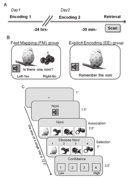
4.2 Experiment Paradigm & Procedure
32 healthy adult participants (20 females) were
randomly assigned of one to two groups (EE or FM).
On day 1 the participants learned 50 new unfamiliar
picture-word associations. On day 2 (24 hours later)
they learned another set of 50 new picture-word
associations. A retrieval memory test for all the 100
new picture-word associations took place 30 minutes
after the acquisition of a second set of associations.
During the retrieval, brain activity was scanned
(Figure 1A). Therefore, the participants were tested
on both recently and remotely encoded information.
The two learning tasks (EE / FM) were designed
differently due to different nature of both learning
procedures (Figure 1B).
The retrieval task was designed as an event
related fMRI experiment in which memory for all
100 items was assessed via an associative four-
alternative forced choice recognition task. The
retrieval procedure was identical for EE and FM as it
was performed inside the scanner (Figure 1C). Each
retrieval trial of an item was 12.5 seconds long and
contained the following intervals: blank screen (1
sec), target label as text and auditory input (1.5 sec),
4 choice pictures appeared on screen, below the
target label (2.5 sec), the word "choose" appeared
onscreen and participants had to respond by
selecting the appropriate key (5 sec), confidence
rating (2.5 sec).
The experiment was designed intentionally to
have participants perform either EE or FM, rather
than perform both EE and FM tasks. It was
important that learning through FM will be implicit
and unintentional, so participants should not know
that the task is a mnemonic task (i.e., requires
memory). However, in EE, participants are explicitly
asked to remember the name of the item.
4.3 Data Acquisition & Pre-processing
The participants were scanned using the Siemens
Trio 3 T scanner, at Baycrest Institute. They
acquired T2*-weighted images, covering the whole
brain using an echo-planar imaging (EPI) sequence
of 50 slices, with repetition time (TR) of 2500 ms,
echo time (TE) of 27 ms, 64 × 64 matrix, slice
thickness of 3.5 mm and a field of view (FOV) of
200 mm. The procedure was designed as an event
related fMRI study.
Figure 1: (A) The experiment structure. (B) Examples of
acquisition through FM (left) and through EE (right). (C)
Retrieval test design which took place inside the fMRI
scanner.
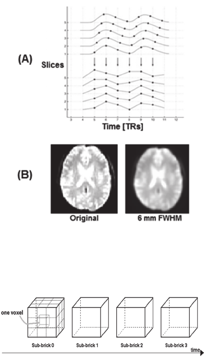
The pre-processing steps included conversion to
4-dimensional AFNI format (Cox, 1996), slice
timing correction using the first slice as a reference
(Figure 2A), movement correction for unintended
head motions and spatial smoothing with 6mm
FWHM Gaussian kernel to increase signal-to-noise
ratio (Figure 2B). Finally, individual participant's
NCTA 2015 - 7th International Conference on Neural Computation Theory and Applications
116
data was converted to a standard coordinate system
(Talairach) to allow data analysis across individuals.
The scanning of each participant was done
during four consequent runs creating a joint dataset
out of four time-series datasets with approximately
150 data volumes each of size 109x91x91, resulted
as a dataset with approximately 600 data volumes.
Therefore, each data volume (data point) contained
1490580 different voxels. We demonstrate the
structure of the collected data in Figure 3.
Figure 2: Examples for pre-processing steps on fMRI data.
(A) Correction of individual's hemodynamic responses
slices acquired aligned to the exact same time (Sladky et
al., 2011). (B) Performance of spatial smoothing on fMRI
volume taken from single participant.
Figure 3: 4-Dimensional structure of AFNI format BRIK
(Cox, 1996) file including 3-dimensional dataset over time
sequence.
5 METHODS
The data points used for analysis were constructed
using scan data obtained for TR=2. This temporal
cut was selected after performing pre-test
classification as suggested in Atir-Sharon et al. work
(Atir-Sharon et al., 2015), taking into consideration
the accordance to the expected HRF response.
We performed further pre-processing over the
time-series data. At first, all non-brain voxels were
removed using a mask. This was done by selecting
voxels from the fMRI dataset that correspond to
non-zero elements in the mask (creating data points
of approximately 200,000 voxels). Afterwards,
linear detrending was performed on each
participant's data set and for each run separately.
Then, normalization over all scans was
conducted. The normalization was done voxel-wise
using z-score for each participant separately. In our
case, the combined dataset involved scans from
different groups and participants taken from
different distributions. Therefore, transformation of
features from different scales to a single scale, with
consideration to the original distributions, was
needed. The z-score method considers the different
distribution characteristics of every group (Wiesen,
2006), hence, it was chosen as the normalization
procedure. The z-score formula is presented in (1),
where z-val is the new z-scored value, f-val is the
original feature value and (μ, σ) are the mean and
standard deviation values:
z-val = (f-val – μ) / σ (1)
For the mean and standard deviation computation in
the z-score equation, several assignments were
tested: (i) from all scans in the dataset; (ii) from
individual participants' scans and (iii) from the
distribution of scans marked as control (baseline) in
the training set. Best classification results were
achieved by using the mean and standard deviation
computed from the distribution of baseline scans
(option (iii)).
Each volume was represented as an individual
data point in the dataset (i.e. each voxel was
considered as a feature). Since the amount of scans
from EE and FM groups was not equal, counter-
balancing of the dataset was performed. This was
done by randomly sampling data points from the
smaller group. This method was applied only on the
training set. Otherwise, more weight would have
been given to prediction accuracies of duplicated
data points against weight of accuracies for data
points that were not duplicated. Therefore, testing
set was left untouched.
Machine learning classification techniques were
used for data analysis. Considering the high
dimensionality of data used in the current study,
feature selection procedure was performed in order
to reduce the number of features used for
multivariate classification analysis. There are several
generic methods for selecting informative features.
Machine Learning Techniques and the Existence of Variant Processes in Humans Declarative Memory
117
We aimed to select the features that best
discriminate between conditions based on their
activation values. It was achieved by ranking the
importance of each feature according to the ANOVA
F-score value obtained for between-group (EE vs.
FM) comparisons.
To find the optimal subset of features for
analysis, we performed exhaustive search for
different sizes of features sets starting from 10
features to full brain features in exponential manner.
Finally, the top 1000 features with highest F-scores
were selected. This relatively large number of
features was chosen to take advantage of inclusion
of weakly informative voxels which can contribute
to an increase in classification rates (Gonzalez-
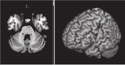
Castillo et al., 2012). In Figure 4, we illustrate the
extracted features in the form of a brain map. In this
example, we display in red selected subset of
features for recollection (correct vs. Incorrect)
classification. This was performed on individual's
fMRI data that belongs to the FM group. Note, that
not all the selected features can be depicted in a
single brain map, but it can be seen that they
concentrated in a specific areas.
A cross-validation classification scheme using
Support Vector Machine classifier (Vapnik, 1998)
with RBF (Radial Basis Function) kernel (Vert,
Tsuda and Schölkopf, 2004) was applied to the
selected features.
Parameters that are not learnt directly within
estimators can be set by searching a parameter space
for the best cross-validation score. Grid search for C
and gamma parameters was performed in the ranges
of 2
-5
to 2
15
and 2
-15
to 2
3
respectively. Grid search
was executed before training on a training portion of
the dataset to achieve increase in accuracy rates. A
pseudo-code for the performed grid search is
presented in Figure 5. In all runs parameters C and
gamma were set to 1 and 2
-3
respectively.
Figure 4: Brain map displaying features selected for
classification analysis of FM recollection (correct vs.
Incorrect).
In cases where the testing set consisted of scans that
for c in [2
-5
, 2
-3
,...,2
15
]:
for g in [2
-15
, 2
-13
, ..., 2
5
]:
for train, test in partition:
model = svm_train(train, c, g)
score = svm_predict(test, model)
cv_list.insert (score)
scores_list.insert(mean(cv_list),c,g)
print max(scores_list)
Figure 5: Pseudo-code for grid search procedure.
were taken from one group only (i.e. all scans were
EE or all scans were FM), a decision making
function was applied. We used majority voting
method as a decision making function, defined as
follows: if the majority of the scans were rated
correctly per participant, the accuracy was set to 1,
otherwise, the accuracy was set to 0.
The software used for the classification was
developed using Python programming language and
based on LibSVM (Chang and Lin, 2011) and
PyMVPA software packages (Hanke et al., 2009). In
Figure 6 we present a complete analysis flow
diagram including all the relevant pre-processing
and processing stages.
6 RESULTS
6.1 Memory Performance
In the information retrieval test, correct response
rates for the recent and for the remote associations
were significantly above chance (binomial tests, p <
0.0001, for both times-of-acquisition, in both
learning groups). Overall, participants from the FM
group were less successful in retrieval, compared to
those from the EE group, in both the recent and the
remote conditions (F(1,30) = 12.2, p < 0.005).
In both groups, recent items were better
recognized than remotely presented items (F(1) =
9.12, p = 0.005) with no significant interaction
between the time of acquisition and the learning
mode (F(1,15) = 0.334, p = 0.565).
6.2 Classification
First, we addressed the question of classifying scans
obtained during correct and incorrect recollection.
Using the proposed classification scheme, we
performed 4-fold (leave one run out) cross-
validation within participants. The mean values of
classification accuracy were close to the chance
level for both groups (EE and FM). We theorized the
NCTA 2015 - 7th International Conference on Neural Computation Theory and Applications
118
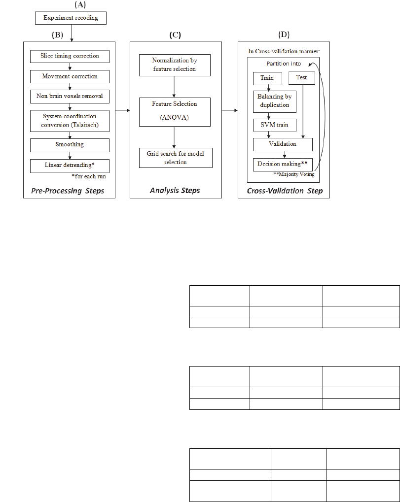
Figure 6: Schematic diagram of the steps performed for whole brain analysis procedure. It consist the following stages: (A)
The initial stage representing the neuroimaging data delivery. (B) The pre-processing stage. (C) Data reduction stage:
reducing data variability efficiently by feature selection. (D) Learning stage: performing multiple times by cross validation
procedure.
reason for that is the existence of two additional
different sub-groups, recent and remote word
acquisition, within each of the initial groups.
Therefore, we classified correct and incorrect scans
within each possibility: EE recent, EE remote, FM
recent, FM remote. For each possibility we chose
10% of all data points randomly as a testing set. The
rest of the data points were used for training. Then,
10-fold cross validation was performed. We report
the values for mean and standard deviation of
classification accuracy over 10 cross-validation folds
for EE in Table 1 and for FM in Table 2.
These results show that a trained classifier was
able to distinguish scans obtained during correct and
incorrect word recollection within each group. The
accuracy is higher for classification of scans for
words learned recently, rather than for words learned
remotely. Furthermore, the discriminating ability is
better within EE group rather than within FM group.
Next, we classified whether the process used for
information acquisition was EE or FM using only
scans from the successful recollection attempts in
the behavioural experiment. We chose randomly
10% from all the scans of all participants as a testing
set. The rest of the scans were used as a training set.
The values and standard deviations for classification
accuracy are presented in Table 3. The results show
that using the neuroimaging data from each one of
the participants for training, we could distinguish
between EE and FM scans very well.
Table 1: Correct vs. Incorrect classification within Explicit
Encoding (EE) using 10-fold cross validation.
Mean Accuracy Standard
Deviation
Recent
0.708 0.09
Remote
0.584 0.067
Table 2: Correct vs. Incorrect classification within Fast
Mapping (FM) using 10-fold cross validation.
Mean Accuracy Standard
Deviation
Recent
0.599
0.063
Remote
0.55
0.068
Table 3: EE vs. FM (using only correct recollection scans)
across participants using 16-fold cross-validation.
Testing set
selection method
Mean
Accuracy
Standard
deviation
Random selection
0.937
0.069
Leave one
participant out
0.638
0.07
These results raise the question of whether the
representation of all the participants in the training
set is crucial to the classification success. That is,
can a machine learning classifier, trained over the
collected data, can successfully distinguish which
label to assign to a new person scan, despite the fact
that the classifier has never seen data from this
participant. To answer this question, we performed a
leave-one-participant-out classification. This was
Machine Learning Techniques and the Existence of Variant Processes in Humans Declarative Memory
119
done across all 16 participants in a cross-validation
manner (leave one participant out). Note that per
iteration, the scans in the testing set are all EE or all
FM. Therefore, we were able to use the majority
voting method for this analysis. The results averaged
across all participants presented in Table 3.
7 DISCUSSION & CONCLUSIONS
In this work, we showed that it is possible to identify
correct and incorrect recollection of memories
acquired through two learning mechanisms: either
Explicit Encoding (EE) or Fast Mapping (FM)
directly from neuroimaging data using machine
learning techniques. The findings suggest that it is
easier to identify recollection success and failure for
information acquired recently rather than for
information after a period of time through EE
mechanism. It may indicate that the newly gained
information, acquired through EE, has started to take
part in consolidation process. At the same time, no
significant change between recollection results of
recent and remote acquisition was seen within the
FM mechanism. This may indicate that FM does not
engage consolidation processes. Further
classification experiments are required to reach a
more general conclusion.
The current results provide additional evidence
for the existence of two memory formation
processes by successfully classifying scans of
correct retrievals following EE and FM. Note that
the classification results for scans taken from an
individual’s data, which were not used previously
for training, were still significant (although less
accurate when training data from a subject were
included). These findings suggest that associative
learning through FM employs alternative neural
pathways to acquire declarative knowledge, which
bypasses the dominant hippocampal-vmPFC axis.
This also indicates that the FM process is eligible for
therapeutic approach for people with hippocampal
brain injuries.
8 FUTURE WORK
Future work should include mapping of the brain
regions and extraction of functional networks
associated with all four group combinations, EE
recent, EE remote, FM recent and FM remote. A list
of possible implementation approaches includes
constructing brain maps using "searchlight"
techniques (Kriegeskorte et al., 2006).
In addition, future work should include brain
regions correlations tests during the retrieval of
memory through EE and through FM in recent and
in remote modes. Those correlations would provide
information regarding the involvement of the
hippocampus and vmPFC regions in the
consolidation processes. To achieve that, one may
use causality analysis techniques (Hu & Liang,
2012) to reveal the causality influences the brain
regions, which are involved with each learning
procedure, have on each other. This could help
reveal new information regarding the mechanism
involved in memory consolidation processes of FM.
ACKNOWLEDGEMENTS
This work is part of the M.Sc thesis of Ms. Gal Star
at University of Haifa under the supervision of Prof.
Larry Manevitz at the Neuro-Computation
Laboratory at Caesarea Rothschild Institute (CRI),
Haifa, Israel.
The research is based on data gathered by
Rotman Research Institute at Baycrest, Toronto,
Canada. The examining of this data was suggested
by Dr. A. Gilboa and complements the work of
Merhav, Karni and Gilboa (Merhav et al., 2015).
The authors are listed in alphabetical order.
REFERENCES
Atir-Sharon, T., Gilboa, A., Hazan, H., Koilis, E. &
Manevitz, L. M. 2015. "Decoding the formation of
new semantics: MVPA investigation of rapid
neocortical plasticity during associative encoding
through Fast Mapping.". Neural Plasticity, vol. 2015,
Article ID 804385, 17 pages.
Bauer, P. J., 2008. "Toward a neuro-developmental
account of the development of declarative memory".
Dev Psychobiol, vol. 50, no. 1, pp. 19-31.
Chang, C. C. & Lin, C. J., 2011. "LIBSVM: a library for
support vector machines". ACM Transactions on
Intelligent Systems and Technology, available from:
<http://www.csie.ntu.edu.tw/~cjlin/libsvm>.
Cox, C., 1996. "AFNI: software for analysis and
visualization of functional magnetic resonance
images", Computers and Biomedial Research, vol. 29,
pp. 126-173.
Frankland, P. W., and Bontempi, B., 2005. "The
organization of recent and remote memories". Nature
Review: Neuroscience, vol. 6, pp. 119-130.
Gais, S., Albouy, G., Boly, M., Dang-Vu, T.T., Darsaud,
A., Desseilles, M., Rauchs, G., Schabus, M.,
Sterpenich, V., Vandewalle, G., Maquet, P., Peigneux,
P., 2007. "Sleep transforms the cerebral trace of
NCTA 2015 - 7th International Conference on Neural Computation Theory and Applications
120
declarative memories". Proceedings of the National
Academy of Sciences of the USA, vol. 104, no. 47, pp.
18778-18783.
Gilboa, A., Hazan, H., Koilis, E., Manevitz, L. and
Sharon, T, 2011. "Two memory systems: identifying
human memory encoding mechanisms from
psychological fMRI data via machine learning
techniques". Proceedings of the International Joint
Conference on Neural Networks (IJCNN), pp. 54.
Gonzalez-Castillo, J., Saad, Z.S., Handwerker, D.A., Inati,
S.J., Brenowitz, N., Bandettini, P.A., 2012. "Whole-
brain, time-locked activation with simple tasks
revealed using massive averaging and model-free
analysis". Proceedings of the National Academy of
Sciences, vol. 109, no. 14, pp. 5487-5492.
Hanke, M., Sederberg, P. B., Hanson, S. J., Haxby, J. V.,
and Pollmann, S., 2009. "PyMVPA: A python toolbox
for multivariate pattern analysis of fMRI data".
Neuroinformatics, vol. 7, no. 1, pp. 37-53.
Hu, S. & Liang, H., 2012. "Causality analysis of neural
connectivity: New tool and limitations of spectral
granger causality". Neurocomputing, vol. 76, no. 1, pp.
44-47.
Kriegeskorte, N., Goebel, R., and Bandettini, P., 2006.
"Information-based functional brain mapping".
Proceedings of National Academy of Science USA,
vol. 103, no. 10, pp. 3863-3868.
McClelland, L., McNaughton, B. L., and O'Reilly, R. C.,
1995. "Why there are complementary learning system
in the hippocampus and neo-cortex: insights from the
successes and failure of connectionist models of
learning and memory". Psychological Review, vol.
102, no. 3, pp. 419-457.
Merhav, M., Karni, A. and Gilboa, A., 2014. "Neocortical
catastrophic interference in healthy and amnesic
adults: A paradoxical matter of time". Hippocampus,
vol. 24, no. 12, pp. 1653-1662.
Merhav, M., Karni, A. and Gilboa A., 2015. "Not all
declarative memories are created equal: fast mapping
as a direct route to cortical declarative
representations". Neuroimage, vol. 117, pp. 80-92.
Mitchell, T., Shinkareva, S., Carlson, A., Chang, K. M.,
Malave, V. L., Mason, R. and Just M. A., 2008.
"Predicting human brain activity associated with the
meanings of nouns". Science, vol. 320, no. 5880, pp.
1191-1195.
Nawa, N. E. & Ando H., 2014. "Classification of self-
driven mental tasks from whole-brain activity
patterns". PLos One, vol. 9, no. 5, e97296.
Norman, K. A., Polyn, S. M., Detre, G. J. & Haxby, J. V.,
2006. "Beyond mind-reading: multi-voxel pattern
analysis of fMRI data". Trends in cognitive science,
vol. 10, no. 9, pp. 424-430.
Sharon. T., Moscovitch, M., and Gilboa, A., 2011. "Rapid
neocortical acquisition of long-tem arbitrary
associations independent of the hippocampus".
Proceedings of the National Academy of Science of the
USA, vol. 108, no. 3, pp. 1146-1151.
Sladky, R., Friston, K. J., Tröstl, J., Cunnington, R.,
Moser, E. & Windischberger, C., 2011. "Slice-timing
effects and their correction in functional MRI".
Neuroimage, vol. 58, no. 2, pp. 588-594.
Squire, L. R., 1992. "Declarative and non-declarative
memory: multiple brain systems supporting learning
and memory", Journal of Cognitive Neuroscience, vol.
4, no. 3, pp. 232-243.
Squire, L. R., and Alvarez, P., 1995. "Retrograde amnesia
and memory consolidation: a neurobiological
perspective". Current Opinion in Neurobiology, vol. 5,
no. 2, pp. 169-177.
Uematsu, A., Matsui, M., Tanaka, C., Takahashi, T.,
Noguchi, K., Suzuki, M. and Nishijo, H., 2012.
"Developmental trajectories of amygdale and
hippocampus from infancy to early adulthood in
healthy individuals". PLos One, vol. 7, no. 10, e46970.
Vapnik, V., 1998. "Statistical learning theory". New York,
NY: Wiley.
Vert, J. P, Tsuda, K. and Schölkopf, B., 2004. "A primer
on kernel methods". Kernel Methods in Computational
Biology.
Warren, D. E. and Duff, M. C., 2014. "Not so fast:
Hippocampal amnesia slow word learning despite
successful fast mapping". Hippocampus, vol. 24, no. 8,
pp. 920-933.
Wiesen J. P., 2006. "Benefits, Drawbacks, and Pitfalls of
z-Score Weighting". 30th Annual IPMAAC
Conference. Available at: "http://annex.ipacweb.org/
library/conf/06/wiesen.pdf" (27 Jun 2006).
Machine Learning Techniques and the Existence of Variant Processes in Humans Declarative Memory
121
