Electromyographic Analysis of the Swim Start
Bilateral Comparison of the Front-weighted and Rear-weighted Track Start from
the OMEGA OSB11 Starting Block
Janna Brit Langholz
1
, Gunnar Westman
1
, Magnus Karlsteen
1
and Christian Finnsgård
1, 2
1
Centre for Sports Technology, Department of Applied Physics, Chalmers University of Technology, Gothenburg, Sweden
2
SSPA Sweden AB, Research, Gothenburg, Sweden
Keywords: Swimming, Swim Start, Electromyography, Muscle Activity, Competition Block.
Abstract: Previous swim start studies involving electromyography (EMG) consistently comprised unilateral
measurements and the attachment of the swimmer via cables to a computer. Therefore the present work
aims for an overall picture of the muscle activation pattern during the swim start by conducting bilateral
measurements with minimal restriction of motion. On that account a multichannel surface EMG device with
a wireless Bluetooth connection and videography is utilized in order to assess the nowadays most common
start dive techniques of competitive swimming events - differently weighted track starts from the OMEGA
OSB11 starting block. The data analysis identified that the normalized muscle activation levels were higher
during the front-weighted than during the rear-weighted start - probably caused by shorter block times and
less contribution of the arms. Furthermore the onset of the muscle activation seems to be different in
between start dive techniques, as for instance the muscles of the rear leg commence contracting earlier while
the muscles of the front leg start later in the rear-weighted compared to front-weighted starts. It is highly
likely that this originates in the position of the center of mass relative to the muscles. A general overview
over the coordination of the different muscles could also be obtained: It became obvious that some muscles
are the main drivers of the swim start (vastus lateralis, soleus) whereas others rather exerted supportive
actions (gluteus maximus, semitendinosus, erector spinae longissimus).
1 INTRODUCTION
Fractions of a second often separate the winner from
the rest in elite swim races. For instance, in 50 meter
butterfly races the finishing times of the first and the
third swimmer are only 0.09 s (male) and 0.05 s
(female) apart (Commonwealth Games Delhi 2010,
butterfly finals) (Slawson, 2012). Therefore each
swimmer should try to improve every movement of
the race. In short distance swimming events the start
off the block is of special importance and may
decide on success or defeat. General training
techniques mainly include time measuring or
perfunctory video analysis. Rule of thumb estimates
widely serve as feedback base for the body position
on the block. However, the fundamental unit which
drives the body forward is often neglected - the
muscle. Voluntary muscle contractions originate in
the motor cortex. Thereafter motor neurons transport
the necessary impulse to the muscle of interest and
cause action potentials which travel along the
muscle membrane. The amplitude and frequency of
these action potentials can be measured by
electrodes so that muscle activity levels and timing
can be analyzed - this process is called
electromyography (EMG) (Christensen, 1989).
Maximal off-the block performance can be achieved
if a beneficial muscle activation pattern is used as
force production on the block and the orientation of
the body segments relative to each other are
determined by the activation of the activation of the
involved muscles. Coaches and athletes should be
able to analyze and react to certain - probably
disadvantageous - activation patterns which can in
return lead to better race times. Overall, this work is
designed to gain an insight into the nowadays most
common swim start techniques utilizing an
electromyographic approach to investigate whether
the principle of EMG and the available EMG
technology can aid swimmers in improving their
start dive technique.
310
Langholz, J., Westman, G., Karlsteen, M. and Finnsgård, C..
Electromyographic Analysis of the Swim Start - Bilateral Comparison of the Front-weighted and Rear-weighted Track Start from the OMEGA OSB11 Starting Block.
In Proceedings of the 3rd International Congress on Sport Sciences Research and Technology Support (icSPORTS 2015), pages 310-320
ISBN: 978-989-758-159-5
Copyright
c
2015 by SCITEPRESS – Science and Technology Publications, Lda. All rights reserved

1.1 Previous Research on the
Competitive Swim Start
The latest changes in the regulations of the
Fédération Internationale de Natation (FINA Rules,
FR 2.7) brought the company Swiss Timing Inc. to
launch their new starting block OMEGA OSB11 in
April 2008 which is now predominantly used in
international competitions (Murrell, 2012). On this
block an additional foot rest (30% incline) is
mounted to the back of the longer and steeper
surface area allowing for an amended track start
called kick start with better performance. Higher
take-off velocities, shorter block times and faster
times at different distances could be recorded
(Honda et al., 2010; Biel et al., 2010; Ozeki et al.,
2012). These facts brought the grab start to mostly
vanish from elite swimming contests (Vint et al.,
2009). Beyond that different studies discovered that
moving the center of mass backwards during the
track start is beneficial compared to the classical
front-weighted track start (Welcher et al., 2008;
Vilas-Boas et al., 2003). A recent study took these
findings and observed the performance of a front-,
neutral- and rear-weighted kick start from the
OMEGA OSB11 over a distance of 15 meters. The
results indicated faster times at 15 meters for the
neutral- and rear-weighted variant of the kick start
from this new starting block (Barlow et al., 2014).
Currently ongoing research takes further parameters
into account including stance width, height of the
center of mass and foot preferences of the swimmer
(Kibele et al., 2015).
In 1964 surface EMG had its debut in the water
environment and introduced a new variable to
swimming movement analysis: muscle activity
(Barthels, 2015). During the last decades
Electromyography found its way into competitive
swimming research. Fatigue analysis of different
swim styles (Conceição et al., 2010; Yasushi Ikuta et
al., 2012) and dry-land exercises (Ganter et al.,
2007; Nazário-de-Rezende et al., 2012) form the
majority of EMG papers in swimming. As most of
the commercially available EMG systems are not
waterproof and rather sensitive to rough under water
movements, measurements have been restricted to
plain swimming motions without regular starts and
turns. Making matters worse, a large number of
EMG systems has to be connected to a computer via
a multitude of wires which may impair the natural
motion of the swimmer.
The swim start move in particular was only
subject to three scientific studies. A first paper
observed electromyographic activity of two different
variants of the backstroke start – feet immerged or
feet emerged. Seven electrodes were placed
unilaterally on arm, trunk and leg muscles (de Jesus
et al., 2011). Another study also focused on the
backstroke start in order to see how different the
muscle activation pattern is in between subjects.
Here eight electrodes were placed on the right side
of the body (Hohmann et al., 2008). A third paper
dealt with the biomechanics of the grab and track
start technique and again eight electrodes were
positioned on upper and lower extremities as well as
the trunk on the right side of the body (Krueger et
al., 2003). All these papers share the fact that the
measurements were conducted unilaterally which
does not allow an overall examination of the
movement. Additionally all subjects were fixed via
cables to the main acquisition unit. This may have
affected the motion during the examined start
technique.
1.2 Purpose of the Paper
Even though EMG measurements are sensitive to
external influences - which especially applies to its
use in the water environment - it has been decided to
advance research regarding the muscular activity
during the block phase of the swim start. It is
assumed that this knowledge may help in optimizing
start techniques as well as suggesting new training
methods and exercises (Clarys et al., 1993). Besides
this the relationship between EMG data and
kinematic variables is said to play a key role in the
evaluation of swimming performance (Conceição et
al., 2013) and should therefore be investigated
further.
2 METHODS
Bilateral measurements with minimal restriction of
motion are to be achieved by carefully selecting the
most important muscles of the swim start and
employing a multichannel surface EMG device
using Bluetooth technology to transfer the measured
data to a computer. In a second step it is analyzed
whether the EMG data can be linked to other
variables of the swim start including angles from the
block and instantaneous horizontal velocities
(kinematic data). It is moreover assessed whether the
results can be connected to the outcome of other
related studies.
The focus is placed on the front- and rear-weighted
kick start (Figure 1) as those techniques are the most
widely applied ones off the OMEGA OSB11 starting
Electromyographic Analysis of the Swim Start - Bilateral Comparison of the Front-weighted and Rear-weighted Track Start from the
OMEGA OSB11 Starting Block
311
block and are subject to the latest swim start studies
(Barlow et al., 2014; Kibele et al., 2015). Due to a
limitation of time, equipment and staff the
observations concentrates on the block phase instead
of including flight and dive phase parameters.
Moreover technology constrains an analysis of the
total start sequence: Bluetooth transmission does not
work under water. This choice is endorsed by the
literature: All subsequent components of the swim
start are influenced by the block phase what gives
each swimmer the task to strive for maximal off-the-
block performance (Mason et al., 2007).
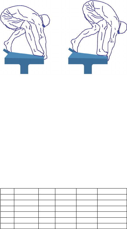
Figure 1: Rear-weighted (left) and front-weighted (right)
initial start position on the block.
2.1 Participants
Three males and three females (five Swedes, one
American) took part in this study. Table 1 shows the
anonymized personal data of each swimmer. All
swimmers were competing on developmental or
national level. Four swimmers preferably had the
right foot on the front edge of the starting block, two
of them the left foot.
Table 1: Personal data of all participating subjects (Lat. =
Laterality, Pref. Pos. = preferred weight distribution on the
block).
No. Gender Age cm/kg Lat. Pref. Pos.
S1 Male 19 179/75 right Neutral
S2 Male 20 192/74 right Front
S3 Female 20 173/65 right Front
S4 Female 19 173/75 right Front
S5 Male 22 177/79 right Front
S6 female 22 171/61 right Neutral
2.2 Equipment
The EMG device of choice is the telemetric unit
MQ16 by Marq Medical which weighs 120 g and
has a data buffer memory of 60 MB that collects the
data and a Bluetooth transmitter sends it to a
computer. Preamplifiers amplify the signal close to
the electrodes and conduct analog to digital
conversion. The noise level of the MQ16 is specified
to be less than 3 µV (Meyland et al., 2014). The
default sampling frequency is 1024 Hz which is
sufficient for EMG signals ranging between 20 and
500 Hz. However, it was decided to set the sampling
frequency of the MQ16 to the widely used value of
2048 Hz (Ali et al., 2014; Barlett, 2007). Surface
electrodes are applied in a bipolar configuration with
a common ground and made of Ag/AgCl. As the
device is to be used under wet conditions it has to be
protected by a waterproof casing (iPad®mini™case
by ECase). Transparent film dressing (Tegaderm™,
3M™) is chosen as covering for the electrodes and
will be additionally fixed with foam tape
(Microfoam, 3M™ ). Furthermore an accelerometer
(mounted to the ankle of the rear foot) is used to
track the swimmer’s first motion on the block after
the starting signal and a pressure mat is used to track
the last motion on the block (foot-off moment).
These two event markers are then used for time
normalization of the movement for successive
averaging of trials. The Casio Exilim EX-FH25
video camera (shutter speed 1/500 s, 120 fps) is
mounted to a rigid tripod to allow the recording of
the swimmer in the sagittal plane.
It was positioned
perpendicular to the plane of motion approx. 5 m
from the center of the swimming lane.
2.3 Measurement Protocol
Personal information of all subjects was captured
and they signed a letter of agreement. Thereafter the
electrode placement procedure commenced
considering the SENIAM guidelines (Surface
Electromyography for the Non-Invasive Assessment
of Muscles, EU project) regarding the electrode
location and positioning process. Nine muscles of
the back and the lower limbs were chosen to
represent the most relevant muscles during the start
dive in swimming: erector spinae longissimus,
gluteus maximus, vastus lateralis, semitendinosus
and soleus. The skin area which was supposed to be
covered by the electrodes was shaved, scrubbed with
sand paper and then cleaned with an alcohol wipe.
This was followed by attaching the MQ16 to the
waist of the swimmer and connecting the cables to
the electrodes. Figure 2 shows that the electrodes
including the pre-amplifiers were covered by
waterproof tape with a size of at least 20 x 10 cm.
All edges were additionally fixed by waterproof
foam tape to prevent water induced detachment of
the sealing. The accelerometer was mounted to the
inside of the rear ankle and covered in the same
WPPDSports 2015 - Special Session/Symposium on Weather, Position and Performance data in Outdoor Sports
312
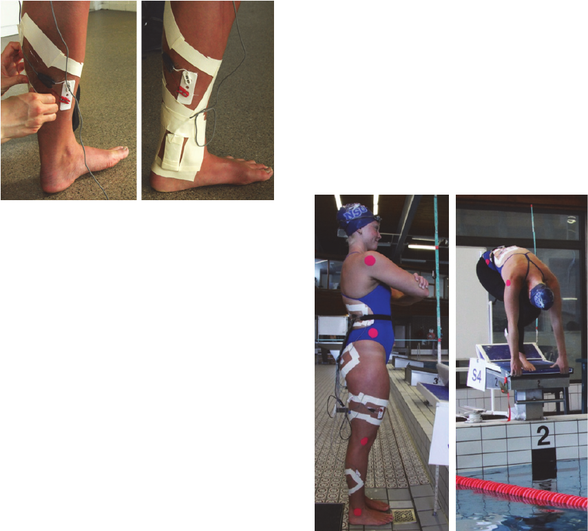
manner as the electrodes.
Figure 2: Covering of the electrodes and placement of the
accelerometer right above the rear ankle.
The movement of the swimmer was supposed to
be as unrestricted as possible. Consequently it was
decided to supply the swimmers with commercial
tights to fix the cables to the lower limbs. This step
had a beneficial secondary effect: minimization of
cable motion. Figure 3 illustrates that pink markers
with a diameter of approximately 4 cm were placed
on the four landmarks defining the body segments:
ankle, knee, hip and shoulder (Krueger et al., 2003).
Maximum voluntary contractions (MVC)
performed on a gym bench are used to normalize the
amplitude of the EMG signal. Normalization makes
it possible to conduct an analysis which is not based
on absolute values. Thereby comparisons between
trials of the same subject or between different
individuals can be made. Three maximal
contractions against an insuperable resistance are
performed for each muscle. A duration of six
seconds and a short period of rest in between is
chosen (Masso et al., 2010; Halaki et al., 2012).
From different papers the most feasible MVC tasks
for the muscles of interest in the present study were
selected (Halaki et al., 2012; Konrad, 2005). For the
reason that varying electrode placement might
change the measured EMG signal, the MVC
recordings for normalization were conducted with
the same configuration as the subsequent EMG start
dive measurements. As the wet pool environment
might impair the electrode fixation, the MVC
measurements were performed beforehand in a dry
environment. Prior to executing the MVC tests, the
swimmers gave feedback on their positioning and
the static resistance they had to work against. Only if
the positioning was suitable for the measurement
and comfortable for the swimmer, the recording was
started.
After all preparatory procedures were
undertaken, the swimmer was ready to conduct three
front-weighted and three rear-weighted start dives.
Figure 3 sows subject S4 in a rear-weighted starting
position. With respect to various existing start jump
techniques the swimmers included a few additional
start jumps of the requested techniques into their
regular training during the previous weeks. In the
front-weighted position they were asked to distribute
the majority of their weight on the front foot whilst
still preserving a stable stance on the block and in
the rear-weighted position they were instructed to
allocate the majority of their weight to the rear foot
and at the same time maintain a stable position on
the block.
Figure 3: Swimmer with fixated electrodes and markers
and the MQ16 on the back. OSB11 cover with pressure
mat placed under the front foot.
The subjects were directed to propel themselves
off the block with maximal effort like they would do
in a competition. In order to prevent the
waterproofing bag and the electrode sealing from
damage, the swimmers were directed to not perform
any powerful swim strokes but return to the edge of
the pool by exerting cautious arm strokes. They were
randomly asked to apply one or the other technique
and after each trial they had approximately five
minutes of rest. All trials were performed in the
same way: When the Bluetooth connection between
the MQ16 and the computer was established and the
swimmer was in readiness, the EMG recording was
started. Right after that the MQ16 transmit trigger
(connected to pressure mat) was enabled. The
Electromyographic Analysis of the Swim Start - Bilateral Comparison of the Front-weighted and Rear-weighted Track Start from the
OMEGA OSB11 Starting Block
313
camera operator then started the video recording,
gave the prestart command "On your mark!" and
then activated the start signal.
2.4 Data Analysis
As the swimmer’s movement off the starting block
only comprises a duration of around a second, it is
assumed that the fatigue effect (frequency analysis)
of the involved muscles can be neglected. Therefore
the subsequent data analysis focused on the analysis
of EMG amplitudes and the timing of the muscle
activation of the different muscles relative to each
other. The MATLAB R2013b software was utilized
for this purpose.
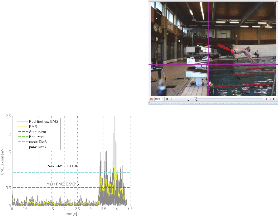
Figure 4: Processed signal (vastus lateralis rear, S3) with
mean and peak RMS values and markers depicting the
start and end of the block phase.
The processing steps included rectification of the
signal, correction of a possible DC offset and
determination of the linear envelope or root-mean-
square (RMS) to specify EMG amplitude
(Lamontagne, 2000). Figure 4 shows an example of
an EMG amplitude analysis. Additionally on- and
offset set times of muscular activity were calculated
and the time stamps from the accelerometer and the
pressure mat on the starting block were found.
The video data was predominantly processed
with the software Tracker (Tracker software, version
4.8x) which helps in calculating times, angles and
velocities after tracking the markers (Figure 5).
Filtering of the digitized landmarks was again done
in MATLAB R2013b. Differences in between the two
start techniques were tested by means of paired t-
tests using SYSTAT 13.
Figure 5: Video scaling & digitizing of the hip marker
(approx. centre of mass) with the software Tracker.
3 RESULTS
Many start dive recordings were accompanied by
intrusion of water under the electrode sealing, which
resulted in erroneous data sets. An averaging of
different trials of the same position and subject
would then lead to unnaturally high or low voltage
values, which do not reflect the actual activation.
Therefore it was decided to neglect the time
normalization and successive averaging of the three
front-weighted and three rear-weighted recordings of
each subject but select the best front-weighted and
the best rear-weighted data set of each subject. For
that reason only five front-weighted and six rear-
weighted data sets enter the final data analysis.
As the subjects have been introduced to the
concept of recording maximum voluntary
contractions prior to the test day, meaningful MVC
recordings were obtained. Additional time during the
MVC tests to adjust the position on the gym bench
or the fixation of the limbs, also contributed to the
positive outcome of the MVC recordings.
Contraction periods could clearly be separated from
rest periods, which facilitated proper RMS
calculations and identification of the maximal
voltage value for each muscle.
However, as also depicted in other papers, MVC
recordings may not be appropriate to normalize
dynamic movements as they represent the ability of
the muscle to contract in a static setting. In dynamic
movements the synchronization of motor units is
different. This constraint must be kept in mind.
WPPDSports 2015 - Special Session/Symposium on Weather, Position and Performance data in Outdoor Sports
314
3.1 Activation Levels
The main actors (vastus lateralis front/rear, soleus
front/rear) predominantly register the highest
activation levels, ranging between 48.6% and
191.0% for mean RMS. Additionally the standard
error is rather high for these strong, rapidly acting
muscles. The gluteus maximus rear and
semitendinosus rear settle just below with an
average activation of 38.1% and 34.1% respectively.
The back muscles (erector spinae longissimus) as
well as the gluteus maximus front and
semitendinosus front show lower mean RMS values
18.9% and 29.4% with smaller standard errors than
the main actors.
3.2 Activation Timing
After the reaction time has passed the muscles begin
to take up their work at different times. It can clearly
be seen that the erector spinae longissimus is on
average active before any motion can be detected
(accelerometer/video). Almost synchronously the
gluteus maximus and semitendinosus of the rear leg
take up their work and also the semitendinosus front
begins contracting almost simultaneously with the
two aforementioned muscles. Shortly after the vastus
lateralis rear and soleus rear show their activation
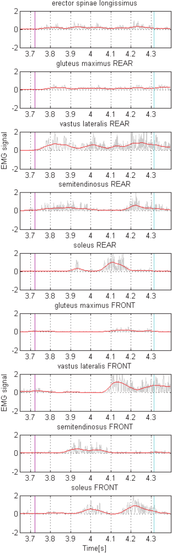
onset. Figure 6 illustrates that, after almost half of
the block time has passed, the gluteus maximus front
comes into the game marking the start of the pushing
phase of the front leg. Lastly the S front and the
vastus lateralis front perform the last powerful action
off the block with an onset at 54.8% and 57.1% of
total block time respectively.
Generally the rear leg comes into action before
the front leg. When observing the two different
initial positions it can be seen that the muscles of the
rear leg during the rear-weighted start generally start
contraction earlier than in front-weighted starts. On
the other hand, the muscles of the front leg seem to
have a later onset during the rear-weighted than
during the front-weighted start.
3.3 Kinematic Parameters
Reaction times (start signal until start of motion) of
0.245 ± 0.013 s have been identified by means of
Tracker from the video recordings. It can be seen
that the reaction times for the rear-weighted start are
often shorter (0.236 s) compared to the front-
weighted start (0.254 s). Beyond that it was
observed that the swimmers remained on average
0.074 s longer on the starting block in rear-weighted
track starts.
Figure 6: EMG signals [V] of all nine investigated
muscles during a front-weighted start dive (S1). The pink
line depicts the start of motion (accelerometer signal) and
the blue line represents the foot-off moment (pressure mat
signal).
Electromyographic Analysis of the Swim Start - Bilateral Comparison of the Front-weighted and Rear-weighted Track Start from the
OMEGA OSB11 Starting Block
315
This leads to a longer average start-to-flight time
(start signal until foot-off) for the rear-weighted start
(0.841 ± 0.034 s) off the OMEGA OSB11 starting
block - even though the detected reaction times were
shorter than for the front-weighted starts. For a
front-weighted start the swimmers required on
average 0.783 ± 0.045 s. Generally the men achieved
faster starting times than the women.
The calculated angles from the block for the six
subjects depict a certain pattern with regard to the
initial position on the block: rear-weighted starts
seem to lead to smaller angles. Yet it cannot be
proven that the angles during the front-weighted
start are significantly higher than during the rear-
weighted start dive (p-value = 0.097). Most angles
measured per subject per position are close together,
but the angle from the block ranges largely in
between subjects depicting values ranging from 25.1
to 48.6 degrees.
The instantaneous, horizontal velocities also
indicate a particular pattern: the velocities off the
block are significantly higher for rear-weighted
starts (p-value = 0.013). The angle also seems to
have a certain influence on the velocity as it is
frequently detected that the smaller the angle, the
higher the horizontal velocity at foot-off becomes.
An average instantaneous, horizontal velocity of
2.166 ± 0.112 s is found for front-weighted and
2.369 ± 0.110 s for rear-weighted starts. The
velocity recorded might however not represent the
maximal velocity throughout the jump. For different
subjects higher horizontal velocities were found
shortly before or after foot-off, depending on the
individual stretching pattern during this phase of the
start.
4 DISCUSSION
The EMG data acquisition during the start dive
formed the core of the present investigation. Each
subject conducted three front-weighted and three-
rear weighted kick starts off the OMEGA OSB11
starting block. As favored the subjects did not report
any impairment of motion due to the cables on their
limbs or the MQ16 on their back. When the
electrodes remained dry under the waterproof tape,
they were able to measure the muscular activation
during the start dive. This leads to the assumption
that the selected electrode positions as well as the
skin preparation chosen, were appropriate for the
current study. The noise of the components has been
found to be negligible and a certain variability in the
data due to variations in between subjects has to be
accepted. However, certain events negatively
influenced the EMG data acquisition in the water
environment – detachment of the electrodes from the
skin and disturbance of clamps and amplifiers by the
water.
It was found that the mean EMG amplitude
during the rear-weighted start is significantly smaller
than the amplitude during the front-weighted start
technique. This is mainly caused by longer block
times in rear-weighted starts as the center of mass
(CoM) needs to travel a longer distance until foot-
off than in the front-weighted version. Additionally
the arms are capable of contributing a large part to
the forward motion by pulling the swimmer out of
the initial rearward position. The standard error was
found to be much higher for the forceful main actors
of the start dive (vastus lateralis, soleus) than for the
other muscles. This may originate from the
dynamics of the start dive leading to high voltage
peaks in the EMG curve of these strong muscles.
A comparison of the onset times for front-
weighted and rear-weighted start dives
predominantly revealed an earlier muscle onset
within the rear leg in rear-weighted starts. Certain
muscles of the front leg, however, start contracting
later than in front-weighted starts. This discrepancy
leads to a non-significant difference between the
muscular onset of the front-weighted and rear-
weighted track start technique when conducting a
statistical analysis. A reason for this might be that
the initial position places a high weight on the rear
foot allowing an immediate force production in the
beginning whereas the CoM has to be moved a long
distance before the muscles of the front leg can start
executing their function.
The parameters obtained from the video
recordings support the assumption that the selected
subjects are suitable representatives for aspiring
swimming professionals. When opposed to the
outcome of other swim start studies, the subjects of
this study show similar results - however, not with
the same magnitude as elite swimmers (Barlow et
al., 2014).
Visually connecting the EMG measurements to
the video recordings ultimately revealed the motion
sequences during which the different muscles are
predominantly active (Figure 7). The interplay of
different muscle groups was identified, for instance
the almost simultaneous activation of the gluteus
maximus and the vastus lateralis muscles and the
dissimilar activation of semitendinosus and soleus in
the same leg. The collaboration of the gluteus
maximus and the vastus lateralis was also identified
previously (de Jesus et al., 2011).
WPPDSports 2015 - Special Session/Symposium on Weather, Position and Performance data in Outdoor Sports
316
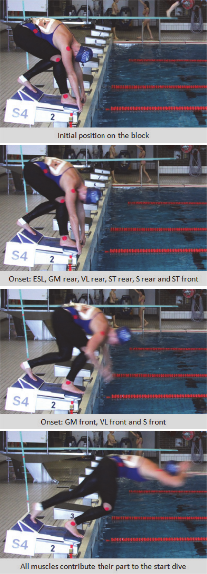
Figure 7: Images of a rear-weighted start dive (S4)
indicating the different onset times of the recorded
muscles.
The mentioned dissimilar activation of
Hamstring and Calf muscles can be explained by the
differing required performance conditions. During
the start dive the Hamstrings influence the
swimmer’s hip and knee extension at smaller knee
angles whereas the Calves seem to take up their
work at larger knee angles conducting the last
powerful movement on the block.
Besides, the semitendinosus of the front leg
counterbalances the forces which are generated
when the soleus of the rear leg pushes off the foot
rest on the back of the block in order to preserve a
secure stance on the block. After a leg has left the
block the semitendinosus of the same leg might
show another peak related to the injury prevention
mechanism of the knee joint; At that point in time
the soleus of that leg already shows strongly
decreasing activation levels.
Moreover the back muscles as well as the gluteus
maximus help in extending the swimmer into the
favored streamline position as they show activation
at the beginning of motion and right before and after
foot-off. This role is confirmed by different swim
start publications which assigned the same
responsibilities to the abovementioned muscles (de
Jesus et al, 2011, Hohmann et al., 2008). As the
upward rotation of the trunk commences first with
the help of the back muscles and leg extensors
causing vertical acceleration of the trunk and
simultaneously a force pointing downwards in the
direction of the lower limbs, the extension of the
legs only starts when the hips begin to be
unweighted. A possible drop of activity in the hip
extensors (gluteal muscles and Hamstrings) after the
first part of the push phase might be related to the
termination of the upward rotation of the trunk.
Furthermore the fact that the muscular activation of
the ST of the front leg peaks later than the one of the
rear leg seems to be linked to the positioning of the
muscle relative to the CoM meaning that the muscle
in the front leg cannot contribute its part to the hip
extension and stance stabilization until the CoM
reached a certain location further to the front of the
starting block.
After foot-off the muscle activation profiles vary
remarkably in between subjects as each swimmer
conducts different corrective and stabilizing
movements during the flight phase (Hohmann et al.,
2008). Some swimmers still show elevated EMG
profiles in their back muscles whereas others
continue to contract their soleus muscles for a
beneficial feet position. It was neither aimed nor
possible to make statements about later swim start
Electromyographic Analysis of the Swim Start - Bilateral Comparison of the Front-weighted and Rear-weighted Track Start from the
OMEGA OSB11 Starting Block
317
phases as it is a highly individual issue and the
Bluetooth connection vanishes upon water entry.
Taking the EMG recordings and the video data
into account one might see that the rear leg is the
initiator and main driver of the observed track start
technique. It sets the body in motion and moves the
CoM to the front of the starting block. Changing the
initial position changes the activation levels and
timing in both legs but the rear leg generally
performs the bigger part of the work which can be
seen in predominantly higher activation levels.
However, without the stabilizing actions of the front
leg, which form the basis for a solid stand on the
block, and the last push off the block, which defines
the positioning of the swimmer in the air, the start
jump would not be as effective and efficient as it is.
5 CONCLUSION
Muscles vary largely in volume and structure
leading to a different base of operation for each
muscle and restricting the comparability of
activation levels and timing in different muscles.
Moreover some of the leg muscles observed belong
to the group of biarticular muscles (e.g. gluteus
maximus, Hamstrings) meaning that they traverse
two joints and thereby have another principle of
operation than monoarticular muscles (Bobbert et
al., 1988). Additionally it has to be noted, that in
complex movements like the swim start which
include all body segments, muscles influence the
joints they cross as well as other joints (Bobbert et
al., 1988). Another issue is the link between EMG
activation and produced tension: After a muscle has
been stimulated, a delay of 20 to 100 ms occurs
between a recorded muscular activity onset and the
resulting mechanical output which can be observed
in video recordings (Bartlett, 2007; Bobbert et al.,
1988).
The complexity of the human body as well as the
ambiguous relationship between EMG and
kinematic data unfortunately limit the information
one can extract from EMG measurements and a
combination of those with video data. Solely activity
levels and timing can be detected, but not the
muscle’s exact task within a movement (Bartlett,
2007). In order to avoid speculative interpretation,
the present work did not assign a particular muscle
to a certain function, body position or joint angle but
gave a suggestion regarding the contribution of a
muscle to a certain motion sequence during the
swim start.
The physiological and mechanical considerations
made within the scope of this work are not the only
ones influencing the muscular activation during the
start jump in swimming. In future investigations
further muscles or muscle properties should be
addressed and the location of the CoM or different
body segments varied (Bobbert et al., 2007). Besides
altering the scope of the investigation, improvements
should be made regarding the equipment, for
instance using wireless electrodes in order to
facilitate better sealing, finding a solution for the
Bluetooth loss between air and water and
strengthening the synchronization of all applied
measurement devices. Additionally trying to supply
the swimmers and coaches with quickly processed,
meaningful data would be a desirable advancement
(Hamill, 2010).
It is assumed that a step in the right direction was
made by pointing out the importance of the
understanding of the muscular tasks exerted by the
swimmers body. This can aid in developing training
methods and prospective start dive techniques.
Unfortunately the process of recording EMG with
the current technology is highly sensitive and
protracted so that an application in everyday swim
training sessions is - for the time being - not feasible.
ACKNOWLEDGEMENTS
The authors wish to acknowledge the support from
all swimmers of MASS and Göteborg Sim who
participated in the measurements.
REFERENCES
Ali, A., Sundaraj, K., Ahmad, R.B., Ahamed, N.U., Islam,
A., 2014. Recent observations in surface
electromyography recording of triceps brachii muscle
in patients and athletes, Applied Bionics and
Biomechanics, 11, (p. 105–118).
Barlow, H. et al., 2014. The effect of different kick start
positions on OMEGA OSB11 blocks on free swimming
time to 15 m in developmental level swimmers,
HumanMovement Science, 34 (2014), (p. 178-186).
Barthels, Katharine M., 2015. Biomechanical Research in
Swimming: Past, Present and Future, Physical
Education Department, California Polytechnic State
University, https://ojs.ub.uni-konstanz.de/cpa/article/
download/906/.
Bartlett, R., 2007. Introduction to Sports Biomechanics –
Analysing Human Movement Patterns, Taylor &
Francis e-Library, Second edition.
WPPDSports 2015 - Special Session/Symposium on Weather, Position and Performance data in Outdoor Sports
318
Biel, K., Fischer, S., Kibele, A., 2010. Zur Effektivität des
neuen Startblocks (OSB 11) beim Schrittstart im
Schwimmen, Sportverlag Strauß (Köln):
Biomechanische Leistungsdiagnostik im Schwimmen
– Erfahrungen im Leistungssport und Ableitungen für
die Ausbildung von Studierenden. Beiträge zum dvs-
Symposium Schwimmen, (p. 113–117).
Bobbert, M. F., Jaspers, R. T., Sijpkens, I. W. T., 2007.
Muscle activation patterns in squat jumps from
different initial positions, Journal of Biomechanics, 40
(S2).
Bobbert, M. F., Van Ingen Schenau, G. J., 1988.
Coordination in vertical jumping, Journal of
Biomechanics, 21(3), (p. 249-262).
Christensen, H., 1989. EMG Analysis, IEEE Engineering
in Medicine & Biology Society, 11th annual
international conference, (p. 1468-1469).
Clarys, J., Cabri, J., 1993. Electromyography and the
study of sports movements: a review, Journal of Sports
Sciences, 11, (p. 379-448).
Conceição, A., Silva, A., Barbosa, T.M., Louro, H., 2013.
Observation and Technical Characterization in
Swimming: 200mBreaststroke, Rev. Bras.Med.
Esporte, Vol. 19, No. 1.
Conceição, A., Silva, A., Palma, S., Silva, H., Gamboa,
H., Louro, H., 2010. Electromyography in Front
Crawl Technique - Case Study, The Open Sports
Sciences Journal, 3 (2010), (p. 67-69).
de Jesus, K., de Jesus, K., Figueiredo, P., Goncalves, P.,
Pereira, S., Vilas-Boas, J. P, Fernandes, R., 2011.
Electromyographic analysis of two different feet
positions in back stroke start, Portuguese Journal of
Sport Sciences, 11 (Suppl. 2), 2011, (p. 191-194).
Ganter, N., Witte, K., Edelmann-Nusser, J., Heller, M.,
Schwab, K., Witte, H., 2007. Spectral parameters of
surface electromyography and performance in swim
bench exercises during the training of elite and junior
swimmers European Journal of Sport Science, 7, 3, (p.
143-155).
Halaki, M., Ginn, K., 2012. Normalization of EMG
Signals: To Normalize or Not to Normalize and What
to Normalize to? Computational Intelligence in
Electromyography Analysis – A Perspective on
Current Applications and Future Challenges, (p. 175-
194).
Hamill, J., 2010. Real-TimeMeasurement using EMG
andMotion Capture Systems, XXVIII International
Syposiumof Biomechanics in Sports.
Hohmann, A., Fehr, U., Kirsten, R., Krueger, T., 2008.
Biomechanical analysis of the backstroke start
technique in swimming, E-Journal Bewegung und
Training, 2 (2008), (p. 28-33).
Honda, K. E., Sinclair, P. J., Mason, B. R., Pease, D. L.,
2010. A Biomechanical Comparison of Elite
Swimmers Start Performance Using the Traditional
Track Start and the New Kick Start, Biomechanics and
Medicine in Swimming XI. Proceedings of the XI th
International Symposium for Biomechanics and
Medicine in Swimming, (p. 94–96).
Kibele, A., Biel, K., Fischer, S., 2015. SWIM START
STANDPOINTS ON THE OSB11 STARTING
BLOCK, Institute for Sports and Sport Science,
University of Kassel, Germany.
Konrad P., 2005. The ABC of EMG - A Practical
Introduction to Kinesiological Electromyography,
Version 1.0, April 2005.
Krueger, T., Wick, D., Hohmann, A., El-Baharawi, M.,
Koth, A., 2003. Biomechanics of the Grab and Track
Start Technique, Biomechancis and Medicine in
Swimmng IX, (p. 219-223).
Lamontagne, M., 2000. Application of Electromyography
in Movement Studies, 18 International Symposium on
Biomechanics in Sports.
Mason, B., Alcock, A., Fowlie, J., 2007. A kinetic analysis
and recommendations for elite swimmers performing
the sprint start, Proceedings of the 25th conference of
the international society of biomechanics in sports
(Brazi), (p. 192–195).
Massó, N., Rey, F., Romero, D., Gual, G., Costa, L.,
Germán, A., 2010. Surface electromyography
applications in the sport, ApuntsMed Esport, 45(165),
(p. 121-130).
Merletti, R., Parker, P., 2004. ELECTROMYOGRAPHY
Physiology, Engineering, and Noninvasive
Applications, JohnWiley & Sons, Inc., Hoboken, New
Jersey, ISBN 0-471-67580-6.
Meyland, J., Heilskov-Hansen, T,, Alkjær, T., Koblauch
Sigurd Mikkelsen, H., Wulff Svendsen, S., Frølund
Thomsen, J., Hansson, G.-Å., B. Simonsen, E., 2014.
Sex differences in muscular load among house
painters performing identical work tasks, European
Journal of Applied Physiology, 114, (p. 1901–1911).
Murrell, D., Dragunas, A., 2012. A Comparison of Two
Swimming Start Techniques from the Omega OSB11
Starting Block, Western Undergraduate Research
Journal: Health and Natural Sciences, Volume 3.
Nazário-de-Rezende, F., da Cunha Sousa, G., Haddad, E.
G., de Oliveira, V. S., da Silva Medeiros, R., de
Agostini, G. G., Marocolo, M., 2012.
Electromyographic study of the Deltoid,
PectoralisMajor and Triceps Brachii Muscles in
swimmers during bilateral contractions performed in
multi-joint exercise with different loads, Rev.
Bras.Med. Esporte., Vol. 18, No. 2.
Ozeki, K., Sakurai, S., Taguchi, M., Takise S., 2012.
Kicking the back plate of the starting block improves
start phase performance in competitive swimming,
Proceedings of the 30th Conference of the
International Society of Biomechanics in Sports, 4, (p.
373–376), Melbourne (Australia),
https://ojs.ub.unikonstanz.de/cpa/article/view/5336.
Slawson, S. E.; Chakravorti, N.; Conway, P. P.; Cossor, J.;
West, A. A., 2012. The Effect of Knee Angle on Force
Production, in Swimming Starts, using the OSB11
Block, Procedia Engineering, 34, (p. 801–806).
Vilas-Boas, J. P. et al., 2003. Biomechanical analysis of
ventral swimming starts: comparison of the grab start
with two track-start techniques, Biomechanics and
Medicine in Swimming IX. Proceedings of the IXth
Electromyographic Analysis of the Swim Start - Bilateral Comparison of the Front-weighted and Rear-weighted Track Start from the
OMEGA OSB11 Starting Block
319
International Symposiumon Biomechanics and
Medicine in Swimming (p. 249–253).
Vint, P. F., Hinrichs, R. N., Riewald, S. A.,Mason, R. K.,
McLean, S. P., 2009. Effects of handle and block
configuration on swim start performance, XXVII
international society of biomechanics in sports
conference, Limerick (Ireland), http://w4.ub.uni-
konstanz.de/cpa/article/view/3172/2976.
Welcher, R. L., Hinrichs, R. N. and George, T. R., 2008.
Front- or rear-weighted track start or grab start:
which is the best for female swimmers?, Sports
Biomechanics,7 (1), (p. 100–113).
Yasushi Ikuta, Yuji Matsuda, Yosuke Yamada, Noriyuki
Kida, Shingo Oda, Toshio Moritani, 2012.
Relationship between decreased swimming velocity
and muscle activity during 200-m front crawl,
European Journal of Applied Physiology, 112 (2012),
(p. 3417–3429).
WPPDSports 2015 - Special Session/Symposium on Weather, Position and Performance data in Outdoor Sports
320
