
MINERVA Project, mid- To near Infrared Spectroscopy
for Improved Medical Diagnostics
Valery Naranjo
1
, Francisco Pe
˜
naranda
1
, Mariano Alca
˜
niz
1
, Bruce Napier
2
,
Mark Farries
3
, Gary Stevens
3
, John Ward
4
, Cestmir Barta
5
, Radek Hasal
5
,
Angela Seddon
6
, Slawomir Sujecki
6
, Samir Lamrini
7
, Uffe Møller
8
, Ole Bang
8
,
Peter M. Moselund
9
, Munir Abdalla
10
, Danny De Gaspari
10
, Rosa M. Vinella
10
,
Hedda Malm
11
, Gavin R. Lloyd
12
, Nick Stone
13
, Jayakrupakar Nallala
13
,
Juergen Schnekenburger
14
, Lena Kastl
14
and Bj
¨
orn Kemper
14
1
Inst. Int. de Investigaci
´
on en Bioingenier
´
ıa y Tecnolog
´
ıa Orientada al Ser Humano,
Universitat Polit
`
ecnica de Valencia, Valencia, Spain
2
Vivid Components, Dr.-R
¨
orig-Damm 22, 33102, Paderborn, Germany
3
Gooch & Housego (Torquay) Ltd., Broomhill Way, Torquay, Devon, TQ2 7QL, U.K.
4
Gooch & Housego (UK) Ltd., Dowlish Ford, Ilminster, Somerset, TA19 OPF, U.K.
5
BBT-Materials Processing SRO, Doubicka 11, Praha 8, 184 00, Czech Republic
6
George Green Institute for Electromagnetics Research, Faculty of Engineering,
University Park, University of Nottingham, Nottingham, NG7 2RD, U.K.
7
LISA Laser Products OHG, Fuhrberg & Teichmann Max-Planck-Str. 1,
37191, Katlenburg-Lindau, Germany
8
DTU Fotonik, Department of Photonics Engineering, Technical University of Denmark,
2800 Kgs. Lyngby, Denmark
9
NKT Photonics A/S, Blokken 84, 3460, Birkerød, Denmark
10
Xenics NV, Ambachtenlaan 44, BE-3001, Leuven, Belgium
11
IR Nova AB, Electrum 236, 164 40, Kista, Sweden
12
Biophotonics Research Unit, Gloucestershire Hospitals NHS Foundation Trust,
Gloucester, U.K.
13
Department of Physics, Exeter University, Exeter, U.K.
14
Biomedical Technology Center, University of Muenster, D-48149, Muenster, Germany
vnaranjo@labhuman.com
Abstract. The main idea behind the MINERVA project is the recognition that
for the first time, through breakthroughs in photonic technology, it is possible to
open the mid-IR electromagnetic spectrum (3-12 µm) for rapid medical imag-
ing. In particular this could greatly improve the chances of early cancer diagno-
sis. MINERVA will exploit and develop the advances in soft glass optical fibres,
acousto-optic (AO) modulator design, crystal growth, fibre lasers, supercontin-
uum sources and detectors in the mid-IR. Two specific high impact applications
will be addressed: high volume pathology screening (i.e. automated microscope-
based examination of routine patient samples) and human skin surface examina-
tion (i.e. non-invasive investigation of suspected skin cancer). In an Integrating
Project of this scale it is possible to pursue several targets in parallel, each of
which alone brings significant benefits. Together they could begin a new branch
of the bio-medical imaging industry.
Naranjo V.
MINERVA Project, mid- To near Infrared Spectroscopy for Improved Medical Diagnostics.
DOI: 10.5220/0006162400530069
In European Project Space on Intelligent Systems, Pattern Recognition and Biomedical Systems (EPS Lisbon 2015), pages 53-69
ISBN: 978-989-758-095-6
Copyright
c
2015 by SCITEPRESS – Science and Technology Publications, Lda. All rights reserved
53
1 Introduction
MINERVA is a project funded by the European Commission through its Seventh Frame-
work Programme (FP7) [1]. It brings together thirteen partners from across Europe with
the common objective of developing mid-infrared (mid-IR) technology to improve the
early diagnosis of cancer (Fig. 1).
Mid-IR radiation is an exciting new area for real-time molecular-sensing with ap-
plications in different areas: medicine and healthcare (e.g. early cancer detection: the
MINERVA application space), environment and energy (e.g. monitoring exhaust gases),
security (e.g. detection of narcotics or explosives, food security), chemical and indus-
trial manufacturing (e.g. process control and quality assurance).
The MINERVA mid-IR range (1.5 to 12 µm) is rich in spectroscopic absorption
peaks of biomolecules such as fats, proteins and carbohydrates. In particular it has been
shown that, through the latest data analysis techniques, this spectral region can be used
to identify the presence of early cancer. Currently there is a lack of practical sources
and components for this spectral region, and so these mid-IR diagnostic techniques are
restricted to laboratory demonstrations.
MINERVA aims to develop fibre, lasers and broadband sources, components, mod-
ulators and detectors to access this important part of the spectrum. In parallel it will
identify analytical techniques using the new photonic hardware to improve early skin
cancer diagnosis and the rapid and automatic assessment of biopsy samples using a
microscope.
Fig. 1. Logos of the thirteen partners of MINERVA’s consortium.
1.1 mid-IR Spectroscopy: A New Tool for Pathologists
The spectral region studied in MINERVA (1.5-12 µm) includes the so-called “finger-
print region” in which many biomolecules have tell-tale absorption peaks. By studying
the pattern of absorbed radiation it is possible to deduce details of the type and dis-
tribution of these molecules, which in turn provides important information for disease
diagnosis.
It is emphasised that this process is not as straightforward as simply spotting certain
chemicals, or “cancer markers”. The information is buried in the inter-related distri-
bution of species and subtle biochemical changes. It requires a powerful mathematical
54
EPS Lisbon January 2015 2015 - European Project Space on Intelligent Systems, Pattern Recognition and Biomedical Systems
54
technique known as multi-variate analysis to extract useful information from the reams
of spectral data in order to spot the warning signs of cancer.
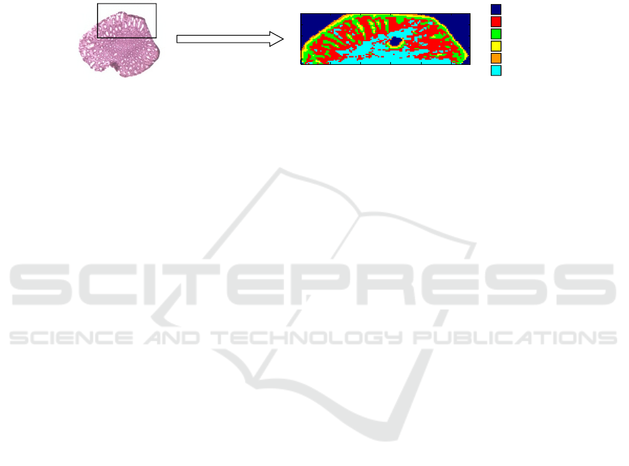
One form of multi-variate analysis is correlation mapping, which enables the visu-
alisation of diseased cells or regions from spectral data (Fig. 2). MINERVA combines
novel mid-IR spectroscopy with correlation mapping and hopes to lead to a break-
through diagnostic technology.
2www.minerva-project.eu
MId- to NEaR infrared spectroscopy
for improVed medical diAgnostics
2
MINERVA newsletter #1 July 2013
Mid-IR spectroscopy: a new tool for pathologists
The spectral region studied in MINERVA (1.5-12 μm) includes the so-called “fingerprint
region” in which many biomolecules have tell-tale absorption peaks. By studying the pattern
of absorbed radiation it is possible to deduce details of the type and distribution of these
molecules, which in turn provides important information for disease diagnosis.
It is emphasised that this process is not as straightforward as simply spotting certain
chemicals, or “cancer markers.” The information is buried in the inter-related distribution of
species and subtle biochemical changes. It requires a powerful mathematical technique
known as multi-variate analysis to extract useful information from the reams of spectral data
in order to spot the warning signs of cancer.
One form of multi-variate analysis is correlation mapping, which enables the visualisation of
diseased cells or regions from spectral data. MINERVA combines novel mid-IR spectroscopy
with correlation mapping and hopes to lead to a breakthrough diagnostic technology.
MINERVA will develop a suite of mid-IR photonic hardware to improve access to this
information. Working in the mid-IR is extremely challenging, and MINERVA will need to break
new ground in several technical areas:
• The project coordinator (G&H) will develop mid-IR components such as fused combiners
(glass devices used to combine or separate signals into different optical fibres), and
acousto-optic modulators (to switch the signals and separate wavelengths at high speed).
• These AO devices will need new types and sizes of calomel crystals from BBT.
Mid-IR spectroscopy
Correlation mapping
CaF
2
Collagen I
Collagen III
DNA
Oleic acid
Albumin
20 40 60 80 100
10
20
30
40
Images courtesy of
Gloucestershire Hospitals
NHS Foundation Trust
• Mid-IR glass fibre to carry the radiation efficiently and conveniently
is being produced at University of Nottingham.
• Novel pump lasers at 2.9 μm and 4.5 μm from LISA Laser will be
used by DTU and NKT to generate a range of supercontinuum
sources in ZBLAN, indium fluoride and chalcogenide glasses,
spanning the MINERVA range from 1.5 μm to 12 μm.
• Xenics and IRnova are advancing the state-of-the-art in Type II
superlattice detectors, which offer a cost effective route to highly
efficient detection in the mid-IR.
• University of Exeter and Gloucestershire Hospitals NHS Trust will
develop the multivariate algorithms and techniques for high volume
screening of human samples.
• WWU Muenster will develop a skin cancer diagnostic process.
• UPVLC (Valencia) is working on the user interface and visualisation.
• The project is managed and administrated by Vivid Components.
Fig. 2. Correlation mapping enables the visualisation of diseased cells or regions from spectra.
MINERVA will develop a suite of mid-IR photonic hardware to improve access to
this information. Working in the mid-IR is extremely challenging, and MINERVA will
need to break new ground in several technical areas:
– Gooch & Housego (G&H), the project coordinator, will develop mid-IR compo-
nents such as fused combiners (glass devices used to combine or separate signals
into different optical fibres), and acousto-optic (AO) modulators (to switch the sig-
nals and separate wavelengths at high speed).
– These AO devices will need new types and sizes of calomel crystals from BBT-
Materials Processing SRO (BBT).
– Mid-IR glass fibre to carry the radiation efficiently and conveniently is being pro-
duced at University of Nottingham (NOTT).
– Novel pump lasers at 2.9 µm and 4.5 µm from LISA Laser Products OHG (LISA)
will be used by the Technical University of Denmark (DTU) and NKT Photonics A/S
(NKT) to generate a range of supercontinuum sources in ZBLAN, indium fluoride
and chalcogenide glasses, spanning the MINERVA range from 1.5 µm to 12 µm.
– Xenics and IRnova are advancing the state-of-the-art in Type II superlattice detec-
tors, which offer a cost effective route to highly efficient detection in the mid-IR.
– University of Exeter and Gloucestershire Hospitals NHS Trust (GHNT) will de-
velop the multivariate algorithms and techniques for high volume screening of hu-
man samples.
– Westfaelische Wilhelms-Universitaet Muenster (WWU) will develop a skin cancer
diagnostic process.
– Universitat Polit
`
ecnica de Val
`
encia (UPV) is working on novel algorithms for the
analysis of histopathological images and the recognition and classification of hiper-
spectral data of cancer samples.
– The project is managed and administrated by Vivid Components.
In the next sections it will be presented an overview of the expectancies of the
project and the main preliminary advances reached by the different groups.
55
MINERVA Project, mid- To near Infrared Spectroscopy for Improved Medical Diagnostics
55
2 Mercurous Halides: Unique Acousto-Optic Materials for IR
from BBT
BBT is a world leader in the growth and processing of Mercurous Chloride (Hg
2
Cl
2
,
Calomel) single crystals with excellent AO properties and is thus in favourable posi-
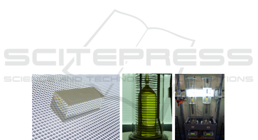
tion to address this question. Calomel single crystals (Fig. 3.a) exhibit a wide range of
optical transmission, high indices of refraction, extremely high value of acousto-optic
figure of merit M2, very low velocity of shear acoustic wave, high value of birefrin-
gence (four times higher than Calcite), etc. The Calomel crystals are well adapted to
fabricate acousto-optic devices operating in the mid and far IR (3 to 20 µm).
At the beginning of the MINERVA project the production technology enabled the
growth of calomel crystal boules with a diameter of 26 to 29 mm (Fig. 3.b) and length
of 45 to 60 mm (typically 55 mm). Within the MINERVA project the new technology
is being developed enabling the growth of cylindrical crystal boules with a diameter up
to 35 mm, which is necessary for the further manufacturing of acousto-optical tuneable
filters of new design proposed by G&H.
The Calomel crystal growth process is highly demanding, difficult and complex, es-
pecially in case of bigger 35mm boules. The growing process is powered by a dynamic
temperature field and corresponding axial and radial temperature gradients. The whole
process has to be carefully maintained within narrow physical condition limits. All the
equipment and accessories have to be newly developed by BBT and adjusted to the spe-
cific conditions for growing of the 36mm diameter crystals including the temperature
controllers. These controllers are equipped with brand new cultivation programs with
respect to the bigger material mass. A total of six crystallizers will be built within the
project. Currently, four units are operational and tested (Fig. 3.c).
Fig. 3. (a) Polished Calomel AOTF substrate. (b) Growing Calomel crystal, diameter 28mm. (c)
Two cultivation crystallizer units with Calomel crystals.
56
EPS Lisbon January 2015 2015 - European Project Space on Intelligent Systems, Pattern Recognition and Biomedical Systems
56
3 Er:ZBLAN Fibre Laser at 2.9 µm from University of
Nottingham and LISA
The partners NOTT and LISA will develop a 2.9 µm laser based on Er-doped ZBLAN
fibres diode-pumped at 976 nm. This fibre laser will be used as pump source for ultra-
long wavelength supercontinuum generation (3-9 µm). The first step is the develop-
ment of a fibre laser in an external cavity configuration. For that purpose a simulation
model based on the rate equation and signal propagation equations is implemented by
the NOTT group (Fig. 4). Different parameters will be studied, e.g. absorption cross-
sections, emission cross-sections, and gain cross-sections, to predict the optimum laser
performance.
3www.minerva-project.eu
MId- to NEaR infrared spectroscopy
for improVed medical diAgnostics
3
MINERVA newsletter #3 May-2014
Er:ZBLAN fibre laser at 2.9 µm
The partners LISA Laser Products OHG (LISA) and Dr.
Slawomir Sujecki‘s team at the University of Nottingham
(NOTT) will develop a 2.9 µm laser based on Er-doped
For more info contact Dr. Samir Lamrini
SLamrini@lisalaser.de
Above: Set-up of the Er:ZBLAN fibre for
absorption studies. The green
fluorescence results from up-conversion
processes.
Above: Set-up of the Er:ZBLAN fibre in an external
cavity configuration pumped with high-power fibre-
coupled diodes. First experiments showed a good
agreement with the simulations carried out at
Nottingham.
Right: Modelling scheme of the
Er:ZBLAN fibre laser with an external
cavity configuration. For the exact
prediction both the 2.9 µm and the
1.6 µm laser signal were analysed in
forward and backward propagation.
ZBLAN fibres diode-pumped at 976 nm. This fibre laser will be used as pump source for ultra-
long wavelength supercontinuum generation(3-9µm).Thefirststepisthedevelopment of a
fibre laser in an external cavity configuration. For that purpose a simulation model based on the
rate equation and signal propagation equations is implemented by the NOTT group (see sketch
above). Different parameters will be studied, e.g. absorption cross-sections, emission cross-
sections, and gain cross-sections, to predict the optimum laser performance.
In parallel, LISA will carry out experiments for the handling (stripping, cleaving, splicing) of the
soft glass fibre and target both high-power and high-energy laser operation with different
resonator configurations. Coated focussing and collimating optics have to be designed and
manufactured for the laser studies. After the evaluation of the first tests in CW operation LISA‘s
scientists and engineers will design a compact and robust cooled housing for the 2.9 µm laser.
Regarding high-energy operation special acousto-optic modulators (AOM) based on TeO
2
will
be designed and built by G&H and delivered to LISA.
Fig. 4. Modelling scheme of the Er:ZBLAN fibre laser with an external cavity configuration. For
the exact prediction both the 2.9 µm and the 1.6 µm laser signal were analysed in forward and
backward propagation.
In parallel, LISA will carry out experiments for the handling (stripping, cleaving,
splicing) of the soft glass fibre and target both high-power and high-energy laser oper-
ation with different resonator configurations (Fig. 5).
Fig. 5. (a) Set-up of the Er:ZBLAN fibre for absorption studies. The green fluorescence results
from up-conversion processes. (b) Set-up of the Er:ZBLAN fibre in an external cavity configura-
tion pumped with high-power fibre-coupled diodes. First experiments showed a good agreement
with the simulations carried out at Nottingham.
Coated focussing and collimating optics have to be designed and manufactured for
the laser studies. After the evaluation of the first tests in CW operation LISA’s scientists
and engineers will design a compact and robust cooled housing for the 2.9 µm laser.
57
MINERVA Project, mid- To near Infrared Spectroscopy for Improved Medical Diagnostics
57
Regarding high-energy operation special acousto-optic modulators (AOM) based on
TeO2 will be designed and built by G&H and delivered to LISA.
Further information about MINERVA’s fibre laser can be found in [2–10].
4 Extreme IR Supercontinuum Modelling at DTU
DTUs team has the task of fibre modelling in MINERVA in close collaboration with
the fibre manufacturing group at NOTT. The DTU group also models dynamic super-
continuum generation along the fibres using both measured material data and calculated
fibre properties. This advanced modelling requires extensive computational resources
in order to accurately follow the rapid spectral broadening, which covers over four oc-
taves (from 1 µm to 16 µm); made possible by the strong non-linearity of chalcogenide
glasses and the extremely high numerical aperture (NA) of the Nottingham fibres. Fig-
ure 6 shows two graphics with some results of the numerical modeling of mid-IR su-
percontinuum generation. Thorough analysis of the modelling has been presented in
[11–13].
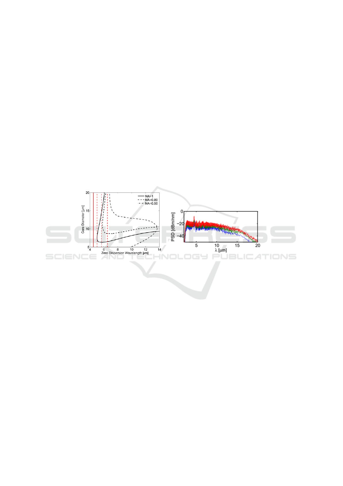
Fig. 6. (a) Zero dispersion wavelengths versus core diameter for step-index fibres (based on fibres
fabricated at the University of Nottingham) with NA as given in the legend. (b) Modelling shows
that a fibre with core diameter 10 µm and NA = 1.0 exhibiting no second zero dispersion is
optimum. Super-continuum generation beyond 12 µm is observed numerically.
5 MINERVA Supercontinuum Sources from NKT
It has been mentioned that the mid-IR region contains a wealth of spectral data which
can yield important information on the chemical composition of samples from gases
and liquids to living cells. However, the investigation of this topic has been limited by
the available photonic sources. Researchers had to choose between a very low intensity
broadband source such as a “globar” thermal source, or a high intensity but narrowband
source, such as a laser diode.
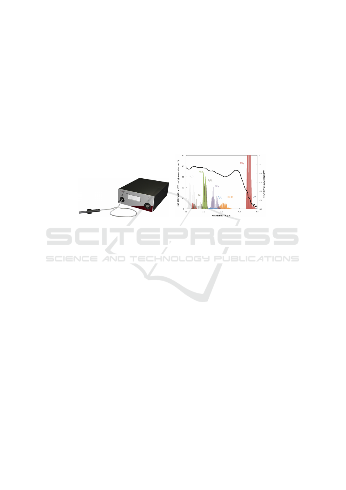
NKT Photonics is dedicated to providing flexible sources of high intensity light in
an easy to use format (Fig. 7.a). It has already established commercial supercontin-
uum systems which can deliver any wavelength from 400 to 2000 nm on demand. It
has recently launched the EXTEND-UV accessory which can extend the wavelength
58
EPS Lisbon January 2015 2015 - European Project Space on Intelligent Systems, Pattern Recognition and Biomedical Systems
58
coverage to cover the 270-400 nm UV region. The company would now like to push
the limits of supercontinuum sources at longer wavelengths, reaching into the mid-IR
region.
In MINERVA NKT is developing zirconium fluoride (ZrF
4
) glass fibre supercon-
tinuum sources to cover the 1.5-4.5 µm spectrum. Subsequently it will investigate even
longer wavelengths by utilising newly developed indium fluoride (InF
3
) fibres to extend
the spectrum beyond the transmission band of ZrF
4
glasses.
These sources could detect changes in cells by monitoring absorption in the 2.6-
3.8 µm region which relates to the balance between lipids and proteins (Fig. 8.a). The
increase in wavelength from 4.5 µm up to >5 µm would make it possible to interrogate
additional important gas absorption lines such as carbon monoxide.
Fig. 7. (a) Schematic of a typical NKT supercontinuum source. (b) Graph showing absorption
spectra of some key bio-molecules in the lower end of the “fingerprint region”.
In the first 2 years of the MINERVA project NKT has already developed several
supercontinuum sources with output power of up to 2.5 W. These sources are more
than a million times brighter than most thermal light sources and even brighter than a
Synchrotron. We have shown the limits of zirconium-fluoride based systems by setting a
new record for the longest wavelength supercontinuum generated at 4.75 µm. However,
the chemometric specialists in MINERVA found that the main region of interest was the
2.5-3.8 µm region so we have also shown how the main power in the output spectrum
can be shifted down to the main region of interest by altering the design of the nonlinear
fiber. These first Supercontinuum sources are already at work in the development of the
MINERVA-lite microscopy setup which will soon be applied to bio sample imaging.
Meanwhile NKT is pushing onward in the development of the mid-IR supercon-
tinuum sources. The initial sources were based on rather long pulse nanosecond pump
lasers with relatively low pulse repetition frequencies. This made the sources incompat-
ible with most of the Fourier transform spectrometers (FTIRs) that many researchers
use in the mid-IR region. In addition, the low repetition rate made it time consuming to
counter any noise in the source by averaging over many pulses. NKT is therefore now
developing sources based on much shorter pump pulses and with higher repetition rate
in order to reduce noise and make the sources compatible with standard FTIRs.
As these mid-IR supercontinuum sources become available and known in the field,
the MINERVA consortium expects the emergence of new markets. For example, an
59
MINERVA Project, mid- To near Infrared Spectroscopy for Improved Medical Diagnostics
59

important spectroscopic application in the petrochemical industry is to monitor single
wavelengths in the 3-3.5 µm band in order to optimise the refining processes. Monitor-
ing the whole spectrum simultaneously would allow a full real-time chemical analysis
of the output chemicals.
Some relevant references concerning the supercontinuum sources within the MIN-
ERVA framework have been already published [14–24]
6 MINERVA type-II Superlattice IR Detectors from IRnova
Type-II superlattice (T2SL) is a material/technology that can be used for high quality
cooled photon detectors, with tailorable bandgap from 2 µm and upwards. The name
comes from the fact that the conduction and valence bands display a so-called “bro-
ken type-II” (sometimes also called “type-IIb” or even “type-III”) alignment between
the constituent materials, which can be InAs/GaSb/AlSb, or alloys thereof (Fig. 8.a).
In contrast to typical quantum well devices, e.g. the active regions of semiconductor
lasers, the superlattice layers in the T2SL material are so thin (typically 3 nm) that
mini-bands are formed in the material. These mini-bands resemble the conduction and
valence bands of a bulk semiconductor material. By carefully selecting the superlat-
tice layer thicknesses and compositions, novel materials can be defined to meet widely
different needs.
irn054479-2
Utskrift: 2013-05-20
(Mallutgåva 2011-02-02)
Date/Datum
2013-05-20
Rev
3
Page/Sida
1
Approved/Godkänd
Type of document/Dokumenttyp
REPORT
Issued by/Utfärdare
Carl Asplund / Henk Martijn
Distribution
For information only/För kännedom
IR detection using type-II superlattice photodiodes
T2SL (Type II SuperLattice) or sometimes also called SLS (Strained Layer Superlattice) is a material / technology
that can be used to make high quality cooled infrared photon detectors with a cut-off wavelength ranging from 2 µm
to 30 µm. This covers the SWIR, MWIR, LWIR and VLWIR wavelength bands, loosely defined as 2-3 µm, 3-5 µm,
8-12 µm and >12 µm, respectively.
A superlattice is a system made of a repeating sequence of thin layers of different materials. If the layer thicknesses
are small enough in a quantum mechanical sense, minibands are formed in the material. The result is an artificial
material with properties that can be engineered; in the detector case the bandgap energy corresponding to the desired
cut-off wavelength. When two semiconductors are brought in contact, there are several ways the valance and
conduction bands can align. If both the valence and the conduction band edge of the second material are above the
band edges of the first material, it is called a broken type II band alignment.
Ec
Ev
GaSb
InAs
GaSb GaSb
InAs
Ec
Ev
GaSb
InAs
GaSb GaSb
InAs
Figure 1 Band alignment of InAs GaSb and the forming of minibands.
The III/V compound materials InAs and GaSb form such a band alignment (see Figure 1). As can be seen in Figure 2,
InAs has a lattice mismatch of less than 1% on GaSb. Starting with GaSb substrates alternating layers of InAs and
GaSb with atomic layer precision can be deposited using MBE (Molecular Beam Epitaxy). By interface engineering
(create an interface layer of InSb) or using more complicated superlattices like Ga
x
In
1-x
Sb/InAs, thick strain
compensated structures with high crystal quality can be grown. If doping in the form of trace amounts of Be, Te or Si
is incorporated, photovoltaic p-i-n structures that can be used to detect IR radiation of the desired wavelength are
formed.
Fig. 8. (a) Schematic of T2SL band-gap structure. (b) Detector/Dewar/cooler assembly for T2SL
from IRnova.
Compared with a traditional bulk material for the 3-5 µm range, such as InSb, T2SL
requires less cooling and thus draws less power, which allows for longer cooler lifetime
and consequently lower life-cycle cost. For the 8-12 µm range, the traditional alloy bulk
material HgCdTe (or “MCT”) is difficult to fabricate with high yield, partly due to the
extreme sensitivity of the bandgap to composition (particularly the HgTe:CdTe alloy
ratio). Here T2SL materials have distinct advantages in fabrication.
Focal plane arrays comprising hundreds of thousands of T2SL detector pixels are
flip-chip bonded to a CMOS read-out-circuit and then mounted on a ceramic carrier,
which in turn is glued to a cold finger in a vacuum Dewar housing, complete with an IR
window. The cold finger is cooled to detector operating temperature by a Stirling rotary
cooler. IRnovas detector-Dewar-cooler assembly for T2SL can be seen in Fig. 8.b.
IRnova has recently worked on improving the quantum efficiency (QE) of the de-
tection by applying anti-reflective coatings to the detector surface. By this method, the
60
EPS Lisbon January 2015 2015 - European Project Space on Intelligent Systems, Pattern Recognition and Biomedical Systems
60
QE was increased from approximately 50% to 80% in the wavelength region of interest.
This improves the signal-to-noise ratio and allows a reduced integration time for each
image frame.
Apart from MINERVA applications, IRnova plans to use T2SL technology for gas
detection of key greenhouse gases with absorption lines in the atmospheric transmission
bands, such as methane and perhaps also sulphur hexafluoride (SF6).
More information about the superlatice IR detectors can be found in [25]
7 Infrared Megapixel Camera Development at Xenics and IRnova
The sensing unit for MINERVA is being developed in a joint effort between Xenics and
IRnova. From the start of the project Xenics has been working on the design of a Read-
Out IC (ROIC), to be integrated through flip-chip technology with the T2SL (Type-2
Super Lattice) photodiode material, which is being developed by IRnova.
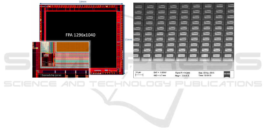
Fig. 9. (a) Global and zoomed view of the designed ROIC, currently in manufacturing. (b) Mi-
croscopic view of manufactured array of T2SL photo diodes.
To provide sufficient resolution for reliable data analysis, a 1280×1024 pixel array
was chosen, on an aggressive pitch of 12 µm. Project requirements including frame
rate, sensitivity and noise level were taken into account in the design process. After
extensive simulations and test sample manufacturing, the final design was taped out to
a manufacturing foundry (Fig. 9.a). The use of advanced 0.18 µm CMOS technology
is required to allow all necessary functionality within the available space of 144 µm
2
per pixel. The first wafers are currently available for post-processing to be followed by
wafer-level verification of the electrical functionality.
In parallel, IRnova has been working on the optimisation of the design and pro-
cessing of the T2SL material, towards cut-off wavelength matching and dark current
minimisation (Fig. 9.b). Once the ROIC chips become available later this year, they
will be hybridized to the sensor chip, and IRnova will integrate the resulting hybrid in
a Sterling-cooled Dewar. The so-called engine will in its turn be integrated into a full
camera by Xenics, to be supplied to the other MINERVA project partners to be used for
capturing spectroscopic images of prepared tissue samples and live cell phantoms.
61
MINERVA Project, mid- To near Infrared Spectroscopy for Improved Medical Diagnostics
61
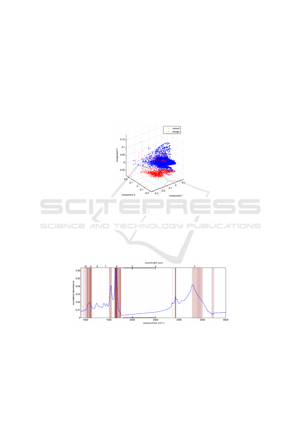
8 Pattern Recognition and Data Analysis at GHNT
The first task of GHNT was to provide supporting evidence for the MINERVA instru-
ment specifications. This was achieved by analysing an existing dataset and applying
pattern recognition techniques to discriminate between benign and cancerous samples
from human colon tissue biopsies (Fig. 10). Sensitivity and specificity of up 86-99%
can be achieved with the existing dataset. Using this study as a baseline GHNT was
able to assess the impact of various factors that will affect the quality and speed of the
MINERVA instrument.
Fig. 10. Partial Least Squares (PLS) scores plot showing the separation between benign and can-
cer samples in baseline study.
Reducing the number of data points per spectrum is one way to potentially speed
up the system; measuring fewer wavenumbers means a faster total acquisition time.
Multivariate pattern recognition algorithms were used to identify potential wavenum-
ber targets for the MINERVA instrument. Figure 11 shows the wavenumber regions
identified as ‘important’ for the baseline study.
Fig. 11. VIP identified wavenumber targets for the MINERVA system (red). Reference spectrum
(blue).
62
EPS Lisbon January 2015 2015 - European Project Space on Intelligent Systems, Pattern Recognition and Biomedical Systems
62
A minimum acquisition time per spectrum means that the MINERVA instrument
will be able to rapidly assess samples in a clinical timeframe. However, reducing acqui-
sition time also increases the amount of noise. To determine what level of noise can be
tolerated by the pattern recognition algorithms GHNT simulated the addition of noise to
the baseline study until it was no longer able to discriminate between pathology groups.
This allowed a minimum target signal-to-noise ratio (SNR) to be determined for the
MINERVA instrument whilst maintaining an acceptable ability to discriminate between
pathology types.
9 High Resolution mid-IR Imaging at University of Exeter
One of the main objectives of Exeters group within the MINERVA project is large scale
pathology screening using mid-infrared (mid-IR) spectroscopy. Currently FTIR spectral
histopathology, which has the potential to develop as a cancer diagnostic tool, is carried
out using a heated silicon carbide rod (“Globar”) as the mid-IR light source and focal
plane array (FPA) based detectors. This technology is limited by the low flux of the
light source and the limited tissue area that can be measured in a given amount of time.
The novel technologies being developed in the MINERVA project; consisting of
mid-IR super-continuum light source (instead of a “Globar”) and new generation mega-
pixel (FPA) detectors (instead of a 128×128 pixel FPA), will be tested on pathological
samples at the University of Exeter.
Currently the base instrument, a commercially available Agilent FTIR imaging sys-
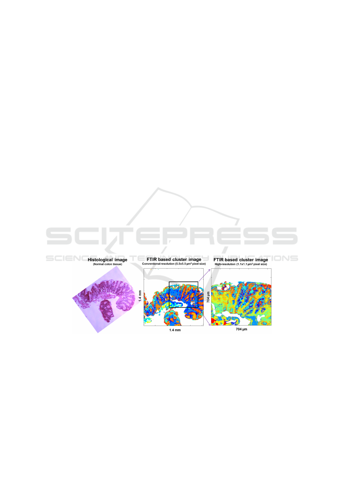
tem, in addition to the conventional Globar source coupled to FPA based imaging, has
been retro-fitted with a new high-resolution imaging capability. The FTIR images ac-
quired using this set-up provided a five-fold improvement in image resolution from 5.5
µm
2
of the current technology to 1.1 µm
2
using the high magnification optics (Fig. 12).
Fig. 12. FTIR based K-means cluster images obtained using conventional and high-resolution
imaging compared with the histological image. Histological features based on the bio-molecular
composition are partitioned in the cluster images. In the high-resolution imaging, tissue and cel-
lular features are more apparent.
Future work in MINERVA will combine these novel technologies for large scale
pathology screening, and also high-resolution imaging in tissue regions of interest, with
63
MINERVA Project, mid- To near Infrared Spectroscopy for Improved Medical Diagnostics
63
the aim to develop faster and accurate cancer diagnostic tools. Initially this will integrate
with a 4.5 µm NKT MINERVA source, and later in the project it will be extended to
very long mid-IR wavelengths: possibly out beyond 10 µm.
10 Development of Standardised Samples for mid-IR
Spectrometer Instruments Testing at WWU
A key task of WWU is to transfer the MINERVA technologies to skin diagnostics and to
use mid-IR spectroscopy for the fast screening of human body surfaces and identifica-
tion of patho-physiologically altered cells and tissue lesions. This requires standardised
cell and tissue sample standards with marker spectra for technology performance test-
ing of the novel optical components and systems and for training of novel approaches
for advanced data analysis.
The work of WWU in the first MINERVA project period thus focused on the estab-
lishment of standard samples with representative spectral information of human skin
and skin cancer cells. WWU has established cell culture models which represent major
cellular skin constituents and skin cancer cell types. Furthermore, sample preparation
procedures on mid-IR compatible substrates have been developed that allow long-term
storage of cell lines without significant losses in the quality of the spectral properties.
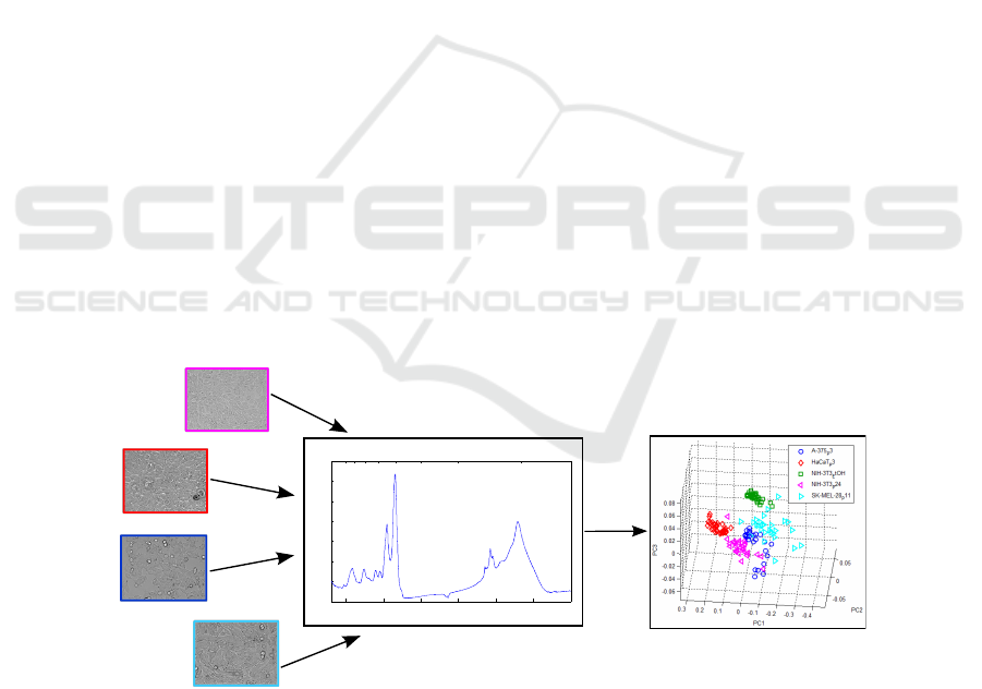
In order to identify suitable marker spectra of human skin, sample sets with different
preparations and cell types were analysed with mid-IR spectroscopy in collaborative
work with GHNT to retrieve reference data for technology performance testing and
for the evaluation novel algorithms for sample analysis and classification developed
by UPV. The principle component analysis (PCA) of mid-IR spectra from different
cell types shows an excellent distinct grouping of skin components as fibroblasts and
keratinocytes and cancer cells.
10 9 8 7 6 5 4 3
wavelength (µm)
1000 1500 2000 2500 3000 3500 4000
0
0.1
0.2
0.3
0.4
0.5
0.6
wavenumber (cm
−1
)
absorbance
fibroblasts (NIH-3T3)
keratinocytes (HaCaT)
melanoma cells (A-375)
melanoma cells (SK-MEL-28)
mid-IR spectroscopy
numerical analysis
Fig. 13. Principle component analysis (PCA) of mid-IR spectral data from fibroblasts (NIH-3T3),
keratinocytes (HaCaT) and skin cancer cells (A-375, SK-MEL-28) illustrates the differentiation
between different cell types.
64
EPS Lisbon January 2015 2015 - European Project Space on Intelligent Systems, Pattern Recognition and Biomedical Systems
64
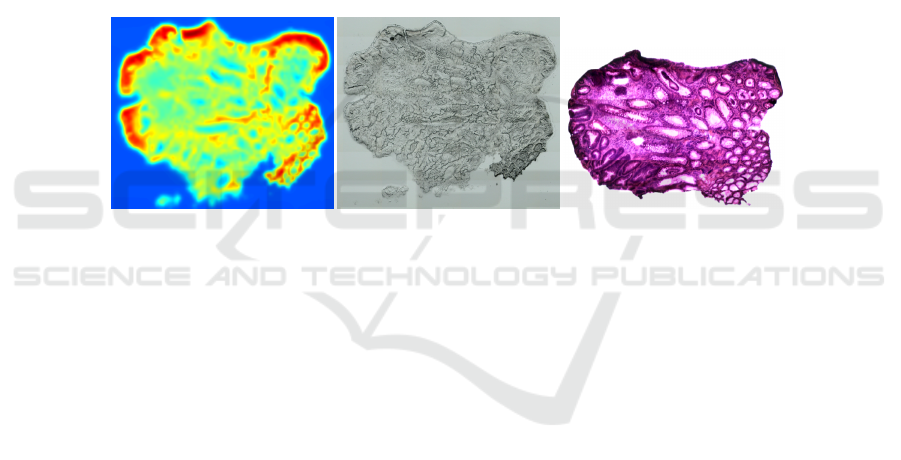
Figure 13 illustrates the analysis and differentiation of different cell types (cancer/
non-cancer) that have been prepared at WWU for the example of results PCA of mid-IR
spectral data from fibroblasts (NIH-3T3), keratinocytes (HaCaT), and skin cancer cells
(A-375, SK-MEL-28). Based on these results, current and future activities at WWU in
MINERVA focus on the development of novel mid-IR standards models for skin cancer
detection that are based on 3D human skin equivalents in vitro. Further information
about the standardization of the cell samples was presented in [26].
11 First Steps with MINERVA Image Processing at UPV
The first objectives of the image and signal processing group at UPV are focused on
segmentation and registration of different kinds of images (Fig. 14): infra-red spectral
images (IR), white light (WL), and those most used by clinicians at present, the haema-
toxylin and eosin (H&E) stained images. The latter is the current “gold standard” used
to distinguish between a healthy or pathological patient sample.
Fig. 14. (a) Infra-red (IR) image. (b) White light (WL) image. (c) Haematoxylin and eosin (H&E)
image.
The objective within MINERVA is to automatically segment regions of interest
(healthy and pathological) in the H&E images and look for their features in the in-
frared spectrum. To achieve this goal the H&E image must be registered with the WL
image (which is already registered with the infrared volume). So, the work is focused
on two interactive steps: registration and segmentation.
Registration allows the matching of elements that clinicians considered important
in the H&E images with the spectral images. A successful registration task would allow
users to learn, and later identify, the areas from which diseased and healthy cells and
patients can be distinguished (Fig. 15.a).
Segmentation concerns the accurate extraction of the cell contours (Fig. 15.b). This
would reduce the huge amount of data to be analysed looking for subtle biochemical
changes (“cancer markers”). Once the contours have been identified, the regions must be
classified as healthy or cancerous depending on subtle features including shape, texture
and clustering. This is an extremely difficult task, but the use of the spectral information
in the mid-IR should eventually aid clinicians to improve on the current gold standard.
More details about the work on image processing in MINERVA project was pre-
sented in [27, 28].
65
MINERVA Project, mid- To near Infrared Spectroscopy for Improved Medical Diagnostics
65
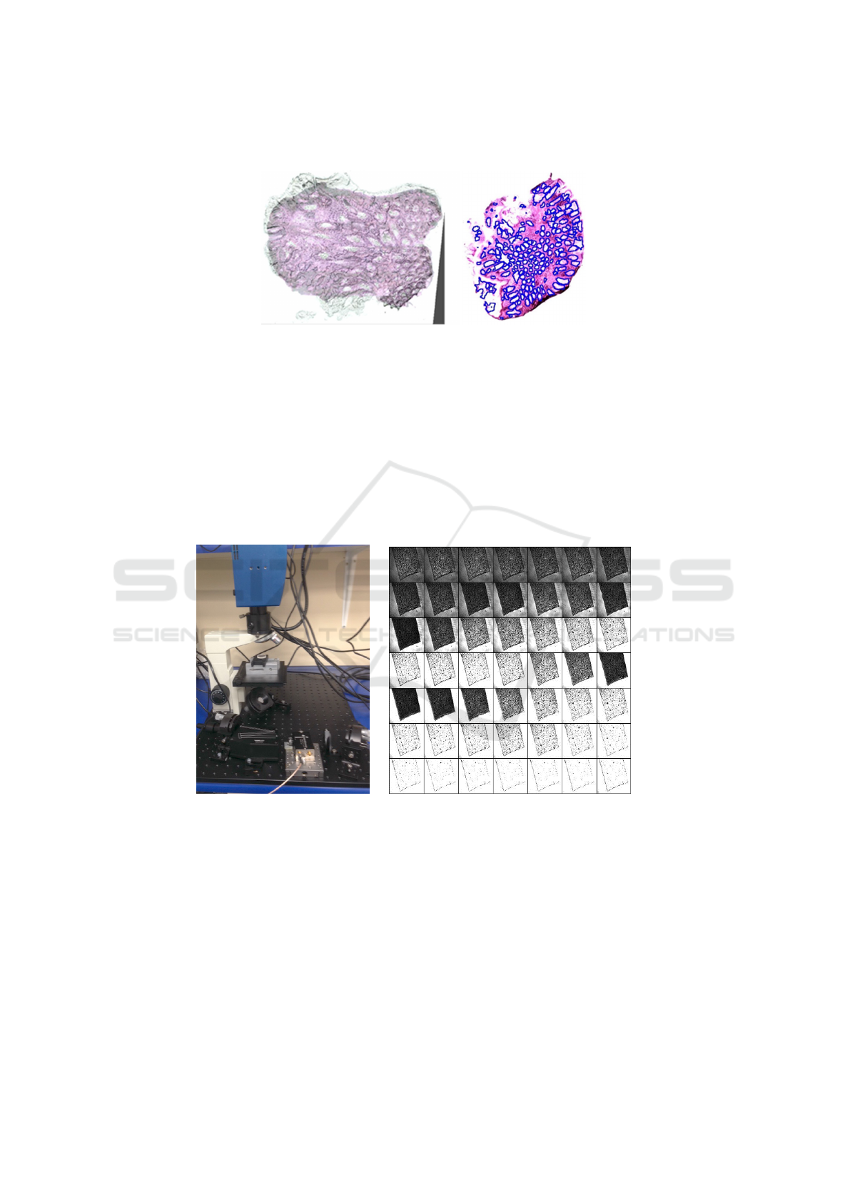
Fig. 15. (a) Projective registration test. (b) Segmentation sample test.
12 MINERVA Lite
A prototype MINERVA system that operates in the 2-4.5 µm wavelength band has been
assembled so that the individual parts being developed by partners in the project can
be evaluated together. The final MINERVA system will operate at even longer wave-
lengths. The key components in the integrated system are: NKT supercontinuum source,
G&H acousto-optic tunable filter, Xenics IR camera, commercial microscope and IR
optics and control electronics.
Fig. 16. (a) Photo of part of the “MINERVA Lite” laboratory set-up. (b) 0.3 Mpixel images at 49
wavelengths which can be used to form an (x, y, λ) image cube in 0.6 s.
The breadboard system is shown on Fig. 16.a. This system can take 0.3 megapixel
images with 20 µm spatial resolution at a rate of 85 frames per second. Each image is
taken at a different wavelength so that a set of 49 spectral images can be built up in
0.6 s (Fig. 16.b). These images can form an (x, y, λ) “image cube”. Each pixel records
a spectrum and this has enabled MINERVA researchers to identify a polymer film in
66
EPS Lisbon January 2015 2015 - European Project Space on Intelligent Systems, Pattern Recognition and Biomedical Systems
66

the sample image. This important preliminary result will be extended in MINERVA to
identify spectra from cancerous cells in tissue samples and in real time on live patients.
More related work in MINERVA project has been published in [29–31]. In addition,
the research done in MINERVA has been mention and reviewed in [32, 33].
References
1. Minerva | MId- to NEaR infrared spectroscopy for improVed medical diAgnostics.
(http://minerva-project.eu/) Accessed: 2015-05-22.
2. Seddon, A.B.: Mid-infrared (IR)–A hot topic: The potential for using mid-IR light for non-
invasive early detection of skin cancer in vivo. Physica Status Solidi (B) 250 (2013) 1020–
1027
3. Oladeji, A., Sojka, L., Tang, Z., Furniss, D., Phillips, A., Seddon, A., Benson, T., Sujecki, S.:
Numerical investigation of mid-infrared emission from Pr
3+
doped GeAsGaSe fibre. Optical
and Quantum Electronics 46 (2014) 593–602
4. Sakr, H., Tang, Z., Furniss, D., Sojka, L., Moneim, N., Barney, E., Sujecki, S., Benson, T.,
Seddon, A.: Towards mid-infrared fiber-lasers: rare earth ion doped, indium-containing, se-
lenide bulk glasses and fiber. In: SPIE BiOS, International Society for Optics and Photonics
(2014) 89380V
5. Sojka, L., Tang, Z., Furniss, D., Sakr, H., Oladeji, A., Bere
´
s-Pawlik, E., Dantanarayana, H.,
Faber, E., Seddon, A., Benson, T., et al.: Broadband, mid-infrared emission from Pr
3+
doped
GeAsGaSe chalcogenide fiber, optically clad. Optical Materials 36 (2014) 1076–1082
6. Sujecki, S.: An efficient algorithm for steady state analysis of fibre lasers operating under
cascade pumping scheme. International Journal of Electronics and Telecommunications 60
(2014) 143–149
7. Dantanarayana, H.G., Abdel-Moneim, N., Tang, Z., Sojka, L., Sujecki, S., Furniss, D., Sed-
don, A.B., Kubat, I., Bang, O., Benson, T.M.: Refractive index dispersion of chalcogenide
glasses for ultra-high numerical-aperture fiber for mid-infrared supercontinuum generation.
Optical Materials Express 4 (2014) 1444–1455
8. Seddon, A.: Mid-infrared photonics for early cancer diagnosis. In: Transparent Optical
Networks (ICTON), 2014 16th International Conference on, IEEE (2014) 1–4
9. Sakr, H., Furniss, D., Tang, Z., Sojka, L., Moneim, N., Barney, E., Sujecki, S., Benson, T.,
Seddon, A.: Superior photoluminescence (PL) of Pr
3+
-In, compared to Pr
3+
-Ga, selenide-
chalcogenide bulk glasses and PL of optically-clad fiber. Optics express 22 (2014) 21236–
21252
10. Sujecki, S., Oladeji, A., Sojka, L., Phillips, A., Seddon, A., Benson, T., Sakr, H., Tang,
Z., Furniss, D., Scholle, K., et al.: Modelling and design of mir chalcogenide glass fibre
lasers. In: Numerical Simulation of Optoelectronic Devices (NUSOD), 2014 14th Interna-
tional Conference on, IEEE (2014) 111–112
11. Agger, C., Kubat, I., Møller, U., Moselund, P.M., Petersen, C., Napier, B., Seddon, A., Su-
jecki, S., Benson, T., Farries, M., et al.: Numerical demonstration of 3-12µm supercontinuum
generation in large-core step-index chalcogenide fibers pumped at 4.5 µm. In: Nonlinear Op-
tics, Optical Society of America (2013) NW4A–09
12. Møller, U., Yu, Y., Petersen, C.R., Kubat, I., Mechin, D., Brilland, L., Troles, J., Luther-
Davies, B., Bang, O.: High Average Power Mid-infrared Supercontinuum Generation in a
Suspended Core Chalcogenide Fiber. In: Nonlinear Photonics, Optical Society of America
(2014) JM5A–54
13. Møller, U., Yu, Y., Kubat, I., Petersen, C.R., Gai, X., Brilland, L., M
´
echin, D., Caillaud, C.,
Troles, J., Luther-Davies, B., et al.: Multi-milliwatt mid-infrared supercontinuum generation
in a suspended core chalcogenide fiber. Optics express 23 (2015) 3282–3291
67
MINERVA Project, mid- To near Infrared Spectroscopy for Improved Medical Diagnostics
67

14. Thomsen, C.L., Nielsen, F.D., Johansen, J., Pedersen, C., Moselund, P.M., Møller, U.,
Sørensen, S.T., Larsen, C., Bang, O.: New horizons for supercontinuum light sources:
from UV to mid-IR. In: SPIE OPTO, International Society for Optics and Photonics (2013)
86370T
15. Moller, U., Bang, O.: Intensity noise of normal-pumped picosecond supercontinuum gener-
ation. In: Lasers and Electro-Optics Europe (CLEO EUROPE/IQEC), 2013 Conference on
and International Quantum Electronics Conference, IEEE (2013) 1
16. Kubat, I., Agger, C., Moselund, P., Bang, O.: Mid-infrared supercontinuum generation in ta-
pered ZBLAN fiber with a standard Erbium mode-locked fiber laser. In: Lasers and Electro-
Optics Europe (CLEO EUROPE/IQEC), 2013 Conference on and International Quantum
Electronics Conference, IEEE (2013) 1
17. Kubat, I., Agger, C., Moselund, P.M., Bang, O.: Optimized ZBLAN fiber for efficient and
broadband mid-infrared supercontinuum generation through direct pumping at 1550nm. In:
1st International Workshop on Spatio-Temporal Complexity in Optical Fibers. (2013)
18. Kubat, I., Agger, C.S., Moselund, P.M., Bang, O.: Mid-infrared supercontinuum generation
to 4.5 µm in uniform and tapered ZBLAN step-index fibers by direct pumping at 1064 or
1550 nm. JOSA B 30 (2013) 2743–2757
19. Moselund, P., Petersen, C., Leick, L., Seidelin Dam, J., Tidemand-Lichtenberg, P., Pedersen,
C.: Highly Stable, All-fiber, High Power ZBLAN Supercontinuum Source Reaching 4.75
µm used for Nanosecond mid-IR Spectroscopy. In: Advanced Solid State Lasers, Optical
Society of America (2013) JTh5A–9
20. Møller, U.V., Sørensen, S.T., Petersen, C.R., Kubat, I., Moselund, P.M., Bang, O.: Supercon-
tinuum generation from ultraviolet to mid-infrared. (In: 15th Conference on Optical Fibers
and Their Applications (OFTA 2014))
21. Kubat, I., Rosenberg Petersen, C., Møller, U.V., Seddon, A., Benson, T., Brilland, L., M
´
echin,
D., Moselund, P.M., Bang, O.: Thulium pumped mid-infrared 0.9–9µm supercontinuum
generation in concatenated fluoride and chalcogenide glass fibers. Optics express 22 (2014)
3959–3967
22. Kubat, I., Petersen, C.R., Møller, U., Seddon, A., Benson, T., Brilland, L., M
´
echin, D.,
Moselund, P., Bang, O.: Mid-infrared supercontinuum generation in concatenated fluoride
and chalcogenide glass fibers covering more than three octaves. In: CLEO: Science and
Innovations, Optical Society of America (2014) STh3N–1
23. Kubat, I., Agger, C.S., Møller, U., Seddon, A.B., Tang, Z., Sujecki, S., Benson, T.M., Furniss,
D., Lamrini, S., Scholle, K., et al.: Mid-infrared supercontinuum generation to 12.5 µm in
large na chalcogenide step-index fibres pumped at 4.5 µm. Optics express 22 (2014) 19169–
19182
24. Petersen, C.R., Møller, U., Kubat, I., Zhou, B., Dupont, S., Ramsay, J., Benson, T., Sujecki,
S., Abdel-Moneim, N., Tang, Z., et al.: Mid-infrared supercontinuum covering the 1.4–13.3
µm molecular fingerprint region using ultra-high NA chalcogenide step-index fibre. Nature
Photonics 8 (2014) 830–834
25. Martijn, H., Asplund, C., von W
¨
urtemberg, R.M., Malm, H.: High performance MWIR
type-II superlattice detectors. In: Proc. of SPIE Vol. Volume 8704. (2013) 87040Z–1
26. Kastl, L., Rommel, C.E., Kemper, B., Schnekenburger, J.: Standardized cell samples for
midir technology development. In: SPIE BiOS, International Society for Optics and Photon-
ics (2015) 931507
27. Naranjo, V., Villanueva, E., Lloyd, G.R., Stone, N., Lopez-Mir, F., Alcaniz, M.: Stained
and infrared image registration as first step for cancer detection. In: Biomedical and Health
Informatics (BHI), 2014 IEEE-EMBS International Conference on, IEEE (2014) 420–423
28. L
´
opez-Mir, F., Naranjo, V., Morales, S., Angulo, J.: Probability density function of object
contours using regional regularized stochastic watershed. In: Image Processing (ICIP), 2014
IEEE International Conference on, IEEE (2014) 4762–4766
68
EPS Lisbon January 2015 2015 - European Project Space on Intelligent Systems, Pattern Recognition and Biomedical Systems
68

29. Stevens, G., Woodbridge, T.: Development of low loss robust soft-glass fiber splices. In:
Workshop on Specialty Optical Fibers and their Applications, Optical Society of America
(2013) W3–21
30. Markos, C., Kubat, I., Bang, O.: Hybrid polymer photonic crystal fiber with integrated
chalcogenide glass nanofilms. Scientific reports 4 (2014)
31. Valle, S., Ward, J., Pannell, C., Johnson, N.: Acousto-optic tunable filter for imaging ap-
plication with high performance in the ir region. In: SPIE OPTO, International Society for
Optics and Photonics (2015) 93590E–93590E
32. Maragkou, M.: Supercontinuum: Reaching the mid-infrared. Nature Photonics 8 (2014)
746–746
33. Steinmeyer, G., Skibina, J.S.: Supercontinuum: Entering the mid-infrared. Nature Photonics
8 (2014) 814–815
69
MINERVA Project, mid- To near Infrared Spectroscopy for Improved Medical Diagnostics
69
