
Remote Heart Rate Determination in RGB Data
An Investigation using Independent Component Analysis and Adaptive Filtering
Christian Wiede, Julia Richter, Andr
´
e Apitzsch, Fajer KhairAldin and Gangolf Hirtz
Department of Electrical Engineering and Information Technology,
Chemnitz University of Technology, Reichenhainer Str. 70, 09126 Chemnitz, Germany
Keywords:
Heart Rate Detection, rPPG, Vital Parameters, Image Processing, Independent Component Analysis, Adaptive
Filtering.
Abstract:
An emerging topic in the field of elderly care is the determination and tracking of vital parameters, such as
the heart rate. This parameter provides important information about a person’s current health status. Within
the last years, various research focussed on this topic. The recognition of vital parameters is increasingly
relevant for our ageing society. This paper presents a method to remotely determine the human heart rate with
a camera. At this point, we suggest to use independent component analysis (ICA) and adaptive filtering for a
robust detection. In our processing chain, we used different image processing techniques, e. g. face detection,
and signal processing techniques, e. g. FFT and bandpass filtering, in this study. An evaluation with several
probands, illuminations, frame rates and different heart rate levels showed that we could achieve a mean error
of 4.36 BPM, which corresponds to CAND of 94.45 %, and a speed of 35 fps.
1 INTRODUCTION
In a steadily ageing society, taking care for el-
derly plays a major role. In the last years, several
technical assistance systems have been developed to
help elderly people in their daily activities (Meinel
et al., 2015) and to call help in case of emergencies
(Wohlrab et al., 2015). These works show that it is
possible to detect a fall in the home environment and
inform relatives or caregivers. However, these sys-
tems only act after the emergency occurred. The goal
of our project is to detect a person’s current health
status and act pre-emptively in case of indications for
possible emergencies. Thus far, current assistant sys-
tems are not able to perform this. They are not able to
decide whether a person is sleeping in an armchair or
has suffered a circulatory collapse and is unconscious.
One possibility to overcome this problem is to de-
tect human vital parameters by means of optical sen-
sors. These so-called physiological parameters (e. g.
heart rate, breath rate and oxygen saturation) are de-
tectable with normal cameras due to the fact that the
spatial and temporal resolution of the cameras in-
creased in the last years. Especially the heart rate de-
tection drew attention in recent research (Poh et al.,
2010; van Gastel et al., 2014). Inspired by these
works, this paper presents a novel possibility to ro-
bustly detect the heart rate. We used ICA and an
adaptive filtering to robustly detect the heart rate from
remote. Furthermore, we realised a real-time imple-
mentation, so that this method is suitable for elderly
care applications.
A high benefit of this method is its contact-less
working mode. This method proved to be very con-
venient for patients, because they do not have to wear
any devices. In that way, effects such as skin irri-
tations and discomfort can be avoided. Measuring
the heart rate allows the detection of bradycardia and
tachycardia at a very early stage, so that emergencies
can be avoided. Besides helping elderly people, such
a system would be most beneficial for detecting the
health status of neonatals to avoid sudden infant death
syndrome, for monitoring a driver’s well-being or for
triage in hospitals.
This paper is organised as follows. Firstly, there
will be an overview of related studies in this field.
Secondly, we introduce our method for remote heart
rate detection. Thereupon, we present our results,
which is followed by a discussion. Finally, we sum-
marise our findings and give an outlook on further de-
velopments.
240
Wiede, C., Richter, J., Apitzsch, A., KhairAldin, F. and Hirtz, G.
Remote Hear t Rate Determination in RGB Data - An Investigation using Independent Component Analysis and Adaptive Filtering.
DOI: 10.5220/0005694002400246
In Proceedings of the 5th International Conference on Pattern Recognition Applications and Methods (ICPRAM 2016), pages 240-246
ISBN: 978-989-758-173-1
Copyright
c
2016 by SCITEPRESS – Science and Technology Publications, Lda. All rights reserved

2 RELATED WORK
Measuring the human heart rate is an active field of
research in the last century. The oldest and most used
approach is the electrocardiography (ECG) developed
by Willem Einthoven in 1901. An electrocardiograph
measures the electric potential between two points on
the skin of a human body. The resulting character-
istic curves originates from the conduction system of
the heart. By simply counting the existing spikes, it
is possible to determine the human heart rate. It is the
gold standard even nowadays.
Another method evaluates the volumetric changes
of the tissue which are caused by the blood flow. This
method is called plethysmography. The heart rate
can be measured by the variations of air pressure,
impedance or strain. In 1937, Hertzman and Speal-
man recognised the potential of an optical method that
later was called photoplethysmography (PPG) (Hertz-
man and Spealman, 1937). When light is transmitted
through a tissue, it changes its wavelength (modula-
tion) depending on the blood flow. The crucial part is
the pulsatile fraction of the arterial blood flow, which
is only one percent of the overall transmitted light.
With this method, thin body parts, such as fingers or
earlobes, are penetrated by light emitted by a photo
diode (Allen, 2007). The light that was not absorbed
is measured on the other side of the body part. This
method is also called transmissive PPG, and it is of-
ten used for pulse oximetry. Next to the transmissive
PPG there is the reflectance PPG that measures the
reflected light that is emitted from the tissue. The
SNR of the reflective PPG is one dimension smaller
than the SNR of the transmissive PPG. However, the
reflective PPG has the disadvantage of being contact-
based. Moreover, it requires external light sources.
An alternative way is the remote photoplethys-
mography (rPPG), which is contact-less and does not
need any external light source. The basic concept
goes back to the year 2005 and was described by
Humphrey et al. (K. Humphreys and Ward, 2005).
In 2007, Garbey et al. showed a possibility to solve
this challenge with a thermal camera (Garbey et al.,
2007). The first idea to measure the human heart rate
with rPPG in the visible light spectrum was published
by Verkruysse et al. in 2008 (Verkruysse et al., 2008).
They recorded videos of the human face with a RGB
camera and did not use any other light sources about
from daylight and normal artificial light. This ambi-
ent light could be considered as an additional source
of noise. The distance between camera and propositi
was 1-2 m. The propositi were instructed not to move
while sitting to avoid movement artefacts. A ROI was
selected in the persons’ faces and a spatial averaging
was applied to all three colour channels. They deter-
mined the heart rate in an image sequence by using
Fast Fourier Transform (FFT). The heart rate is repre-
sented by the change of illumination in face.
Although these publications have a basic function-
ality, they suffered from a low accuracy and artefacts
in the signal (e. g. moving artefacts). Poh et al. pro-
posed a more advanced method (Poh et al., 2010; Poh
et al., 2011). They used independent component anal-
ysis (ICA) as a blind source separation for the three
colour channels. This resulted in a more stable heart
rate determination that is robust against small motion
artefacts. Later, this general idea was improved by
van Gastel et al. (van Gastel et al., 2014) by us-
ing only the forehead region and by applying several
temporal filters. Instead of ICA, Lewandowska et al.
(Lewandowska et al., 2011) suggested to use prin-
cipal component analysis (PCA), which gives accu-
rate results while less computational power is needed.
In another publication, chrominance-based rPPG was
introduced. At this point, colour difference signals
were used to eliminate specular reflections on the skin
(de Haan and Jeanne, 2013). A different way to solve
the problem of rPPG is the Eulerian video magnifi-
cation (Wu et al., 2012; Rubinstein, 2014). With this
method, small movements in images can be visualised
to the human eye. He et al. showed how to detect
rPPG with this method (He et al., 2014). An alterna-
tive way that uses Newtonian reaction is presented by
Balakrishnan 2013 (Balakrishnan et al., 2013). They
detected certain interest points in the images, applied
PCA and determined small movements caused by the
cyclic blood movement. Other methods demonstrated
the possibility to make rPPG invariant against motion
as well (Li et al., 2014; Wang et al., 2015).
In 2015, van Gastel et al. proved that rPPG also
works in the near infrared spectrum (van Gastel et al.,
2015). They showed that the same methods are appli-
cable for these wavelengths.
On the basis of pulse detection, it is possible to ex-
tract other important medical parameters such as the
morphology of the signal or the heart rate variability
(HRV).
In our paper, we propose an approach to combine
ICA and adaptive filtering for a robust heart rate de-
termination.
3 METHODS
3.1 System Overview
The following section describes the methods we ap-
plied in order to design a system for rPPG. Figure 1
shows the major steps. At first, images are acquired.
Remote Heart Rate Determination in RGB Data - An Investigation using Independent Component Analysis and Adaptive Filtering
241
t=0
t=1
t=2
(1) Face Detection and
Forehead Extraction
(2) Temporal Signal Extraction
(3) Bandpass Filtering
f
(4) Independent Component Analysis
(5) Fast Fourier Transform
(6) Adaptive Filtering
Heart Rate
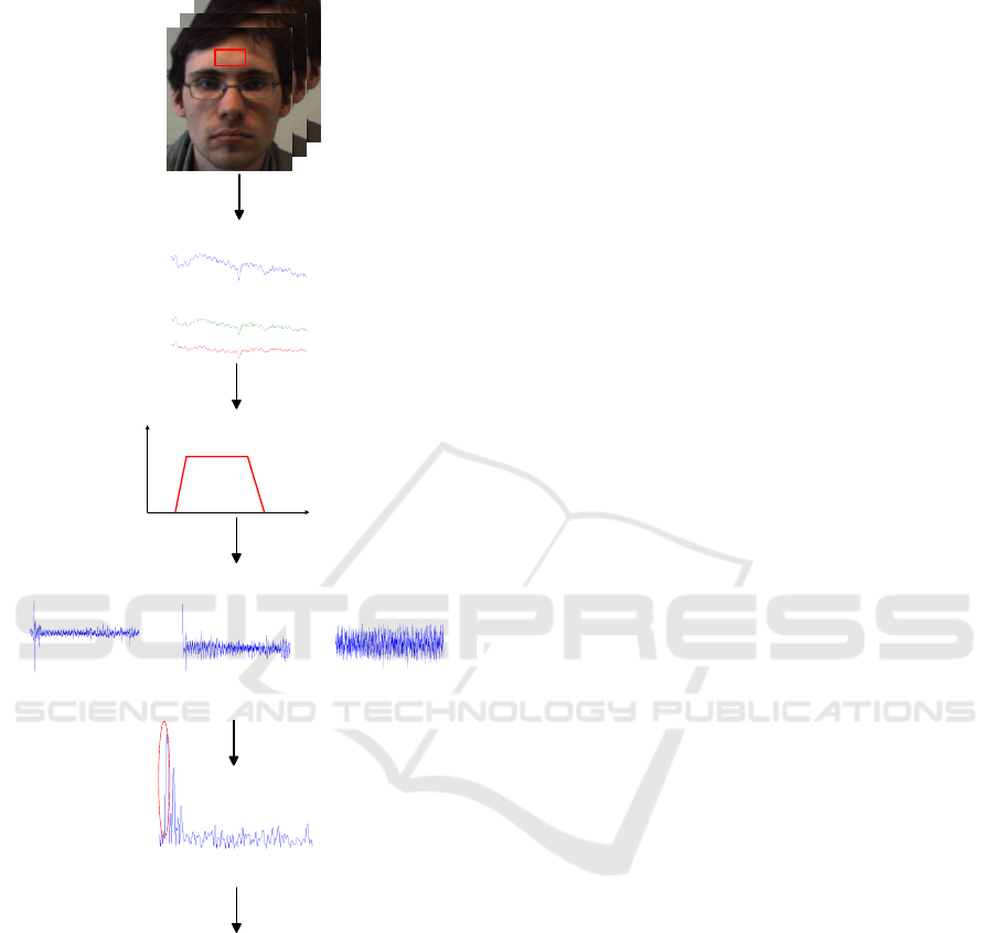
Figure 1: This overview shows the system functionality.
First of all, the face is detected and a forehead region is
defined in the image (1). Then, three mean temporal sig-
nals are extracted from the RGB channels (2). This step
is followed by a bandpass filter (3). The ICA splits the
three colour channels into three independent components
(4). By using Fast Fourier Transform, existing frequencies
in these components are determined and the highest peak in
the spectrum is detected as the preliminary heart rate (5).
With an adaptive filtering, quick changes in the signal can
be compensated (6), so that as final result, the heart rate can
be obtained.
This step is followed by a face detection, the selection
of the forehead region and the extraction of the three
colour channels. A bandpass limits the frequencies to
the natural limits of the human heart rate. Afterwards,
an ICA is applied to the three channels. A frequency
analysis determines the final heart rate. With the aid
of an adaptive filtering, outliers can be eliminated.
A normal RGB camera (Basler ac640-100gc) was
used for image acquisition. It provides a VGA reso-
lution and alterable frame rates can be adjusted. For
each recording, the frame rate was set to a fixed value
(30 fps or 50 fps) to have equidistant time steps for
the signal processing. The automatic controls for ex-
posure time and white balancing were switched off to
avoid invalid changes in the signal.
3.2 Face Detection and Forehead
Region Extraction
Due to the fact that our method works colour-based,
we need regions in the image where skin pixels are
visible. At this point, the face is the most suitable re-
gion. Normally, the face is not occluded by garments
and usually has a vertical orientation, which simpli-
fies the detection.
In the last decades, face detection is a well-studied
topic. We used the Viola-Jones Detector (Viola and
Jones, 2004) in OpenCV, which classifies Haar-like
features in cascades. The face detection itself gener-
ates bounding boxes with the face inside. There are
several face regions that are not suitable for heart rate
determination, because they do not provide signifi-
cant information, e. g. hair and eyebrows, or because
they show strong movement artefacts, e. g. eyes and
mouth. Therefore, only a part of the face region was
taken into account. For a better signal quality, a re-
gion of interest (ROI) was selected that provides a rel-
atively low noise-affected heart rate signal. For this,
the forehead region has been chosen, since it disposes
sufficient superficial vessels because of the thin skin.
Moreover, it shows a good light reflection character-
istic with a low light absorption of the tissue. Further-
more, movement artefacts are significantly less fre-
quent than in other face regions. The ROI is defined
by a static window within the face region that has al-
ways the same position with respect to bounding box
of the face.
3.3 Temporal Signal Extraction and
Band Pass Filtering
After having selected the forehead region, all pixels
inside the ROI are summarised and averaged. These
ICPRAM 2016 - International Conference on Pattern Recognition Applications and Methods
242
0 1,000 2,000
−10
0
10
Time [Samples]
Magnitude
ch1(t)
0 1,000 2,000
−5
0
5
10
Time [Samples]
ch2(t)
0 1,000 2,000
−5
0
5
Time [Samples]
ch3(t)
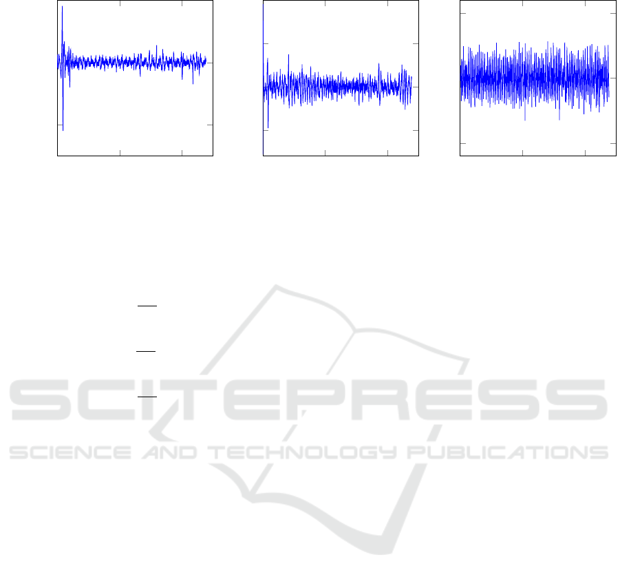
Figure 2: In result of the ICA, there are three independent components. The third component ch3(t) showed the best result in
our implementation.
operations are performed for all three RGB colour
channels:
R
mean
(t
0
) =
1
n
roi
roi
∑
i
roi
∑
j
R
i, j
(t
0
) (1a)
G
mean
(t
0
) =
1
n
roi
roi
∑
i
roi
∑
j
G
i, j
(t
0
) (1b)
B
mean
(t
0
) =
1
n
roi
roi
∑
i
roi
∑
j
B
i, j
(t
0
) (1c)
R
i, j
, G
i, j
and B
i, j
denote a single pixel in the ROI
of the corresponding channel. n
ROI
is the number of
all pixels in the ROI. Consequently, for every specific
moment in time t
0
, there is one value for each colour
channel that represents the mean value R
mean
, G
mean
and B
mean
.
In order to exclude implausible frequencies that
cannot represent the human heart rate, a bandpass fil-
ter BP is applied, see Equation (2a) to Equation (2c).
Only frequencies higher than 0.5 Hz (30 BPM) and
lower than 3 Hz (180 BPM) are considered during
further computations, i. e. the signals R
BP
, G
BP
and
B
BP
. For our practical implementation, we used a FIR
filter with 201 filter components. This filter has a lin-
ear phase response, which means that all frequencies
have the same group delay. Moreover, the filtering
has the effect of a pre-whitening, which is necessary
for the ICA.
R
BP
(t) = BP(t) ∗ R
mean
(t) (2a)
G
BP
(t) = BP(t) ∗ G
mean
(t) (2b)
B
BP
(t) = BP(t) ∗ B
mean
(t) (2c)
3.4 Independent Component Analysis
The three colour channels contain several sources of
image noise and artefacts, e. g. motion. The objective
is to find the underlying, original signals and extract
the pulse signal by decomposing the colour channels.
One possibility for decomposition is the ICA. This
method assumes that our observations are a linear
combination of the independent sources. Hence, it
is called blind source separation. In the general equa-
tion~x = A
~
s,~x denotes the vector of the observed com-
ponents, A is the so-called mixing matrix with lin-
ear concatenated elements and
~
s represents the inde-
pendent source components. For this application, the
equation can be formulated as:
R
BP
(t)
G
BP
(t)
B
BP
(t)
=
a
1,1
a
1,2
a
1,3
a
2,1
a
2,2
a
2,3
a
3,1
a
3,2
a
3,3
ch
1
(t)
ch
2
(t)
ch
3
(t)
(3)
For this implementation, we used the FastICA ap-
proach of Hyv
¨
arinen (Hyv
¨
arinen, 1999). It is accu-
rate, fast and available for several programming lan-
guages.
In Figure 2, all three components are displayed.
The component with the highest periodicity in the sig-
nal, which is visible in the harmonics of the spec-
trum, is most likely the component we are looking
for. In our study, the third independent components
ch
3
(t) shows the highest periodicity. In order to re-
move the high-frequent noise that was caused by the
ICA, we applied a smoothing window, which aver-
ages over three samples. This lowpass is denoted as
LP.
ch
3smooth
= LP(t) ∗ ch
3
(t) (4)
The smoothed result ch
3smooth
is shown in
Figure 3.
Remote Heart Rate Determination in RGB Data - An Investigation using Independent Component Analysis and Adaptive Filtering
243
0
500
1,000
1,500
−5
0
5
Time [samples]
Magnitude
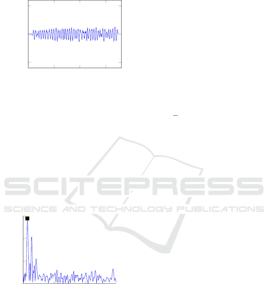
Figure 3: This plot shows the smoothed third independent
component.
3.5 Frequency Analysis
Finally, the frequencies of the signal ch
3smooth
are
determined using the Fast Fourier Transform (FFT).
Since the heart rate can change over time, we per-
formed the FFT in a small window of three seconds.
In this short interval, the human heart rate will not
change considerably. In frequency domain, the most
prominent peak is selected. This peak represents the
heart rate HR at time t
0
.
HR(t
0
) = max(
|
FFT(ch
3smooth
)
|
) (5)
0
500
1,000
1,500
0
20
40
60
BPM
Magnitude
Spectrum of the third smoothed component
Figure 4: The Fast Fourier Transform provides the fre-
quency spectrum of the third smoothed component. To in-
crease the frequency resolution, zero padding was applied.
The maximum peak is for this example at 72.5 BPM.
In order to increase the frequency resolution, we
applied zero padding to the signal. This guarantees a
more accurate quantisation without changing the sig-
nal information.
3.6 Adaptive Filtering
In order to eliminate false detections appearing in the
form of sudden changes in the heart rate from one
window to the next, we suggest the usage of an adap-
tive filter. If the absolute difference between the cur-
rent heart rate value HR(t
0
) and the previous heart rate
value HR(t
0
− ∆t) is higher than a threshold T) of 10
BPM, a sliding average is computed using the last 10
values according to Equation (6) and Equation (7). ∆t
denotes the time interval between to samples.
T =
|
HR(t
0
− ∆t) − HR(t
0
)
|
(6)
HR
ad
=
(
HR(t
0
) if T < 10 BPM
1
10
∑
t
0
t=t
0
−9
HR(t) otherwise.
(7)
4 EXPERIMENTAL RESULTS
AND DISCUSSION
4.1 Setting
For our experiments, we used a Basler RGB cam-
era (acA640-100gc). This camera records data with a
fixed frame rate. Both automatic exposure time con-
trol and automatic white balancing are switched off.
As far as lighting sources are concerned, we tested
multiple subjects under different lighting conditions,
whereas the only lighting sources were daylight and
the illumination of the interior lights in the room. The
distance between camera and the probands was be-
tween one and two meters. The probands were in-
structed to sit still with the face directed to the cam-
era. The recorded sequences have a length between
30 seconds and 2 minutes.
As reference we used a Polar FT1 heart rate mon-
itor. This system is composed of a heart rate sen-
sor, which is mounted on a chest strap, and a display,
which shows the current heart rate. The display is
visible in the corresponding videos as well, with the
result that the current reference heart rate can be com-
pared with our results.
In order to generate more variability in the heart
rate, the probands were asked to perform squats be-
fore the recording of some sequences. This results in
higher heart rates and is more representative with re-
spect to the natural range of the human heart rate.
In the next sections the accuracy, the utilization
of other colour channels than RGB and the computa-
tional speed are considered and discussed.
ICPRAM 2016 - International Conference on Pattern Recognition Applications and Methods
244

4.2 Accuracy
In our recordings, the average heart rate ranged from
67 BPM to 114 BPM. The mean error is calculated
for each window from t
0
to t
end
as the absolute dif-
ference between the measured heart rate HR
ad
(t) and
the reference heart rate HR
Re f
(t), see equation 8.
To measure the accuracy, we used this mean error
and the complement of the absolute normalised dif-
ference (CAND) as evaluation criterion, as shown in
equation 9.
Mean =
t
end
∑
t=t
0
HR
Re f
(t) − HR
ad
(t)
(8)
CAND = 1 −
HR
Re f
− HR
ad
HR
Re f
(9)
In our publication, the CAND is expressed in per-
cent. The higher this value, the better is the algorithm.
In our overall results, the mean error of the heart rate
detection is 4.36 BPM and the CAND is 94.45 %.
This shows that we can accurately detect the human
heart rate with our method. For our addressed appli-
cation fields, these errors are reasonably small.
Table 1: Mean error and the CAND for the different image
sequences.
Sequence Mean Error in BPM CAND in %
Sequence 1 5.63 92.34
Sequence 2 2.59 96.27
Sequence 3 3.36 95.24
Sequence 4 5.54 94.77
Sequence 5 4.85 93.64
We demonstrated that the ICA and the adaptive
filtering reduce the error significantly. However, the
problem of ICA is the selection of the most suitable
component. Methods such as autocorrelation or spec-
tral density could solve this issue.
In another experiment, we tested our algorithm for
another colour space, the HSI. The HSI color space
splits the channels in hue, saturation and intensity.
We expected that the decoupling of intensity and hue
shows better result than RGB. However, the results
were a tenfold worse. That indicates that our pro-
posed method is not suitable for the HSI colour space.
4.3 Computational Speed
In order to increase the speed of our algorithm, we re-
implemented the Matlab algorithm in C++ by using
the computer vision library OpenCV. For the C++ im-
plementation, we measured the computational speed.
For the tests, an Intel core i7 quad core processor with
2.9 GHz and 16 GB RAM was used. For compiling
GCC 5.2 was used.
In the case of computing the ICA in every time
step, the mean processing time, starting from image
acquisition to the derivation of the current heart rate,
is 28 ms. This corresponds approximately to 35 fps,
which demonstrates that our method runs fluently on
the described system. A speed comparison with other
publications was not possible, because this informa-
tion is not provided in literature.
A possibility to increase the speed is to calculate
the mixing matrix of the ICA only every n windows
or using the result of the ICA of the previous time step
for initialisation. Another acceleration option is to use
only R and G components of the RGB channel for
the ICA, because we assume that these two channels
contain the main part of the actual heart rate signal.
As a consequence, the computational effort will be
less with probably the same accuracy.
5 CONCLUSIONS
In this paper, we presented a method for remote heart
rate determination using ICA and adaptive filtering.
With regard to applications in domestic environments
and for elderly care, the obtained results are adequate.
For other use cases, especially in clinical environ-
ments, where highly accurate measurements are re-
quired, accuracy has to be improved.
For future work, the algorithm should also be ro-
bust against motion artefacts. One solution could be a
feature tracking on the forehead region.
By porting the algorithm to an embedded system,
a more flexible and praxis-oriented solution could be
achieved.
With the help of such a system, it could be possi-
ble to pre-emptively detect emergencies in domestic
environments.
ACKNOWLEDGEMENTS
This project is funded by the European Social Fund
(ESF). We would like to thank all probands who took
part in the experiments and supported us with their
video records.
REFERENCES
Allen, J. (2007). Photoplethysmography and its application
in clinical physiological measurement. Physiological
Measurement, 28(3):R1–R39.
Remote Heart Rate Determination in RGB Data - An Investigation using Independent Component Analysis and Adaptive Filtering
245

Balakrishnan, G., Durand, F., and Guttag, J. (2013). Detect-
ing pulse from head motions in video. In Computer
Vision and Pattern Recognition (CVPR), 2013 IEEE
Conference on, pages 3430–3437.
de Haan, G. and Jeanne, V. (2013). Robust pulse rate from
chrominance-based rppg. Biomedical Engineering,
IEEE Transactions on, 60(10):2878–2886.
Garbey, M., Sun, N., Merla, A., and Pavlidis, I. (2007).
Contact-free measurement of cardiac pulse based on
the analysis of thermal imagery. Biomedical Engi-
neering, IEEE Transactions on, 54(8):1418–1426.
He, X., Goubran, R., and Liu, X. (2014). Using eulerian
video magnification framework to measure pulse tran-
sit time. In Medical Measurements and Applications
(MeMeA), 2014 IEEE International Symposium on,
pages 1–4.
Hertzman, A. B. and Spealman, C. R. (1937). Observations
on the finger volume pulse recorded photoelectrically.
American Journal of Physiology, 119:334–335.
Hyv
¨
arinen, A. (1999). Fast and robust fixed-point algo-
rithms for independent component analysis. Neural
Networks, IEEE Transactions on, 10(3):626–634.
K. Humphreys, C. M. and Ward, T. (2005). A cmos camera-
based system for clinical photoplethysmographic ap-
plications. In Proceedings of SPIE, volume 5823,
pages 88–95.
Lewandowska, M., Ruminski, J., Kocejko, T., and Nowak,
J. (2011). Measuring pulse rate with a webcam - a
non-contact method for evaluating cardiac activity. In
Computer Science and Information Systems (FedC-
SIS), 2011 Federated Conference on, pages 405–410.
Li, X., Chen, J., Zhao, G., and Pietikainen, M. (2014).
Remote heart rate measurement from face videos un-
der realistic situations. In Computer Vision and Pat-
tern Recognition (CVPR), 2014 IEEE Conference on,
pages 4264–4271.
Meinel, L., Richter, J., Schmidt, R., Findeisen, M., and
Hirtz, G. (2015). Opdemiva: An integrated assis-
tance and information system for elderly with demen-
tia. In Consumer Electronics (ICCE), 2015 IEEE In-
ternational Conference on, pages 76–77.
Poh, M.-Z., McDuff, D., and Picard, R. (2010). Non-
contact, automated cardiac pulse measurements using
video imaging and blind source separation. Optics Ex-
press, 18(10):10762–10774.
Poh, M.-Z., McDuff, D., and Picard, R. (2011). Ad-
vancements in noncontact, multiparameter physiolog-
ical measurements using a webcam. Biomedical En-
gineering, IEEE Transactions on, 58(1):7–11.
Rubinstein, M. (2014). Analysis and Visualization of Tem-
poral Variations in Video. PhD thesis, Massachusetts
Institute of Technology.
van Gastel, M., Stuijk, S., and de Haan, G. (2015). Motion
robust remote-ppg in infrared. Biomedical Engineer-
ing, IEEE Transactions on, PP(99):1–1.
van Gastel, M., Zinger, S., Kemps, H., and de With, P.
(2014). e-health video system for performance anal-
ysis in heart revalidation cycling. In Consumer Elec-
tronics Berlin (ICCE-Berlin), 2014 IEEE Fourth In-
ternational Conference on, pages 31–35.
Verkruysse, W., Svaasand, L. O., and Nelson, J. S. (2008).
Observations on the finger volume pulse recorded
photoelectrically. Optics Express, 16(26):21434–
21445.
Viola, P. and Jones, M. J. (2004). Robust real-time face
detection. International Journal of Computer Vision,
57(2):137–154.
Wang, W., Stuijk, S., and de Haan, G. (2015). Exploiting
spatial redundancy of image sensor for motion robust
rppg. Biomedical Engineering, IEEE Transactions on,
62(2):415–425.
Wohlrab, D., Heß, M., Apitzsch, A., Langklotz, M.,
Schwarzenberger, A., Bilda, S., Schulz, H., Hirtz,
G., and Mehner, J. (2015). Hom-e-call - an en-
hanced fall detection system based on accelerometer
and optical sensors applicable in domestic environ-
ment. In Jaffray, D. A., editor, World Congress on
Medical Physics and Biomedical Engineering, June 7-
12, 2015, Toronto, Canada, volume 51 of IFMBE Pro-
ceedings, pages 1461–1464. Springer International
Publishing.
Wu, H.-Y., Rubinstein, M., Shih, E., Guttag, J., Durand, F.,
and Freeman, W. T. (2012). Eulerian video magnifi-
cation for revealing subtle changes in the world. ACM
Trans. Graph. (Proceedings SIGGRAPH 2012), 31(4).
ICPRAM 2016 - International Conference on Pattern Recognition Applications and Methods
246
