
Brain Activation and Cognitive Load during EEG Measured
Creativity Tasks Accompanied by Relaxation Music
Dalia Papuc
1
, Oana Bălan
2
, Maria-Iuliana Dascălu
1
, Alin Moldoveanu
2
and Anca Morar
2
1
University Politehnica of Bucharest, Faculty of Engineering in Foreign Languages,
Splaiul Independentei, 313, Bucharest, Romania
2
University Politehnica of Bucharest, Faculty of Automatic Control and Computer Science,
Splaiul Independentei, 313, Bucharest, Romania
Keywords: EEG, Creative Task, Alpha Power, Cognitive Load.
Abstract: Creativity tasks require specific imagery, memory and semantic processes, as it has been revealed by
various neuroscientific studies. There is significant evidence that an increase of spectral power in the EEG
alpha band is inter-related with creative ideation as a form of top-down activity. However, any creational
task demands a certain level of cognitive load or workload for memory retrieval, mental schemes design,
semantic processing, image formation and concept construct. This paper aims to measure cognitive load
during a series of divergent thinking creative tasks accompanied by relaxation music, to examine event-
related brain activation and raw power measures in the baseline relaxation, alternate uses creative and
verbalization stages of the proposed experimental procedure, as well as to study task-related
synchronization/desynchronization between these phases. Also, its purpose is to verify whether slow
relaxing music influences creativity, with effects in the brainwaves amplitude variations, especially in the
alpha (8-12 Hz) frequency band. We concluded that relaxing music induces creativity and causes an
increase in the alpha brain waves for innovative ideas generation and verbalization with a diminished level
of cognitive load.
1 INTRODUCTION
Along with mental constructs such as intelligence,
creativity is an important asset of the human
psychological profile, with applicability for
producing original and innovative work in a wide
range of scientific domains, for social
communication and interaction or for achieving
various lifetime proposed goals (Fink & Benedek,
2012, Smith 1995). Creativity has been studied
within many experiments requiring completion of
different types of originality tasks, among which a
notable position is reserved to the divergent
production alternate uses tasks, where participants
are asked to bring original and unconventional
solutions or explications to common notions or
concepts (Arden 2010). This type of task involves
retrieval of existing knowledge from the memory
and (re)combination of it into singular ideas (Paulus
and Brown, 2007), generating diversified approaches
in manifold ways, as opposed to convergent thinking
that brings about only one straight solution. There
are many neuroscientific studies that have analysed
brain activity during creativity tasks, considering
spectral power activation within the EEG bands,
event-related potentials (ERP), event-related
synchronization/desynchronization (ERS/D) or
functional connectivity between different cortical
areas – coherence and phase lag. The alpha band is
the most sensitive band in relation with creativity
demands – there is higher alpha power in the
posterior regions of the brain during activation
periods as compared to baseline resting stages
(Jausovec, 1997), in the central and parietal areas
(Molle, 1999), posterior regions of the right
hemisphere during divergent, free-associative
thinking tasks than in the convergent ones
(Shemyakina, 2007) and in the posterior cortical
areas (parietal and parietoocipital) in the alternate
uses procedure (Fink, 2009). Also, more original
ideas conduct to higher right hemisphere alpha
synchronization (Grabner, 2007, Martindale, 1984).
Increases in the parietal and occipital areas is
associated with higher cognitive load, memory
Papuc D., BÄ
ˇ
Clan O., DascÄ
ˇ
Clu M., Moldoveanu A. and Morar A.
Brain Activation and Cognitive Load during EEG Measured Creativity Tasks Accompanied by Relaxation Music.
DOI: 10.5220/0006511201560162
In Proceedings of the International Conference on Computer-Human Interaction Research and Applications (CHIRA 2017), pages 156-162
ISBN: 978-989-758-267-7
Copyright
c
2017 by SCITEPRESS – Science and Technology Publications, Lda. All rights reserved
search, retrieval and semantic associations, increased
focus on goal-directed tasks, diverting irrelevant
stimuli, internal mnemonic representations or
memory retrieval according to the purposes of the
user – top-down activation in the absence of bottom-
up stimulation (dorsal parietal) (Cabeza, 2008,
Jensen, 2002). The right hemisphere is dedicated to
imagery and association of unrelated semantic
concepts (Bowden, 2003). Alpha synchronization
appears also in the prefrontal cortex, especially in
the right hemisphere (Hietanen, 1998), reflecting
high processing demands (Benedek, 2011) and
increased attention (Knyazev, 2007). The results
from (Punsawad et al, 2014) showed increased
levels of alpha and theta during creational tasks in
the right frontal areas (channel F8).
Cognitive load theory states that working
memory is limited in regard with the quantity of
information that can be kept in mind at a moment of
time and to the ability of processing and combining
novel knowledge (Antonenko et al, 2010, Peterson,
1965). EEG measurements revealed that alpha
desynchronization is associated with increased
attention and cognitive load in the parietal areas
(Gevins, 1997). Also, it is related to searching and
retrieving information from the long-term memory.
Theta synchronization on the other hand is related to
task difficulty and emotional factors, especially in
the frontal midline locations (Klimesch, 2005,
Gevins, 2000) and is associated with episodic and
working memory processes. Delta power increases
also during complex mental tasks in the temporal
regions (channels T7 and T8), demonstrating the
amount of attention given by the user to internal
information processing (Dolce, 1974), while low
beta bands have synchronized powers in the frontal
midline areas (Klimesch, 1999)
Gifted individuals exhibit higher alpha power
changes (high level of relaxation) and activation in
the frontal cortical area, whereas hard working
subjects present low level alpha synchrony,
particularly in the temporal regions (O’Boyle 1995).
In (Antonenko et al, 2010), video and picture
presentation activated the occipital and temporal
lobes (responsible for visualization), while text
presentation, the frontal one (assigned to verbal
processing).
2 PROPOSED METHOD
Our study proposes an alternate uses divergent
thinking set of tasks, in which the subject is required
to think about uncommon uses for 5 different words
- pencil, brick, key, paper clip and shoe, while
listening to slow relaxing music. The first step is
represented by the fixation cross to which the
subjects have to look for a time period of 10
seconds. This step will be referred to as reference
(or calibration, to record baseline brain activity)
because this time interval aims to be reference
interval for the EEG activity. Secondly, a stimulus
word was shown for 5 seconds. The subjects were
given instructions to be as innovative as possible and
to come up with different usages than the common
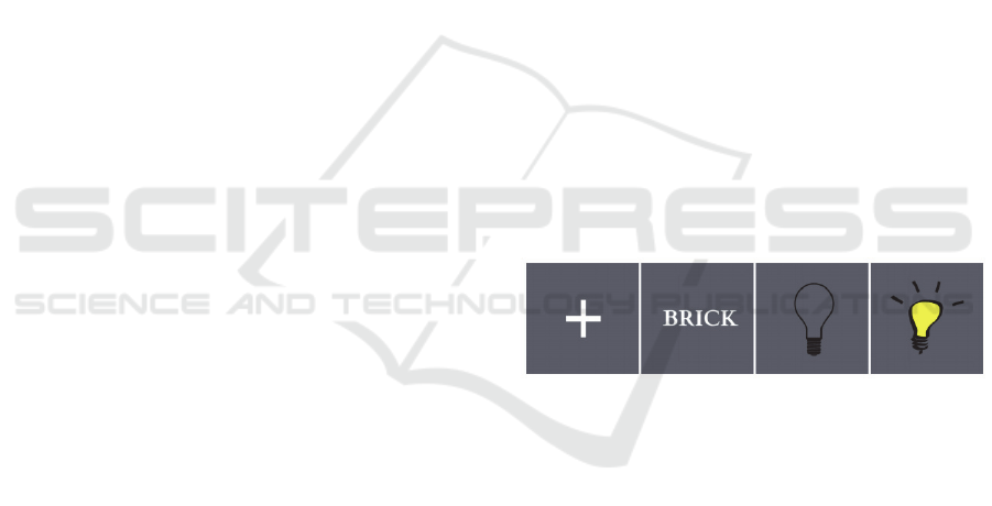
one for the displayed object. The next step is
represented by the uncoloured bulb image, 30
seconds of creative thinking time for the
participants. During this step, the subjects were prior
instructed not to verbalize their answer yet, but only
to think of original uses of the previously seen
stimulus. Simultaneously, the subject was provided
relaxation music via headphones. The final phase is
represented by the coloured bulb image, which lasts
for 10 seconds, time in which the participants had to
verbalize their creative ideas (Figure 1).
Verbalization has been chosen over listing because it
takes less time and is simpler for participants, the
emphasis being on the idea generation process. The
task requires 55 seconds to be completed. Each of
the subjects performed the task five times, once per
stimulus word.
Figure 1: The experimental procedure.
3 THE EXPERIMENT
3.1 Subjects
The study was performed on six subjects (four
females and two males) aged 21-29, five
undergraduate students and a teacher. The subjects
were orally informed about the purpose of the
experiment, were provided instructions and gave
their approval consent before the start of the tests.
3.2 Data Acquisition
The hardware devices used to capture EEG signals
through the non-invasive method were the Brain
Products actiCAP Xpress Bundle set together with
V-Amp 16ch amplifier (Acticap website). The
software used for EEG acquisition was OpenVibe
for Brain Products (Open Vibe website). All the
signals were sampled at a frequency of 512 Hz, as it
is the optimal sampling rate for analysing resting
EEG (Jing & Takigawa, 2000). The used electrodes
positions were: fronto-parietal left (FP1) and right
(FP2), fronto-central left (FC1) and right (FC2),
central left (C3) and right (C4), temporal left (T7)
and right (T8), parietal left (P3) and right (P4) and
occipital left (O1) and right (O2). Furthermore, 4
midline electrodes were used (Fz, Cz, Pz, Oz).
Electrodes impedances were kept below 10 kΩ in
order to minimize the noise and artefacts in the data.
The recorded data was bandpass filtered between
0.5-100 Hz.
3.3 Data Acquisition
The term EEG artefact refers to recorded electrical
activity whose origin is non-cerebral. Depending on
its origin, these artefacts are split into physiological
and non-physiological artefacts. The first category is
generated from the body (other than the brain), while
the second one from outside the body. Blinking, eye
movements, muscle activity, body or head
movement, cardiac, pulse waves, they all represent
examples of the physiological artefacts. Some of
these can be recognized. Non-physiological artefacts
are caused by either electrical interference with other
power sources or the abnormal functioning of the
recording equipment. A typical electrical
interference artefact present in EEG recordings has
as source power lines and equipment. This artefact
has a 50/60 Hz frequency. Our signals were DC-
offset corrected in order to prevent the influence of
voltage imbalance issues and powerline
contamination was removed using the notch filter.
Blinking, cardiac and muscle activity artefacts,
together with the bad channels were automatically
removed using the Brainstorm software (Tadel et al,
2011). Independent Component Analysis has been
performed in EEGLAB (EEG LAB website) in order
to separate independent sources, reject artefacts and
identify brain related activity in coloured
topographic maps.
3.4 Power Spectrum Analysis in
Frequency Bands
The information was extracted for the last step
which is statistical analysis. Firstly, the data that is
to be statistically analysed requires having the same
length. Currently, most of the recordings have
around 55 seconds, -4/+15 seconds, according to the
number of inputs the subjects were having in the
verbalization phase. Given the interest in studying
how the brain waves change in the divergent
thinking phase, as stated in the hypothesis, the signal
was split such as the reference phase may be
compared with the other phases. Therefore, the
signal was split as follows - from a time window
perspective: [0 10] for the reference phase, [15 25],
[25 35] & [35 45] for the creative thinking phase and
[45 55] for the verbalization phase. Extracting all
these intervals was done manually in EEGLAB.
After being split into intervals, the EEG recordings
were imported in Brainstorm where further
processing steps were taken.
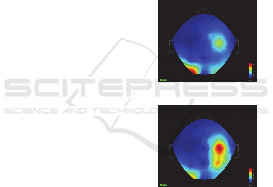
Figure 2: Power spectral analysis for Resting period -
averaged response across all subjects.
Figure 3: Power spectral analysis for Creative Thinking
period - averaged response across all subjects.
Considering the averaged responses for all the
subjects, alpha activity was prominent as follows: in
the resting phase - in the left occipital region (Figure
2), in the creative thinking phase – in the right
parietal area (Figure 3), while in the verbalization
stage – in the right parietal and left occipital regions
(Figure 4). Similar patterns are observed in the theta
band, with the only difference that in the
verbalization phase, activation is prominent only in

the right parietal cortical area and to a lower extent
in the left occipital region, demonstrating the slight
influence of visualization in verbal tasks. Alpha
synchronization occurs in the right parietal area and
desynchronization is evident in the occipital region
in the thinking stage, as compared to resting
baseline.
Figure 4: Power spectral analysis for Verbalization period
- averaged response across all subjects.
3.5 Statistical Analysis
Even though fluctuations over time in the alpha
waves can be noticed by examining the time-series
for a signal time-frequency decomposition, in order
to formalize the results and to be able to actually
draw conclusions, a statistical analysis needs to be
performed. Another reason for performing this
analysis is represented by the number of data that
would require a thorough individual analysis to
reach a global study result.
Taking into consideration the hypothesis: “There
is a significant change in alpha activity during a
creative activity as compared to a non-creative one”,
the study aims to evaluate whether creativity causes
an increase in the alpha brain waves through a given
divergent thinking task and a statistical hypothesis
test is applied to check whether there is a significant
difference between the two phases. The data was
analysed in a parametric Student t-test and 2D
topography of the results was plotted.
30 files (10 seconds duration) resulting from
resting baseline recording were compared to 90 files
(10 seconds duration each) from the creativity task
recording. The results show significant differences
in the left fronto-central cortical area at p<0.13
where the mean of the alpha power in the creative
thinking condition is higher than in the resting
phase. Taking the first test scenario, reference (A)
vs. thinking (B) phase and the previous explanations,
the results are statistically significant in the direction
of condition B (blue) represented by the thinking
phase in this scenario. The confidence level for these
tests is close to 90%. The results of this study prove
that indeed creative ideas generation is associated
with the growth of the alpha activity in comparison
with the reference period (Figure 5).
Figure 5: Student t-test. Reference (A) vs Creative
Thinking (B), p < 0.13.
Figure 6: Student t-test. Verbalization (A) vs Creative
Thinking (B), p < 0.01.
30 files (10 seconds duration) resulting from
verbal recordings were compared to 90 files (10
seconds duration each) from the creativity task
recording. The results show significant differences
in the left fronto-central and right parietal and
temporal cortical areas at p<0.01 where the mean of
the alpha power in the verbal condition is higher
than in the creative thinking phase. Given the fact
that the critical level for the verbalization vs.
reference test is lower (p < 0.01) than the one of
thinking (B) vs. reference (A) phases (p < 0.13), and
that the regions where alpha activity presents
significant changes are identical, we would expect
that in the verbalization phase the alpha activity is
increased as compared to thinking phase. Figure 6
proves a remarkable increase in the alpha activity in
the frontal and temporal lobes as well as in the

central region, to be more specific, in the frontal left
hemisphere - especially frontopolar (FP1) and in the
temporal (T8) and central (C4) right hemisphere
(Figure 6).
The values displayed in the 2D test result views
(Figure 5 & 6) represent the significant t-values, the
sensors that have the value of p > α being set to 0.
We may state that for those ‘white’ brain regions the
hypothesis is likely to occur → the null hypothesis
(H0) is not rejected, thus no significant changes
occur in the alpha waves. The two-tailed tests
establish whether the difference between the two
groups, A and B, is statistically significant in either
the positive or negative direction (Frost, 2016) In the
figures, the red values represent a higher amplitude
for condition A, while the blue values for the
condition B.
Quantitative EEG (QEEG), as the measurement
of the electrical brain activity and connectivity
between different areas, offers substantial and
valuable information about the dynamic changes of
brain activation, interrelation, engagement and
overload of various areas. Brain connectivity refers
to coherence analysis (correlation between different
brain areas) and phase lag (speed of information
transfer on the cortex and rate of data transfer).
The results showed high correlation in the frontal
area (FP1 x FP2), parietal and temporal regions (P3
x P4, P3 x T7, Pz x T7, Pz x P3). High rates of data
transfer (high values of phase lag) occur in the
frontal regions for the theta band (FP1 x FP2) and in
the left frontal, parietal and central areas for the
alpha band (FP1 x C3, C3 x Pz, T7 x C3, FC1 x C3,
FC1 x FP2, P3 x P4, P3 x T7).
4 DISCUSSION
In a creativity related brain activity study, the
answers of subjects that were more innovative
positively correlated with lower alpha (8 - 10 Hz)
amplitudes mainly in the left hemisphere. The same
study also shows desynchronization in the central
and posterior regions, but not in the frontal one
(Razumnikova, 2007). Taking into account the
results of the aforementioned study which also state
that highly original responses generated increased
event-related synchronization in the left hemisphere
for the low alpha band (Haarmann et al, 2013), we
may make the assumption that the increased alpha
activity in the left hemisphere is correlated with
higher originality in the subjects’ answers.
Beta synchronization in the frontal and midline
regions are indicators of cognitive load. In our study,
alpha synchronization occurs for all the frequency
bands and beta desynchronization is visible in the
frontal and midline areas. Also, there is no
significant difference between the resting and
creative task for the theta and delta bands. Thus, we
conclude that the level of cognitive workload is
reduced during alternate uses divergent thinking
tasks as far as it concerns the delta, theta and beta
bands and that a sign of activation is visible only for
the alpha band, indicating memory retrieval and
attentional demands. However, as far as it concerns
the averaged responses across all subjects and trials
(Figure 3), alpha synchronization is visible in the
creative thinking phase in the parietal area and less
visible in occipital area.
One aspect to discuss is the value of the p-value
threshold. To increase the accuracy of the statistical
tests, the p-value threshold could have been
decreased, mainly for the reference vs thinking
groups test, however, the results would not have
shown this significant information if done so.
Moreover, a larger sample size will normally lead to
a better estimate too. The eventual presence of
physiological artefacts, the possibility of not having
the phases according to such strict time intervals as
defined in the experimental task, and not having the
ability to check precisely whether the subjects are
following the instructions for the first 45 seconds
(especially thinking creatively for the whole 30
seconds interval) of the EEG recordings, they all
contribute to eventual discussions on test’s accuracy.
5 CONCLUSIONS
The aforementioned findings allow us to validate the
study hypothesis which states that there is a
significant change in alpha activity during a creative
activity as compared to a non-creative one. Through
giving the subjects an alternate uses task which aims
to measure divergent thinking while listening to
relaxing music and recording their brain activity, it
was evaluated and concluded that creativity causes
an increase in the alpha brain waves in the
innovative ideas generation as well as in their verbal
expression phases with a diminished level of
cognitive load.
Further research concerns encompass increasing
the number of subjects and trials per experiment in
order to obtain higher statistical significance and
designing an experiment where the subjects will be
required to listen to different music genres or

semantically significant ambient sounds connected
to the meaning of the word they have to creatively
think about.
ACKNOWLEDGEMENTS
This project has received funding from the European
Union’s Horizon 2020 research and innovation
programme under grant agreement No 643636
“Sound of Vision. This work has been funded by
University Politehnica of Bucharest, through the
“Excellence Research Grants” Program, UPB –
GEX 2017. Identifier: UPB- GEX2017, 18 from
8.09.2017/2017 SAFE-VR.
REFERENCES
Fink, A., Benedek, M., 2012. EEG alpha power and
creative ideation. Neurosci. Biobehav.
Smith, S.M., Ward, T.B., Finke, R.A., 1995. The Creative
Cognition Approach. MIT Press,Cambridge.
Arden, R., Chavez, R.S., Grazioplene, R., Jung, R.E.,
2010. Neuroimaging creativity: a psychometric
review. Behavioural Brain Research 214, 143–156.
Paulus, P.B., Brown, V.R., 2007. Toward more creative
and innovative group idea generation: a cognitive-
social-motivational perspective of brainstorming.
Social and Personality Psychology Compass 1, 248–
265.
Jausovec, N., 1997. Differences in EEG activity during the
solution of closed and open problems. Creative
Research Journal 10, 317–324.
Mölle, M., Marshall, L., Wolf, B., Fehm, H.L., Born, J.,
1999. EEG complexity and performance measures of
creative thinking. Psychophysiology 36, 95–104.
Shemyakina, N.V., Danko, S.G., Nagornova, Z.V.,
Starchenko, M.G., Bechtereva, N.P. ,2007. Changes in
the power and coherence spectra of the EEG rhythmic
components during solution of a verbal creative task of
overcoming a stereotype. Human Physiology 33, 524–
530.
Fink, A., Graif, B., Neubauer, A.C., 2009. Brain correlates
underlying creative thinking: EEG alpha activity in
professional vs. novice dancers. NeuroImage 46,854–
862.
Grabner, R.H., Fink, A., Neubauer, A.C., 2007. Brain
correlates of self-rated originality of ideas: Evidence
from event-related power and phase-locking changes
in the EEG. Behavioral Neuroscience 121, 224–230.
Martindale, C., Hines, D., Mitchell, L., Covello, E., 1984.
EEG alpha asymmetry and creativity. Personality and
Individual Differences 5, 77–86.
Cabeza, R., Ciaramelli, E., Olson, I.R., Moscovitch, M.,
2008. The parietal cortex and episodic memory: an
attentional account. Nature Reviews Neuroscience 9,
613–625.
Jensen, O., Gelfand, J., Kounios, J., Lisman, J.E., 2002.
Oscillations in the alpha band (9-12 Hz) increase with
memory load during retention in a short-term memory
task. Cerebral Cortex 12, 877–882.
Bowden, E.M., Jung-Beeman, M., 2003. Aha! Insight
experience correlates with solution activation in the
right hemisphere. Psychonomic Bulletin & Review
10,730–737.
Hietanen, J.K., Surakka, V., Linnankoski, I., 1998. Facial
electromyographic responses to vocal affect
expressions. Psychophysiology 35, 530–536.
Benedek, M., Bergner, S., Könen, T., Fink, A., Neubauer,
A.C., 2011. EEG alpha synchronization is related to
top-down processing in convergent and divergent
thinking. Neuropsychologia 49, 3505–3511.
Knyazev, G.G., 2007. Motivation, emotion, and their
inhibitory control mirrored inbrain oscillations.
Neuroscience & Biobehavioral Reviews 31, 377–395.
Punsawad, Y., Chathong, W., Wongsawat, Y., 2014. The
use of quantitative EEG in creativity study with simple
task,
APSIPA, Asia-Pacific.
Antonenko, P. D., Paas, F., Grabner, R., & Van Gog, T,
2010. Using electroencephalography to measure of
cognitive load. Educational Psychology Review, 22,
425-438.
Peterson, L., & Peterson, M., 1959. Short-term retention
of individual verbal items. Journal of Experimental
Psychology, 58, 193–198.
Gevins, A., Smith, M. E., McEvoy, L., & Yu, D., 1997.
High-resolution EEG mapping of cortical activation
related to working memory: Effects of task difficulty,
type of processing, and practice. Cerebral Cortex, 7,
374–385.
Klimesch, W., Schack, B., & Sauseng, P., 2005. The
functional significance of theta and upper alpha
oscillations for working memory: A review.
Experimental Psychology, 52, 99–108.
Gevins, A., & Smith, M. E., 2000. Neurophysiological
measures of working memory and individual
differences in cognitive ability and cognitive style.
Cerebral Cortex, 10, 829–839.
Dolce, G., Waldeier, H., 1974. Spectral and multivariate
analysis of EEG change during mental activity in man.
Clinical neurophysiology, 577-584.
Klimesch, W., 1999. EEG alpha and theta oscillations
reflect cognitive and memory performance: a review
and analysis. Brain Research Reviews, 169-195.
O'Boyle, M. W., Benbow, C. P., & Alexander, J. E., 1995.
Sex differences, hemispheric laterality, and associated
brain activity in the intellectually gifted.
Developmental Neuropsychology, 4, 415–443.
Acticap Express Bundle. Available at:
http://www.brainproducts.com/actiCAPXpress.
Open Vibe. Available at: http://openvibe.inria.fr/
Jing, H., & Takigawa, M. 2000. Low sampling rate
induces high correlation dimension on
electroencephalograms from healthy subjects.

Psychiatry and Clinical Neurosciences, 54 (4), 407-
412. doi:10.1046/j.1440-1819.2000.00729.x.
Tadel, F., Baillet, S., Mosher, J.C., Pantazis, D., Leahy,
R.M., 2011. Brainstorm: A User-Friendly Application
for MEG/EEG Analysis. Computational Intelligence
and Neuroscience, vol. 2011, ID 879716.
EEG LAB. Available at: https://sccn.ucsd.edu/
eeglab/index.php.
Frost, J., 2016. Understanding t-Tests: t-values and t-
distributions. Retrieved June 21, 2017, from
http://blog.minitab.com/blog/adventures-instatistics-
2/choosing-between-a-nonparametric-test-and-a-
parametric-test.
Razumnikova, O. M., 2007. Creativity related cortex
activity in the remote associates task. Brain Research
Bulletin, 73 (1-3).
