
Study of the Neuro-electrostimulation Influence on the Head Skin
Capillary Blood Flow
Vladimir Kublanov, Mikhail Babich, Anton Dolganov, Evgenii Shleymovich,
Boris Zhilkin and Evgenii Plesniaev
Ural Federal University, 620002, Mira 19, Yekaterinburg, Russian Federation
Keywords: Neuro-Electrostimulation, ‘SYMPATHOCOR-01’, Blood Perfusion, Pennes Equation, Thermal Imaging.
Abstract: The pilot study of the ‘SYMPATHOCOR-01’ neuro-electrostimulation device influence on the head skin
capillary blood flow is described. The infrared thermographic camera was used for the head skin capillary
blood flow registration. The analysis of the registered thermograms was performed for mean head skin
temperature evaluation. The experiment has shown that application of the neuro-electrostimulator in the
blocking mode of the sympathetic nervous system caused the decrease of the head surface temperature. The
temperature decrease is associated with the perfusion rate increase on the capillary level, which is in
agreement with the neuro-electrostimulation application techniques.
1 INTRODUCTION
Many technologies of the physical fields application
are aimed to improve performance of the circulatory
system (Mesquita et al. 2013, Morishita et al. 2014,
Yamabata et al. 2016, Jin et al. 2017). The most
promising among them control the autonomic
nervous system (ANS) to provide constrictive
management of the blood vessels tone.
The ‘SYMPATHOCOR-01’ neuro-electrostimu-
lation device is capable of performing such control.
The medical techniques of the ‘SYMPATHOCOR-
01’ device application implement the methodology of
dynamic correction of the sympathetic nervous
system correction (DCASNS) and provide correction
of the autonomic balance, defined by the relation of
the sympathetic and parasympathetic departments of
the ANS (Kublanov, Shmirev, et al. 2010, Kublanov
et al. 2017).
The design process of the ‘SYMPATHOCOR-01’
device medical application was accomponied by the
experimental studies on laboratory animals
(Kublanov, Danilova, et al. 2010, Kublanov et al.
2012) and the single-photon emission computed
tomography imaging. (Kublanov et al. 2004). In
clinical practice, the device has been successfully
applied for treatment of the vascular dystonia;
headaches of different origin, including migraine;
hypertension; neurosensory hearing loss; degenera-
tive sight deficits and atrophy of the visual nerve;
neurosis-like syndromes and neuropathies of the
various origins (Kublanov, Shmirev, et al. 2010).
However, in the previous works the changes in the
capillary blood flow were not studied. In most cases
the capillary blood flow define the skin’s temperature
fluctuations. The goal of the present work is to
conduct pilot study for investigation of the
‘SYMPATHOCOR-01’ device influence on the head
skin capillary blood flow by means of the infrared
thermography.
2 MATHERIALS AND METHODS
2.1 Experiment Description
The neuro-electrostimuation procedure was perfor-
med by the modern implementation of the
‘SYMPATHOCOR-01’ device. The key targets of the
‘SYMPATHOCOR-01’ devices are the neural
formations in the neck region. The device is included
in the register of medical equipment products of the
Russian Federation – registration certificate № FCR
2007/00757.
The modern device implementation consists of
two blocks. The first block is used for generation of
the spatially distributed rotating field of current
pulses and has two multi-element electrodes which
Kublanov V., Babich M., Dolganov A., Shleymovich E., Zhilkin B. and Plesniaev E.
Study of the Neuro-electrostimulation Influence on the Head Skin Capillary Blood Flow.
DOI: 10.5220/0006592803510355
Copyright
c
2018 by SCITEPRESS – Science and Technology Publications, Lda. All rights reserved
are placed on the neck. The first block is supplied by
the built-in accumulator, has dimensions of 90 х 50 х
18.5 mm, and weighs less than 200 g. The second block
is used for management and control of the neuro-
electrostimulation procedure. At the moment, the
second block is realized as the application for the
Android operation system. Bluetooth low energy
channel exchange information between the two blocks.
The pilot study was conducted in the Research
Medical and Biological Engineering Centre of High
Technologies, Ural Federal University. The
experimental program of the study had an approval
№8 from 16 October 2015 of local ethics committee
in Ural State Medical University. The one relatively
healthy volunteer – male, 27 years old, doesn’t have
any health complains – has participated in single
experiment. Prior to the study volunteer has signed
the participation consent. The whole experiment was
supervised by the physician. The experiment layout is
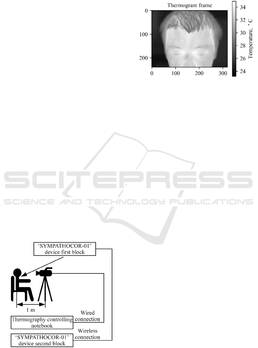
shown on Fig. 1.
During the whole study the volunteer was sitting
on the chair. The infrared thermographic camera NEC
Thermo Tracer TH9100WL was used for registration
of the skin surface temperature. The infrared
thermographic camera was placed on the tripod at the
distance of 1.0 meter from the volunteer’s head. The
thermogram frame center was projected on the
volunteer’s forehead. In the bottom the thermogram
was limited by the nose, to prevent the breathing
artifacts. Example of the registered thermogram
frame is shown on Fig. 2.
The camera’s output was connected to the note-
book, which controlled the thermograms registration
and storage. The resolution of the registered
thermograms was 320 x 240 pixels; 3 frames per
second. The thermal resolution was 0.1°С.
Figure 1: Experiment layout.
Figure 2: Thermogram frame example.
The neuro-electrostimulation device was used in
the blocking mode of the Sympathetic Nervous
System (SNS). For that, the following values of the
biotropic field features were set: the partial impulse
length – 30 us, modulation frequency – 50 Hz. The
amplitude value set in a way, that volunteers had a
subjective vibration feeling in the ear lobe.
2.2 Study Timeline
The pilot study consisted of 5 stages, each lasting 5
minutes:
1st stage – the baseline record (without neuro-
electrostimulation);
2nd stage – stimulation of the left neck
ganglion of the SNS;
3rd stage – rest (without neuro-
electrostimulation);
4th stage – stimulation of the right neck
ganglion of the SNS;
5th stage – aftereffect (without neuro-
electrostimulation).
The thermogram frames were registered during
the whole study. The whole study data was stored as
4500 *.csv files, each containing information about
single thermogram frame.
2.3 Thermogram Frame Processing
The block-diagram of the thermogram processing is
presented on Fig. 3.
The processing algorithm is a cycle; each cycle
iteration process single thermogram frame. At the
first step, the next thermogram frame is selected. On
the second step, the threshold temperature value
(T
threshold
) is evaluated. For the T
threshold
evaluation the
histogram distribution of the thermogram frame is
analyzed. The histogram of the thermogram frame,
shown on Fig. 2, is presented on Fig. 4.
Figure 4: Thermogram frame histogram.
Figure 3: Thermogram processing block-diagram.
Histogram, presented on Fig. 4, has a polymodal
distribution. Each thermogram frame consists of three
normal distributions, they are B – background tempe-
rature, H – hair temperature and S – skin temperature.
The expectation maximization algorithm is used to
evaluate the probability densities of each distribution
(Moon 1996). The equality temperature of probability
densities for distributions B and H was used as the
T
threshold
.
On the third step of the iteration the mask M is
constructed using the T
threshold
value. For each
horizontal (x) and vertical (y) indexes of the original
thermogram each element of M is defined as
followed:
,
=
1,
,
0,
(1)
The mask allows separating image of the head
from the background. The mask, constructed for the
thermogram frame, shown on Fig. 2, is presented on
Fig. 5.
Figure 5: Thermogram frame mask.
On the fourth step of the iteration, the mean
temperature value of the head is evaluated in
accordance with the following formula:
=
∑
,
∘
,,
∑
,,
(2)
On the fifth iteration step the T
mean
value is saved
to the output array.
The algorithm was written on Python 3.6.0 with
Anaconda 4.3.1 distribution. As the result of the
algorithm running, the plot of the mean temperatures
for all thermogram frames was created.
3 RESULTS AND DISCUSSION
The human body thermoregulation is organized by
the variety of the physical process that includes
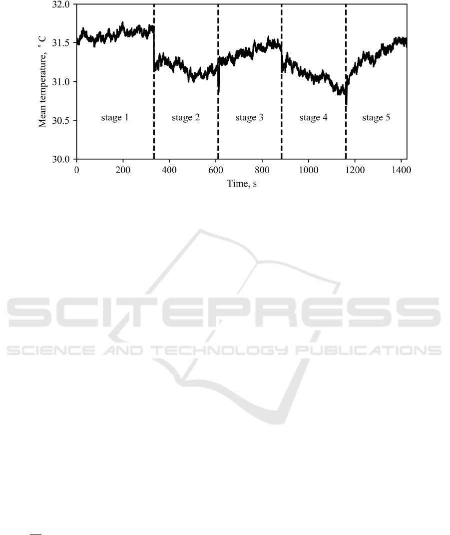
Figure 6: Mean temperature plot for whole study timeline.
metabolic heat generation, change of the thermal
insulation features of the tissues and sweating. One of
the metabolic heat production components is the non-
contracting thermogenesis. The short-term control of
the non-contracting thermogenesis is done through
the ANS. The suppression of the nervous system
activity leads to the decrease of the non-contracting
thermogenesis. The temperature regulation effects,
which are associated with the blood supply, vary for
different functional areas. For the human head there
are two types of the functional areas: acral areas,
which includes hears, lips and nose. The second type
includes the remaining skin surface of the head
(Hensel et al. 1973).
The blood suply of the acral areas is controlled by
the noradrenalin sympathetic nerves. Increase of the
sympathetic tone causes shrinking of the vasculars.
Shrinked vasculars significantly deacrease
convention.
The sweating process is only regulated through
the holinergetic sympathetic fabrics. The blocking of
the holinergetic synapsis results in the sweating
decrease, which, in turn, increase body temperature.
The heat distribution in living organism tissues is
described by the Pennes bio-heat equation (Bergman
and Incropera 2011):
=∇
∇T
+
−
+
(3)
where: ρ – density of the biological tissue, c – heat
capacity of the biological tissue, k – thermal
conduction of the biological tissue. ω
b
–mass blood
flow per unit volume of the biological tissue,
c
b
– blood heat capacity, q
m
– metabolic heat per unit
volume of the biological tissue, T
a
– arterial blood
temperature, T – biological tissue temperature,
∂T/∂t – temperature variation rate.
According to the Pennes equation the temperature
of the biological tissue is defined by three
components: heat exchange with the surrounding
biological tissues {∇(k∇T)}, heat exchange with the
blood {ω
b
c
b
(T
a
–T)}, metabolic heat of the tissue - q
m
.
Mean temperatures plot for all thermogram
frames, with annotated experiment stages is shown on
figure 6. The plot shows, that mean temperature of the
head surface decreases during the 2nd and 4th stages
– neuro-electrostimulation stages. The difference
between the time-averaged temperature of the first
and second stage was 0.44 °С. The difference
between the time-averaged temperature of the first
and the fourth stage was 0.53 °С.
The measuring accuracy of the thermal imager
was ±2% or 2°С, but the thermal resolution was
0.04°С (Gerlach 2006). Therefore, changes on the
0.44°С and 0.53°С are not associated with the noise
of thermal imager. These changes reflect metabolic
reaction of the subject.
The neuro-electrostimulation device was working
in the blocking mode of SNS activity. Therefore,
stimulation decreases the vascular tone and, as the
results improves perfusion rate in tissues. In
accordance with the Pennes equation, in particular the
second component {ω
b
c
b
(T
a
–T)}, the skin surface
should decrease, on the other hand, the blocking of
the SNS activity leads to the non-contracting
thermogenesis (the q
m
component). Jointly, it results
in the general decrease of the head skin temperature.
When the stimulation process is stopped – stages
3 and 5 – blood perfusion and non-contracting
thermogenesis tends to return to the original values.

This results in the graduate increase of the
temperature to the baseline values.
One can note the different behavior of the thermal
changes in the stages 2 and 4. It can be associated with
the different organization of the left and right upper
ganglia.
4 CONCLUSIONS
Presented in the paper results show that neuro-
electrostimulation of the neck neural conducting path
allows one to regulate skin capillary blood flow of the
head and, as the result, to change skin temperature. If
the blocking mode of the sympathetic nervous system
is selected than it is possible to decrease skin
temperature. It was hypothesized that this physical
phenomena is define by the changes of the blood
perfusion and decrease of the non-contracting
thermogenesis
The development of the described in this work
methodology can result in application of the
‘SYMPATHOCOR-01’ neuro-electrostimulation for
treatment of the skin defects, burns and in cosmetic
tasks by means of the blood flow control.
REFERENCES
Bergman, T. L. and Incropera, F. P., 2011. Introduction to
heat transfer. John Wiley & Sons.
Gerlach, N., 2006. Comparison of Thermal Imaging
Systems Used in Thermography as a Non Destructive
Testing Method for Composite Parts. In: European
Conference on Nondestructive Testing, ECNDT 2006
Proceedings, Tu. 25–29.
Hensel, H., Bruck, K., and Raths, P., 1973. Homeothermic
organisms. Temperature and life. Springer, Berlin
Heidelberg New York, 502–761.
Jin, H.-K., Hwang, T.-Y., and Cho, S.-H., 2017. Effect of
Electrical Stimulation on Blood Flow Velocity and
Vessel Size. Open Medicine, 12, 5–11.
Kublanov, V. S., Babich, M., and Dolganov, A., 2017.
Principles of Organization and Control of the New
Implementation of the‘ SYMPATHOCOR-01’ Neuro-
electrostimulation Device. In: BIOSIGNALS. 276–282.
Kublanov, V. S., Danilova, I. G., Goette, I. F., Brykina, I.
A., and Shaljagin, M. A., 2010. Spatially Distributed
Field of Electric Impulses for Regeneration of Ishemic
Muscules. Biomedical Radioelectronics, (10), 34–39.
Kublanov, V. S., Lavrova, S. A., Shershever, A. S., Telegin,
A. V., and Shmikalov, V. A., 2004. Lechenie epilepsii
s primeneniem prostranstvenno raspredelennich
vrashayushichsya poley impulsov toka [epilepsy
treatment by means of the spatially distributed rotating
fields of current pulses]. Biomedical Radioelectronics,
(5–6), 4–15.
Kublanov, V. S., Porshnev, S. V., Danilova, I. G., Goette,
I. F., Levashkina, A. O., and Syskov, A. M., 2012.
Experimental modeling of the effects of autonomic
regulation in the correction of immobilization stress
rats. Biomedical Radioelectronics, (8), 56–67.
Kublanov, V. S., Shmirev, V. I., Shershever, A. S.,
Kazakov, J. E., and others, 2010. About Innovative
Possibilities of Device ‘SIMPATOCOR-01’ in
Management of Functional Disorders of Vegetative and
Central Nervous System in Neurology, Kremljovskaya
Medicine. Clinichesky Vestnik, 4, 60–64.
Mesquita, R. C., Faseyitan, O. K., Turkeltaub, P. E.,
Buckley, E. M., Thomas, A., Kim, M. N., Durduran, T.,
Greenberg, J. H., Detre, J. A., Yodh, A. G., and
Hamilton, R. H., 2013. Blood flow and oxygenation
changes due to low-frequency repetitive transcranial
magnetic stimulation of the cerebral cortex. Journal of
Biomedical Optics, 18 (6), 067006.
Moon, T. K., 1996. The expectation-maximization
algorithm. IEEE Signal Processing Magazine, 13 (6),
47–60.
Morishita, K., Karasuno, H., Yokoi, Y., Morozumi, K.,
Ogihara, H., Ito, T., Fujiwara, T., Fujimoto, T., and
Abe, K., 2014. Effects of Therapeutic Ultrasound on
Intramuscular Blood Circulation and Oxygen
Dynamics. Journal of the Japanese Physical Therapy
Association, 17 (1), 1–7.
Yamabata, S., Shiraishi, H., Munechika, M., Fukushima,
H., Fukuoka, Y., Hojo, T., Shirayama, T., Horii, M.,
Matoba, S., and Kubo, T., 2016. Effects of electrical
stimulation therapy on the blood flow in chronic critical
limb ischemia patients following regenerative therapy.
SAGE Open Medicine, 4.
