
Pathology and Abnormalities of the Teeth as a Biographical Profile in
Uncovering the Identity of Unknown Skeletal Remains
Eko Prastyo
1
, Ledy Ana Zulfatunnadiroh
1
, Rachmadita Yoga Pratiwi
1
, and Myrtati Dyah Artaria
2
1
Forensic Science Study Program, Post Graduate Studies, Universitas Airlangga, Surabaya
2
The Department of Anthropology, FISIP, Universitas Airlangga, Jl. Airlangga 4-6 Surabaya 60286, Indonesia
Keywords: Individualization, pathology, skeletal remains, dental uniqueness, dentition
Abstract: Proper identification of personal identity is essential in the investigation because mistakes can be fatal in the
judicial process. Identification is based on the peculiar characteristics of the person. The possibility of
positive identification will be increased when considering individualization factors as biographical profile
material. The purpose of this paper is to describe the pathological conditions and abnormalities of teeth and
jaws that can be utilized to increase the completeness of the biographical profile of a person. We use
collections of human skeletons remains in Universitas Airlangga. For this preliminary study, we used 5
crania for observations. The assessment was based on macroscopic observational methods on the teeth and
the alveolar bone. The results show that there are several pathological conditions and disorders that along
with other peculiar characteristics of the individual will add to the individualization of the cranium. Several
dental diseases that are found and can be used for positive identifications are periodontitis, dental caries,
tooth fracture, dental impaction, excessive teeth, malposition and malrotation; and dental stain. Those
characteristics altogether give the uniqueness of the individual. The characteristics should be compared to
images of the person during his living years, and to his known habit-such as smoking and or drinking
coffee/tea. Individualization is needed to enrich biographical profile data in identifying unknown human
skeletal remains, through disease detection and projection of lifelong habit.
1 INTRODUCTION
Proper identification of personal identity is very
important in the investigation because mistakes can
be fatal in the judicial process. Identification is the
determination of the identity of the living or the
dead, based on the typical characteristics of the
person. If any bones and adult skulls are found, there
will be identification of several things. First, race
determination, gender determination, age
identification, height measurement, and facial
reconstruction of the skull also can be done (Byers,
2010).
Dental anthropology is part of the natural
sciences, because this science is part of physical
anthropology. In the process of personal
identification using the tooth many things can be
used as a guide. Each individual has a different
shape or morphology in his teeth. In addition, the
condition of the pathology and abnormalities of teeth
and jaws in each individual is different and can be a
special feature for personal identification (Artaria,
2009).
In this paper the author to describe and explain
the pathological and abnormalities of the teeth and
jaws that can be utilized to increase the
completeness of the biographical profile of a person.
There are not many identification processes that
consider individualization factors as biographical
profile material in terms of uncovering identities of
unidentified skeletal remains. Therefore, the authors
are interested to make a paper that discusses about it.
2 MATERIAL AND METHOD
Materials for this writing is a collection of remaining
human skeletons in Universitas Airlangga. For this
preliminary study, we used 5 cranes for observation.
This assessment is based on macroscopic
observational methods on the teeth and alveolar
bone.
426
Prastyo, E., Zulfatunnadiroh, L., Pratiwi, R. and Artaria, M.
Pathology and Abnormalities of the Teeth as a Biographical Profile in Uncovering the Identity of Unknown Skeletal Remains.
DOI: 10.5220/0007544304260430
In Proceedings of the 2nd International Conference Postgraduate School (ICPS 2018), pages 426-430
ISBN: 978-989-758-348-3
Copyright
c
2018 by SCITEPRESS – Science and Technology Publications, Lda. All rights reserved
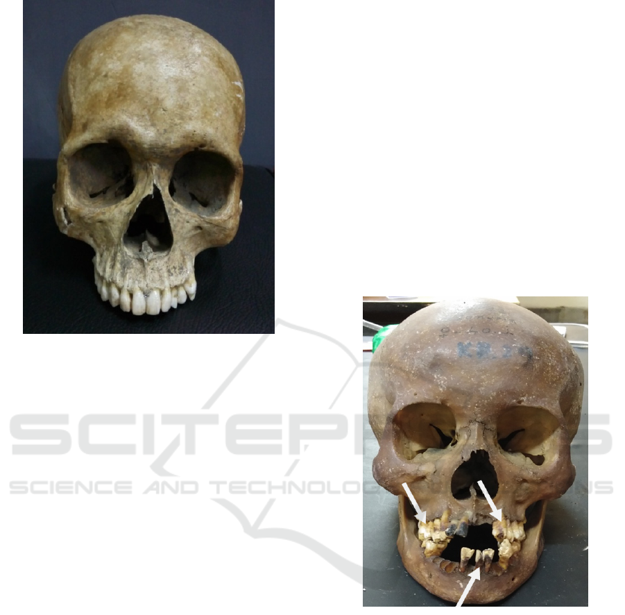
Figure 1: One of the pathological cranium from the rest of
the human skeleton as an object of observation.
3 RESULT AND DISCUSSION
The results show that there are several pathological
groups that signalled dental diseases and disorders in
a person, as well as a person's habit during his
lifetime.
Pathological groups found in cranium, ie;
Periodontitis, Dental caries, Enamel hypoplasia,
Excision. Dental abnormalities found in cranium
include; Fractures on the incisal edge (upper central
incisors), Suspect dental agenesis or impaction of
the upper left canine teeth, Supernumerary teeth or
excess teeth (Parapremolar), Malposition and
malrotation of the supernumerary teeth. Typical
features of specific habits found in cranium are;
Tooth discoloration or tooth staining because of the
bad habit of smoking.
By knowing a pathological state and
abnormalities in the teeth seen in the rest of the
human skeleton we can provide more information as
an attempt to complete the data in order to identify a
person. Moreover, with the discovery of
pathological state and abnormalities in our teeth we
can describe a person's lifetime condition, especially
if the skeletal remains are found in large quantities
and have the same pathological features (Byers,
2010).
3.1 Periodontitis
Periodontitis is a multifactorial disease that causes
infections and inflammation of dental support
tissues, usually resulting in loss of bone and
periodontal ligaments and usually is the cause of
adult tooth loss and edentulousness (Newman et al.,
2018; Ireland, 2006).
Periodontal inflammation has many causes (eg,
bacteria, trauma). However, most of periodontitis
cases are resulted from the accumulation of
microorganisms on the teeth. Risk factors in chronic
periodontitis include the presence of certain
subgingival bacteria, tobacco use, diabetes, age, and
sex. In addition, there is evidence that other factors
may contribute to the pathogenesis of periodontal
disease: environmental, genetic, and systemic (eg,
diabetes) (Gafan et al., 2004; Ireland, 2006).
Figure 2: Alveolar bone resorption and exposed tooth root
that can be seen on the rest of the skeleton, indicating the
occurrence of periodontitis. Alveolar bone resorption is
thorough in all alveolar bone supporting tooth.
3.2 Dental Caries
Caries is the destruction of the crown derived from
bacterial infection. Bacterial metabolic activity
causes acid products that damage the enamel (tooth
enamel). Dental crown caries is caused by aerobic
bacteria. Meanwhile, caries in tooth root is caused
by anaerobic bacteria (Artaria, 2009).
In the remaining skeleton, it can be seen the
presence of caries on the surface of the occlusal, pit
and fissure.
Pathology and Abnormalities of the Teeth as a Biographical Profile in Uncovering the Identity of Unknown Skeletal Remains
427
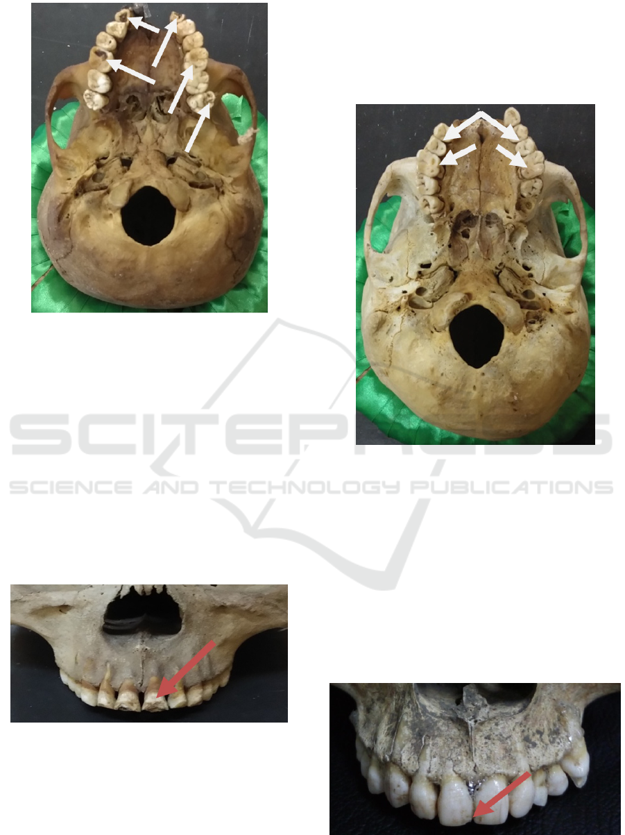
Figure 3: Dental caries can be found on the rest of the
frame. Occurs on occlusal surfaces, pits and tooth fissures.
3.3 Enamel Hypoplasia
Hypoplasia is a dental development disorder that can
occur as a result of trauma or infection prior to
dental eruption in the oral cavity. It is usually
characterized by disturbances on the formation of
enamel matrices. If Turners of hypoplasia is found in
the anterior region of the tooth, the most likely cause
is trauma to the tooth bud. The clinical
characteristics of enamel hypoplasia are highly
unfavorable for aesthetic, dentin sensitivity become
higher, it may lead to malocclusion and dental caries
(Nayak et al., 2016).
Figure 4: Clinical features of enamel hypoplasia are seen
on the labial surfaces of the central incisors.
3.4 Excision
Tooth excision is a physiological change seen in the
morphology of the occlusal anatomy and incisal
tooth due to dental function. Such morphological
changes are characterized by loss of incisal mamelon
or crown cusp on posterior teeth. Tooth excision
occurs because it is widely used for mastication and
Bruxism. The existence of bad habits such as eating
hard foods, eating grains and grinding teeth can also
cause morphological changes in the teeth (attrition)
(Tamril, 2014).
Figure 5: Visible the entire occlusal surface of the
posterior tooth experienced attrition.
3.5 Fracture
Tooth fracture is one of the main causes of tooth
decay after caries and periodontal tissue disease.
Tooth fracture is a condition of the teeth showing the
loss or fragmentation of the fragment of a complete
tooth. This condition is usually caused by trauma to
the face or teeth such as sports that make physical
contact or engage in car accidents (da Silva
Mendonca DH et al., 2012; DiAngelis AJ et al.,
2012).
Figure 6: Fractures on the incisal edge found on the
remaining skeleton.
ICPS 2018 - 2nd International Conference Postgraduate School
428
3.6 Dental Agenesis and Impaction
Dental agenesis on one or more of the tooth
elements is the most common anomaly found in
human teeth. Every tooth, either permanent or
deciduous teeth, has the possibility of having an
agenesis. In the permanent tooth, the third molar is
the most common tooth of the agenesis, followed by
the incisors of both the maxilla and the mandibular
second premolar. Other teeth that are also often
having an agenesis are the mandibular first incisors
and the maxillary second premolar teeth (Jimenez et
al., 2005). A permanent dental agenesis is the
absence of development of one or more permanent
dental elements because they are not formed or may
be due to the non-growth of permanent dental seed
(Vastardis, 2000).
Etiology of impaction teeth can be caused by
local obstruction of hard tissue, local pathology,
impairment of normal development of incisors, and
genetic or hereditary factors. With the exception of
the third molar teeth, the maxillary permanent
canine is the common tooth impaction. Recent
research on maxillary canine impaction frequency
shows a prevalence of 0.27% to 2.4% where women
are more often than men (Becker and Chaushu,
2015).
3.7 Supernumerary Teeth
(Parapremolar)
Supernumerary teeth or extra teeth is a disorder in
which the number of teeth is more than normal.
These additional teeth usually have an abnormal
morphology and shape. Supernumerary teeth that
resemble normal teeth are called supplemental teeth,
whereas more teeth that do not resemble normal
teeth are called accessory. Supernumerary teeth can
be single, multiple, and unilateral or bilateral
eruptions and may be present in one or both jaws.
Supernumerary teeth are more common in the upper
jaw than in the lower jaw. These excess teeth can
also formed in different parts of the jaw, ie in the
area of the upper incisive incisor (also called
mesiodens), next to the molar teeth (also called
paramolars), at the very back of the last molar teeth
(also called disto-molars) or next to premolar teeth
(also called parapremolars). The most common
supernumerary teeth are mesiodens. This disorder is
more common in permanent teeth than in deciduous
teeth (Parolia et al., 2011).
Figure 7: There are excess teeth (Parapremolar) on the rest
of the skeleton. Excessive teeth experience malposition
and malrotation.
3.8 Malposition and Malrotation of
Supernumerary Teeth
Malposition could be teeth crowed, teeth
overlapping, tilted, shifted, and rarely. Malrotation
could be a tooth rotation. The malposition and
malrotation state are often not recorded on the daily
examination (antemortem), so to overcome the
malposition and malrotation conditions, it can be
examined the postmortem data from the mold model
or from roentgen photographs (Harty and Ogston,
1993).
3.9 Tooth Discoloration or Tooth
Staining
Tooth stains can be caused by smoking. Smoking is
an easy thing to meet everyday in the community.
The number of cigarettes greatly affects the rapid
progress of the formation of dental stains. Duration
of smoking can cause thickening of tooth staining
(Sinaga et al., 2014).
Nicotine with decomposition products especially
pyridine is a substance in producing dental stains
which can often be seen in teeth of smokers. This
element will form pigmented deposits attached to the
surface of the teeth and range the colour in brown to
black. Thickened stain deposits can make tooth
surface become rough which will lead to plaque
build up to irritate nearby gums (Sinaga et al., 2014).
Pathology and Abnormalities of the Teeth as a Biographical Profile in Uncovering the Identity of Unknown Skeletal Remains
429
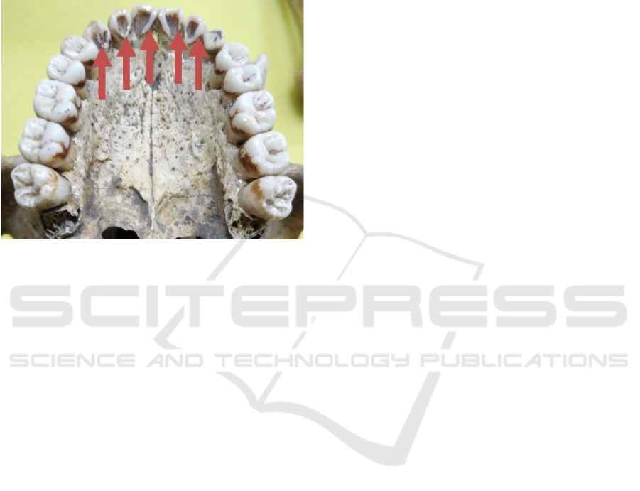
The use and consumption of tobacco and coffee
can cause tooth stains. The use of tea, certain
mouthwashes and pigments in the diet can also
cause the formation of tooth stains and tobacco also
usually cause stains on the enamel surface. Tooth
stains can enter the tooth layer in people who smoke
during their lifetime and are difficult to remove
(Sinaga et al., 2014).
Figure 8: Stains on the tooth surface due to smoking can
be found on the remaining skeleton.
4 CONCLUSIONS
Individualization is needed to enrich biographical
profile data in identifying unknown human skeletal
remains. It should be considered that identification
of pathological conditions and abnormalities in teeth
cannot be determined arbitrarily because
macroscopic determination alone is not sufficient. In
addition to disease detection, individualization can
project lifestyle habits in the form of stress markers,
as in the object of this writing is likely to have a
lifetime smoking habit as clearly visible the typical
features found on the surface of teeth, smoking can
also be a predisposing factor in periodontitis disease
as well as age.
ACKNOWLEDGEMENTS
We thank the Department of anatomy and histology,
physical anthropology section in Universitas
Airlangga for supporting the observation of skeletal
remains. We would like to Acknowledge Prof.
Myrtati Dyah Artaria for support & Co-operation in
preparing this paper.
REFERENCES
Artaria Myrtati Dyah. 2009. Antropologi Dental.
Yogyakarta: Graha Ilmu.
Becker Adrian and Chaushu Stella. 2015. Etiology of
Maxillary Canine Impaction: A Review. American
Journal of Orthodontics and Dentofacial Orthopedics:
Vol 148, issue 4.
Byers Steven N. 2010. Introduction to Forensic
Anthropology, 3th ed. USA: Pearson Education.
da Silva Mendonca DH, da Costa Azevedo ML, Dal Ecco
Leandrini JC, Souza-Gabriel AE. 2012. Functional-
aesthetic treatment of crown fracture in anterior teeth
with severe crowding. RSBO, 9(3):328-33.
DiAngelis AJ, Andreasen JO, Ebeleseder KA, Kenny DJ,
Trope Martin, Sigurdsson Asgeir, Andersson Lars,
Bourguignon Cecilia, Flores MT, Hicks ML, Lenzi
AR, Malmgren Barbro, Moule AJ, Pohl Yango,
Tsukiboshi Mitsuhiro. 2012. Guidelines for the
management of traumatic Dental Injuries:1. Fractures
and luxations of permanent teeth. Dent Traumatol;
28:2-12.
Gafan Gavin P, Lucas VS, Roberts GJ, Petrie Aviva,
Wilson Michael, and Spratt DA. 2004. Prevalence of
Periodontal Pathogens in Dental Plaque of Children. J
Clin Microbiol 42. P.4141-6.
Harty F.J., and Ogston R. 1993. Kamus Kedokteran Gigi.
Alih Bahasa: Narlan Sumawinata dari “Concise
Illustrated Dental Dictionary”. Jakarta: EGC.
Ireland, R. 2006. Clinical textbook of dental hygiene and
therapy. Singapura: Blackwell Munksgaard. p. 57-8
(4).
Jimenez-Castellanos E, Carmona A, Catalina-Herrera CJ,
Cordero E and Jimenez-Castellanos J. 2005.
Variations in the Number of Human Permanent Teeth:
Hypodontia. Eur J Ant. 9:26.
Nayak Vijayendranath, Kini Raghavendra, Baliga
Ashwini, Rao Prasanna Kumar, Bhandarkar Gowri P
and Kashyap Roopashri Rajesh. 2016. A Clinical
Image of Enamel Hypoplasia-Turners Tooth. Austin
Journal of Dentistry: Vol (3) issue 6-2016.
Newman Michael G, Takei Henry H, Klokkevold Perry R,
Carranza Fermin A. 2018. Newman and Carranza’s
Clinical Periodontology, Thirteenth Edition.
Philadelphia: Elsevier. p.346.
Parolia Abhishek, M Kundabala, Dahal Marisha, Mohan
Mandakini, Thomas Manuel S. 2011. Management of
Supernumerary Teeth. Journal of Conservative
Dentistry: Vol 14, issue 3.
Sinaga Putri Amin, Lampus BS, and Mariati Ni Wayan.
2014. Gambaran Pengetahuan Stain Gigi Pada
Perokok Di Kelurahan Bahu Lingkungan V. Jurnal e-
GiGi (eG), Volume 2, Nomor 2, Juli-Desember 2014.
Tamril Rudin. 2014. Pengaruh Bruxism Terhadap
Perubahan Morfologi Gigi Permanen. Fakultas
Kedokteran Gigi Universitas Hasanuddin: Makassar.
Vastardis Heleni. 2000. The Genetic of Human Tooth
Agenesis: New Discoveries for Understanding Dental
Anomalies. Am J Orthod and Dentofac Orthop. 117:
650-6.
ICPS 2018 - 2nd International Conference Postgraduate School
430
