
An Ultra-high Definition and Interactive Simulator for Human
Dissection in Anatomic Learning
Andrei Rafael Brongel
4
, William John Pereira Brobouski
4
, Lucas Murbach Pierin
2
, Carlos Gomes
1
,
Manoel de Campos Almeida
3
and Edson Jos
´
e Rodrigues Justino
3,4
1
Medical School, Pontif
´
ıcia Universidade Cat
´
olica do Paran
´
a - PUCPR, Curitiba, Paran
´
a, Brazil
2
Polytechnic School, Pontif
´
ıcia Universidade Cat
´
olica do Paran
´
a - PUCPR, Curitiba, Paran
´
a, Brazil
3
Centro de Inovac¸
˜
ao em Imagens M
´
edicas, CIIM - PUCPR, Curitiba, Paran
´
a, Brazil
4
Programa de P
´
os-Graduac¸
˜
ao em Inform
´
atica, PPGIa - PUCPR, Curitiba, Paran
´
a, Brazil
Keywords:
Human Anatomy Learning, Dissection, Simulation, Ultra-high Definition, Real Bodies Models.
Abstract:
This paper presents an ultra-high definition simulator for the teaching of human anatomy in class. The simu-
lator in question consists of hardware and software specially developed for this purpose. The hardware seeks
to meet the requirements of real-scale representation of models, visual acuity, color, texture, depth perception
e touch-based interactivity. The software, in turn, offers a set of dissecting tools typically used in anatomical
studies, in addition to allowing connectivity to educational environments and the Internet. The characteristic
that stands out most in this simulator is the fact that it is not an anatomical atlas, but a dissecting table that uses
models from real bodies, which differentiates it from most of the simulators and anatomical atlases developed
to this end.
1 INTRODUCTION
In almost all courses related to life sciences, espe-
cially those related to health such as medicine, nurs-
ing, physiotherapy, dentistry, and others, required
the study of human anatomy. The consensus among
anatomy teachers is that there is currently no replica,
book, or 3D computational tool to replace the direct
manipulation of a real human body. Some features,
such as color, depth, texture, anatomical position, and
detail, are essential for comprehensive learning. It
is also unanimous among anatomy teachers that the
corpse is as close as we can have to a living human
being, even though in dead tissues we cannot observe
some properties. As an example, it is possible to
cite the cerebral activities, vascularisation, heartbeats,
among others. According to the literature (Ghosh,
2015) (Shaikh, 2015) (Singh A.K. and D.M., 2011),
the manipulation of corpses opens the discussion of
three main aspects. The first one concerns ethical is-
sues since many of unclaimed bodies come from do-
nations and are made by forensic laboratories or sim-
ple purchase when the law of the country allows. The
second one concerns health issues. Toxic chemical,
such as formaldehyde, is the main component used
in the preservation of bodies and parts. It makes the
anatomy laboratories unhealthy environments and in-
appropriate for teaching and learning. The third one is
the quality of learning. The permanent manipulations
of the bodies makes them not suitable for teaching and
learning in a short time.
Over the point of view of clinical and surgical
procedures, we can observe the increased use of the
non-invasive procedure, whose supporting instrument
is the images made from cameras coupled to surgical
instruments or from CT (Computerised Tomography)
and MR (Magnetic Resonance) exams. Procedures
such as laparoscopy, endoscopy, and surgeries guided
by neuronavigation have become the most viable so-
lutions for the reduction of the risks of postoperative
infection and the rapid recovery of the patient.
Given the current scenario, researchers all over
the world have been efforts in finding solutions to
minimize the use of cadavers in the teaching of
anatomy and surgical practices. Among another kind
of simulation, the digital models have been showing
promise. There are some excellent examples, such
as the Anatomage table
1
a high-quality artistic at-
las. However, Ackerman (Ackerman, 1998) and Azer
(Azer and Azer, 2016) asserts there are some reasons
1
Anatomage - https://www.anatomage.com/table6/
284
Brongel, A., Brobouski, W., Pierin, L., Gomes, C., Almeida, M. and Justino, E.
An Ultra-high Definition and Interactive Simulator for Human Dissection in Anatomic Learning.
DOI: 10.5220/0007707102840291
In Proceedings of the 11th International Conference on Computer Supported Education (CSEDU 2019), pages 284-291
ISBN: 978-989-758-367-4
Copyright
c
2019 by SCITEPRESS – Science and Technology Publications, Lda. All rights reserved

to restrict the use of this kind of atlas. Among other
things, the diversity of structures found in nature that
is a limiting factor, according to them, because it is
difficult to replicate in computational models still in
the present day. Moreover, it is almost complicated
for a graphic artist to map all the nuances of the hu-
man body.
Based on the last discussion, this paper presents
an ultra-high definition simulator for the teaching of
human anatomy in class. The simulator consists of
hardware and software specially developed for this
purpose. The device seeks to meet the requirements
of high-quality representation of models, visual acu-
ity, color, texture, depth perception e touch-based in-
teractivity. The application (App), in turn, offers a
set of dissecting tools typically used in anatomical
studies, in addition to allowing connectivity to educa-
tional environments and the Internet. The character-
istic that stands out most in this simulator is the fact
that it is not an anatomical atlas, but a dissecting table
that may perform almost all operations that can be ex-
ecuted in a real necropsy table, which differentiates it
from most of the digital anatomical atlases developed
to this end. To simulate a necropsy table, we adopted
the same format for the interactive table. However,
the hardware supports two additional projectors with
the same hardware features.
Five sections make up this paper. The first section
is this introduction. The second section presents the
material used to make the simulator. The third section
contains the method used for building the software of
the simulator. The fourth section presents the results.
Finally, the last section presents a conclusion and fu-
ture works.
2 MATERIAL
We divided the materials used on the simulator into
two groups. The first one belongs to the software and
is made up of the database. The database is the most
relevant material for the characterization of the simu-
lator since it must have its origin in real human bod-
ies and not coming from artistic models. The second
group belongs to the hardware. The hardware must
meet the usability requirements in anatomy classes,
be dynamic and have a user-friendly interface.
2.1 Data Base
In the last four decades, several databases of real hu-
man bodies have emerged, whose objective was to
create a digital database of human bodies, both mas-
culine and feminine, for educational and research pur-
poses. The first database started in the 1990s by
Michael J. Ackerman of the US National Library of
Medicine became known as The Visible Human Body
Project (VHP)(Ackerman, 2002) (Spitzer and Whit-
lock, 1998). Other databases came later, like Korean
(Park et al., 2005) and Chinese (Chen et al., 2018) ,
all using the same cryosection process. However, only
the VHP database became public and available for the
development of new applications under an agreement
with the US National Library of Medicine. The other
databases are private.
The VHP database consists of a 1mm thick sliced
male body, photographed on 70mm colored films and
then scanned in 2k (2048 x 1216 pixels) and 4k (
4096 x 2700 pixels) resolution, totaling 1871 sec-
tions. In addition to the anatomical images, the X-
ray, Computed Tomography and Magnetic Resonance
complete the set. The female body has the same char-
acteristics as the male corpse, with one exception.
The intervals used in the anatomical images were 0.33
mm instead of 1.0 mm. The female set results in more
than 5,000 anatomical images. In Figure 1, we can see
a sample of the male and female database.
(a) (b)
Figure 1: Database samples: (a) Male criosection, (b) Fe-
male criosection.
2.2 Interactive Table
The use of interactive screen has been growing sig-
nificantly in recent years with the advent of the mul-
titouch screens. This kind of screen allows complex
interactions with the user. Two crucial factors were
taken into account to determine the sizing of the ta-
ble and therefore the dimensioning of the screen. The
first one was the screen size. The screen should con-
tain the body of a human with approximately 1.70m.
This feature would ensure the representation of the
organs of the human body in the standard scale (1:1).
The second is the screen resolution. The screen in
question should also support resolutions up to 4k. The
high resolution would guarantee the visual acuity nec-
essary for anatomy studies. Another relevant factor is
the application server, which should support the ma-
nipulation of a high volume of data from the image
databases. The interactive table was designed and
built following the usability parameters in the labo-
ratory for a student group and the teacher. Figure 2
An Ultra-high Definition and Interactive Simulator for Human Dissection in Anatomic Learning
285
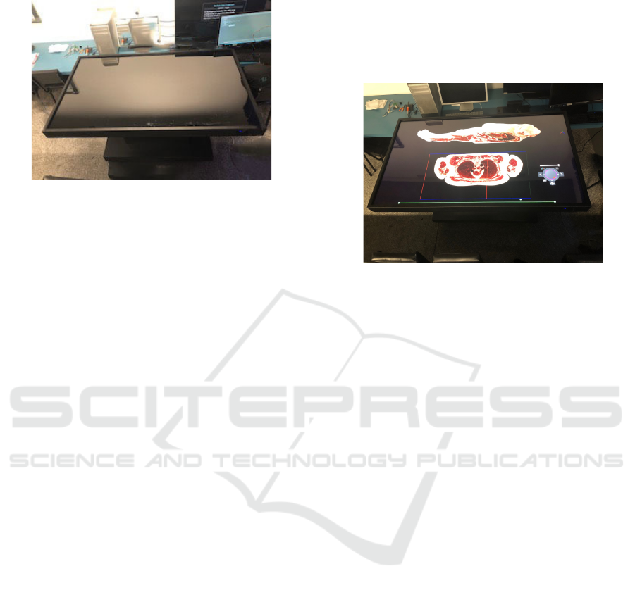
shows the large screen interactive table.
Figure 2: The large screen interactive table.
3 METHOD
The method used to create the simulator took into ac-
count two constructive aspects. The first one dealt
with the adequacy of 2k images databases of the fe-
male and male corpses, to be used by the simulator.
The choice of resolution in 2k was due to the mainte-
nance of the body’s near from the natural scale, one
of the first prerequisites of the project. The second
took care of the interactivity and the user interface, to
make the interface user-friendly.
The choice of VHP occurred because it is a
database of public domain and the existence of both
sexes, the masculine and the feminine. Another im-
portant aspect is in the fact that they are images of
real bodies in ultra-high definition 2k and 4k. Since
its making in the 1990s, numerous works have been
developed using the VHP database images (Acker-
man, 2002) (Spitzer and Whitlock, 1998), many of
them resulted and simulators for different purposes
such as endoscopy (Pflesser et al., 2001). However,
due to the byte size of the 2k and 4k databases, the
generation of synthetic or rendered models was the
most adopted solution. Even with current technology,
it is necessary to use powerful game support compu-
tational resources to manipulate such bases in their
original form, and this has become the great challenge
of this project (the last generation of microprocessor,
high memory capacity, solid state disk, and others).
Another aspect to be highlighted is the resolution of
the images. Rendered models adapt to the display ca-
pabilities, making it easy to create and manipulate 3D
models. Only a few years ago the technology of GPUs
or Video Graphics Cards started to support the reso-
lution in 4k and have memory capabilities and speed
to allow the processing of the VHP base in its orig-
inal form. Based on that features we build the in-
teractive table hardware. Therefore, for this project,
the VHP database was chosen in 2k resolution. The
chosen resolution allows for the maintenance of the
original scale of the slices of cryosection images and
the FULLHD (1920 x 1080 pixels) projection of the
volumetric visualization model. Figure 3 shows an
example.
Figure 3: An Exemple of the 2k (slice) and FULLHD pro-
jection (volumetric projection).
3.1 Database Pre-processing
In the pre-processing of the database, we applied a
classification algorithm based on the SVM (Support
Vector Machine) (Wang et al., 2012) (Tsai et al.,
2006) for the removal of the blue background (ice)
that covered the external part of the bodies and some
internal areas. In Figure 1 it is possible to see such
areas. We apply four different classifiers for the com-
plete removal the ice over the skin, one for each four
different body regions. We do not use any post-
processing.
3.2 Screen Interface
Figure 4 shows the different simulator manipulation
capabilities. We divided the screen area into two sub-
areas. The first contains the anatomical projection
planes chosen for the visualization (coronal in blue,
axial in green and sagittal in red), following the di-
dactic perception in the anatomy study. Each plane
allows navigation between the slices of the body in
a thickness of 0.33 mm (for the female body) and 1
mm (for the male body) (Figure 5, Figure 6, and Fig-
ure 7). This navigation also allows adjusting the other
two planes in a cut position, in order to enable the vi-
sualization of the three cuts in the volumetric model,
in perspective (Figure 4). The second subarea is able
the projection of the volumetric model in perspective
(Figure 4). The model can undergo several geomet-
ric transformations, such as the rotation in the axis
of the current anatomical plane of cut, inversion from
CSEDU 2019 - 11th International Conference on Computer Supported Education
286

the point of view of the body (volumetric model in
perspective, Figure 8), adjustment of the transparency
(Figure 9), stereoscopic visualization for depth per-
ception (Figure 10), among other resources .
3.3 Stereoscopy
The ability to perceive and interact with the structure
of space is one of the fundamental goals of the visual
system. Two retinal images are different because the
retinas are in slightly different places. Stereopsis is
the ability to use binocular disparity as a cue to depth.
The stereoscopy is the technic used to create the stere-
opsis by devices. There are two kinds of stereoscopy
applicable to digital devices. The first and oldest tech-
nic is the anaglyphic. In this kind, two images create
the stereo pair, one in red and the other in cyan. To
view the effect anaglyphic glasses must be used. The
second is the polarized stereoscopy. Its use is more re-
cent, having a wide range of applications, such as 3D
TV’s, 3D cinema and 3D projectors. There are two
kinds of technics of polarization, the linear and the
circular. In both cases, we must use polarized glasses
(Daly et al., 2011).
The use of stereoscopy in medical applications has
been growing in recent years (Livatino et al., 2015)
(van Beurden et al., 2009). In the simulator, we used
anaglyphic stereoscopy initially, in order to evaluate
its performance at the table. Although the results are
promising, as seen in the projection of the body of
Figure 10, its use will be more effective in the vi-
sualization of the inner organs, whose segmentation
process is in progress.
3.4 Interface Objects
The simulator has a graphical interface managed by
an Interface Object called Draggable DIAL (virtual
mouse that concentrates the manipulation tools of the
volumetric model of the bodies such as rotation Fig-
ure 11(f), perspective Figure 11(e), and transparency
Figure 11(a). It also has rotating menu functional-
ity for selecting anatomical pieces available in a li-
brary, and also an option for selecting a type of tis-
sue to be viewed and manipulated from the selected
anatomical piece, Figure 12. It has too a push button
(on/off) for enabling stereoscopic visualization fea-
tures (with anaglyph glasses), Figure 11(b). A push
button (on/off) for visualization of the 2D cut in the
bodies volumetric model, Figure 11(e). A push but-
ton (lock/unlock) the touchscreen to avoid unwanted
touch, Figure 11(c). A push button (on/off) for en-
abling the slices viewer in the volumetric model, Fig-
ure 11(d). A button for activating active blackboard
functions (pens, markers, rubber, save screen, and
colors), Figure 13(c) and Figure 14. A button for acti-
vating the browser for Internet search, 13(b). Finally,
a button for activating accessibility to the Online Ed-
ucation Platforms (OEP), 13(a). In addition to the
DIAL, the simulator has a second control area for the
manipulation of the cuts (1mm slices of the male body
or 0.33mm of the female body). This area has slid-
ing bars of the three anatomical planes (axial, coronal
and sagittal), for each cut of the plane in highlighted.
The displacement of those bars in the 2D visualiza-
tion allows the same cut in the volumetric model. It
should note that such planes orthogonal to the body
can adjust to any angle by merely shifting the bar of
the plane to the desired angular position.
3.5 Reproductive System
As already mentioned in previous sessions, the simu-
lator has two bodies, one male, and one female. The
sex selection is in an initial screen of the simulator,
or later, through the rotating menu of the DIAL. The
different sexes allow the anatomy teachers to explore,
along with the students, the anatomical differences of
both sexes, as well as to present in detail the reproduc-
tive apparatus. The use of real body models provides
biological data that could not be observed in synthetic
models, such as the influence of hormones, testos-
terone, and progesterone, on the formation, growth,
and aging of organs. In Figures 15 and 16 we can see
both male and female sex respectively.
3.6 Imaging Exams
As seen in the sessions where we described the VHP
database, it has two kinds of set of the imaging exams.
The first set is the computed tomography (CT) images
and the second set is the magnetic resonance (MR)
imaging . The male database consists of axial MR
images of the head and neck taken at 4mm intervals
and longitudinal sections of the rest of the body also
at 4mm intervals. The resolution of the MR images
is 256 pixels by 256 pixels, in grayscale. The MR
imaging database consists of three acquisition modes,
T1, T2, and Proton Density Image (PD). CT data are
taken in the axial plane of computed tomography of
the whole body taken at intervals of 1mm at a reso-
lution of 512 pixels by 512 pixels in grayscale. The
female corpse data set has the same characteristics as
the male corpse, with one exception. Axial anatomi-
cal images were obtained at intervals of 0.33 mm in-
stead of 1.0 mm intervals. As seen, CT and MR im-
ages can provide important educational resources for
studying and practice in imaging exams analysis. The
An Ultra-high Definition and Interactive Simulator for Human Dissection in Anatomic Learning
287
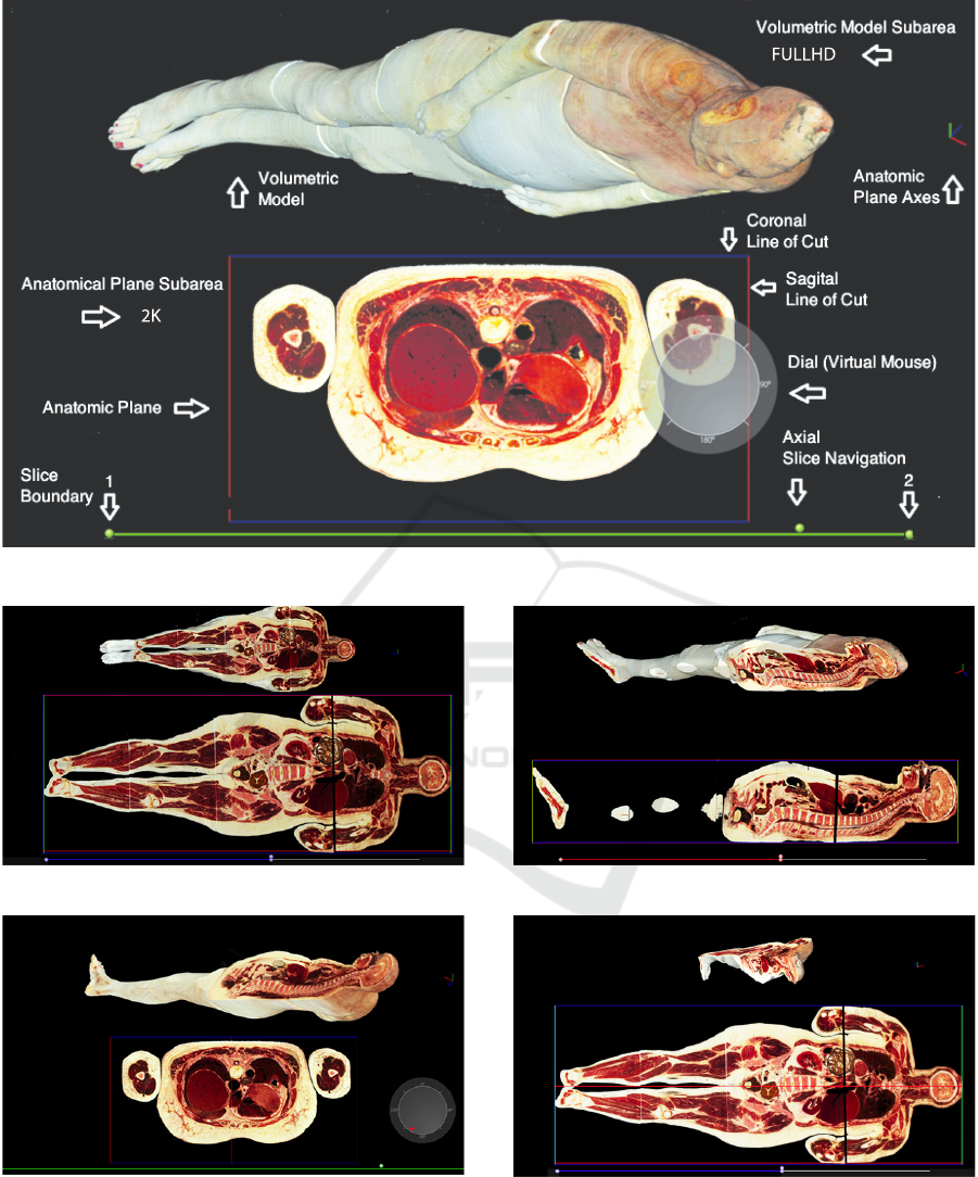
Figure 4: The Interactive Simulator Interface.
Figure 5: Adjustable coronal plane view mode.
Figure 6: Adjustable axial plane view mode.
simulator offers teachers and students the possibility
of associating each CT and MR image with the cor-
related anatomical cut of the body and the volumetric
model. In this way, the student can observe the ex-
ams, such as in real practice, and relating it to the
Figure 7: Adjustable sagittal plane view mode.
Figure 8: Adjustable perspective view mode.
body. Figures 17, 18 and 19 show an example of a
slice, the CT and MR (T1, T2, and PD) images.
CSEDU 2019 - 11th International Conference on Computer Supported Education
288
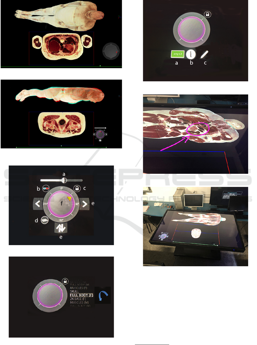
Figure 9: Adjustable transparency view mode.
Figure 10: The full body in stereoscopic view (Anaglyph
Stereoscopy).
Figure 11: The draggable dial in volumetric model tools.
Figure 12: The draggable dial in circular menu library.
Figure 13: The draggable dial in eduction tools.
Figure 14: An exemple of the Active Blackboard.
Figure 15: Male.
4 CONCLUSIONS
This paper presented an ultra-high definition simula-
tor, now called The Visible Human Table (VHT)
2
, for
the dissection and teaching of the human anatomy in
the classroom. The differential to other anatomical
atlases and simulators are in the use of real body im-
ages in ultra-high resolution and non-synthetic body
2
The VHT Project: https://www.youtube.com/watch?
v=yZ-rDkC4UXY
An Ultra-high Definition and Interactive Simulator for Human Dissection in Anatomic Learning
289

Figure 16: Female.
Figure 17: Male: anatomic image.
models which gives it visual acuity, scale compliance
with real human organs, texture, color, and depth per-
ception. Another positive aspect is the diversity of
sex. From teaching and learning, the simulator com-
plies with the precepts used in the study of anatomy,
that is, respecting the anatomical planes of cutting,
volumetric visualization, and dynamics in the dissec-
tion process. Another essential element that charac-
terizes it as a learning tool is the availability of the
imaging exams associated with the anatomical im-
ages in cuts, the connectivity with resources of the
Online education platform and the active blackboard.
As future work, we are planing the inclusion of new
ultra-high-definition segmentations of tissues of spe-
cific organs, such as the skeleton, muscles, circulatory
system, among others. We intend to include the X-ray
exams set, from de VHP databases, in to the simula-
(a) (b)
Figure 18: Database samples: (a) TC image, (b) MR image
T1.
(a) (b)
Figure 19: Database samples: (a) MR image T2, (b) MR
image PD.
tion and support for reading DICOM
3
standard.
ACKNOWLEDGEMENTS
We want to thank the US National Li-
brary of Medicine for providing us the
databases of The Visible Human Body Project
(www.nlm.nih.gov/reseach/visible/), without which
this project would not become possible. The Voxel-
Man for providing the database of segmented inner
organs (Voxel-Man, www.voxel-man.com), which
is assisting us in the process of ultra-high definition
segmentation of the two bodies. To the Financiadora
de Estudos e Projetos - FINEP for the financial
support for the development of this project. Also,
to the Center of Innovation in Medical Imaging of
the PUCPR (CIIM) for the technical and laboratory
support.
3
The Digital Imaging and Communications in Medicine
(DICOM) is a standard for handling, storing, printing, and
transmitting information in medical imaging.
CSEDU 2019 - 11th International Conference on Computer Supported Education
290

REFERENCES
Ackerman, M. J. (1998). The visible human project. Pro-
ceedings of the IEEE, 86(3):504–511.
Ackerman, M. J. (2002). Visible human project
R
: From
data to knowledge. Yearb Med Inform, 11(01):115–
117.
Azer, S. A. and Azer, S. (2016). 3d anatomy models and
impact on learning: A review of the quality of the lit-
erature. Health Professions Education, 2(2):80 – 98.
Chen, D., Zhang, Q., Deng, J., Cai, Y., Huang, J., Li,
F., and Xiong, K. (2018). A shortage of cadav-
ers: The predicament of regional anatomy education
in mainland china. Anatomical Sciences Education,
11(4):397–402.
Daly, S. J., Held, R. T., and Hoffman, D. M. (2011). Per-
ceptual issues in stereoscopic signal processing. IEEE
Transactions on Broadcasting, 57:347–361.
Ghosh, S. K. (2015). Human cadaveric dissection: a histor-
ical account from ancient greece to the modern era. In
Anatomy & cell biology.
Livatino, S., Paolis, L. T. D., D’Agostino, M., Zocco, A.,
Agrimi, A., Santis, A. D., Bruno, L. V., and Lapresa,
M. (2015). Stereoscopic visualization and 3-d tech-
nologies in medical endoscopic teleoperation. IEEE
Transactions on Industrial Electronics, 62(1):525–
535.
Park, J. S., Chung, M. S., Hwang, S. B., Lee, Y. S., Har,
D.-H., and Park, H. S. (2005). Visible korean human:
improved serially sectioned images of the entire body.
IEEE Trans Med Imaging, 24(3):352–360.
Pflesser, B., Petersik, A., Pommert, A., Riemer, M., Schu-
bert, R., Tiede, U., Hohne, K. H., Schumacher, U.,
and Richter, E. (2001). Exploring the visible human’s
inner organs with the voxel-man 3d navigator. Stud
Health Technol Inform, 81:379–385.
Shaikh, S. T. (2015). Cadaver dissection in anatomy: The
ethical aspect. Anatomy and Physiology: Current Re-
search, 5(1):1–2.
Singh A.K., Sharma R.C., S. R. and D.M., M. (2011). Chal-
lenges in cadaver availability for learning and research
in medical sciences. International Journal of Medical
& Clinical Research, 2(2):67.
Spitzer, V. M. and Whitlock, D. G. (1998). The visible hu-
man dataset: The anatomical platform for human sim-
ulation. The Anatomical Record, 253(2):49–57.
Tsai, C.-F., McGarry, K., and Tait, J. (2006). Claire: A mod-
ular support vector image indexing and classification
system. ACM Trans. Inf. Syst., 24(3):353–379.
van Beurden, M., Van Hoey, G., Hatzakis, H., and Ijssel-
steijn, W. (2009). Stereoscopic displays in medical
domains: A review of perception and performance ef-
fects. volume 7240, page 72400.
Wang, X., Wang, S., Zhu, Y., and Meng, X. (2012). Im-
age segmentation based on support vector machine.
In Proceedings of 2012 2nd International Conference
on Computer Science and Network Technology, pages
202–206.
An Ultra-high Definition and Interactive Simulator for Human Dissection in Anatomic Learning
291
