
Effective Image Processing Procedure for Skin Lesion Recognition in
Contactless Skin Diagnosis Devices
Hansoo Kim
Department of Information Security, Seowon University, 377-3, Mushimseoro, Seowongu, Cheongjusi, Choongbuk, Korea
Keywords: Skin Diagnosis, Lesion Recognition, Image Processing, Contactless Skin Diagnosis Device.
Abstract: An image analysis procedure for recognizing various skin lesions under contactless skin diagnosis
environment is proposed. The proposed procedure is composed of five stages, and experimental results
show that issues such as uneven distribution of light are properly addressed, and various skin lesions are
effectively discriminated according to their characteristics using the image processing technology and the
shadow analysis.
1 INTRODUCTION
Composed of epidermis, dermis, and subcutaneous
tissues, skin is the largest organ of human body (Wei
et al., 2018). Containing blood vessels, lymphatic
vessels, nerves, and muscles, which can perspire,
perceive the external temperature, and protect the
body. Covering the entire body, the skin can protect
multiple tissues and organs in the body from
external invasions including artificial skin damage,
chemical damage, adventitious viruses, and
individuals’ immune system. Besides, skin can also
avoid the loss of lipids together with water within
epidermis and dermis so that skin barrier function
can be stabilized (Hu and Yu, 2013). Skin is the first
defender of human body, from various external
hazards.
It is of great theoretical significance and practical
value to study how to extract symptoms of diverse
skin diseases on the basis of modern science and
technology. Under this circumstance, effective and
accurate identification of the types of skin diseases
can be achieved to prescribe treatment according to
patients’ symptoms (Vezhnevets et al., 2003).
Television, social media, and advertising have
had a tremendous impact on consumers’ paying
increased attention to physical appearances and
aesthetics. This has also raised interest in a variety
of cosmetic and aesthetic surgeries and procedures.
This, supported by surge in consumer disposable
income and introduction of technologically
advanced solutions, has had a major impact on
market demand (Market Research Report, 2018). As
a standard of beauty, skin increases its importance as
society develops. However, various skin disease
have emerged due to air pollution, micro dust, and
unnecessary UV exposure due to the ozone depletion.
With the development of dermatology and skin
medicine, the skin analysis technology with image
processing and computer vision gains its importance
recent years. In addition, research has been
conducted to measure skin condition quickly, easily,
and accurately.
In this paper, an image processing procedure for
recognizing skin lesions over contactless skin
imaging device is proposed. Experiments show that
analyzing the shadow of the skin lesions can
effectively detect and classify the lesion of concern.
The rest of this paper is as follows. In Section 2 ,
recent studies and technologies for skin analysis are
introduced. Details of the proposed procedure are
shown in Section 3 . Experimental Results are
shown in Section 4 and Section 5 concludes with
future work.
2 RELATED WORKS
2.1 Skin Diagnosis Device
With the development of semiconductor technology
and small electronic devices industry, devices for
measuring skin using various computerized and
electronic technologies have emerged.
Kim, H.
Effective Image Processing Procedure for Skin Lesion Recognition in Contactless Skin Diagnosis Devices.
DOI: 10.5220/0007948403370342
In Proceedings of the 16th International Joint Conference on e-Business and Telecommunications (ICETE 2019), pages 337-342
ISBN: 978-989-758-378-0
Copyright
c
2019 by SCITEPRESS – Science and Technology Publications, Lda. All rights reserved
337
The global dermatology devices market size was
valued at over USD 9.6 billion in 2017 and is
expected to showcase lucrative growth over the
forecast period, registering a CAGR of 13.5%.
Increasing prevalence of skin cancer and other skin
diseases has significantly contributed to high
demand for various dermatological diagnostic and
treatment procedures in recent times, thereby
spurring product demand (Market Research Report,
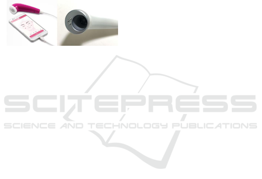
2018). Figure 1 shows one of the representative
personal skin diagnosis devices (Ahn, 2019).
Figure 1: Sample of skin diagnosis device.
To get a clear image, most of these devices make
contact with the skin. Contacting the cylindrical
body with the skin enables to get a clear skin image
with fixed focal length and stable light source.
However, contact with the skin can cause
possible skin contamination due to uncleanness and
corrosion of the contact part. To prevent this, the
contact part needs to keep clean and sterilized which
lead to additional costs and actions.
2.2 Skin Analysis Technology
There have been many researches on skin analysis
using image processing. Various statistical methods
for segmentation and classification of skin lesions in
dermoscopic images is developed (Zaqout, 2016).
Texture and color features of skin diseases are
analysed (Wei et al., 2018; Yang et al., 2018).
Also, applying neural networks including the
Artificial Neural Network (ANN) and the
Convolutional Neural Network (CNN) are
conducted for effective skin recognition (Abbadi et
al., 2010; Menaka and Rohini, 2014; Yang et al.,
2018).
2.3 Object Detection and Recognition
For a long time, algorithms for finding objects in
images have been studied extensively. Numerous
researches have been done in object detection,
recognition, classification and discrimination.
Scale-Invariant Feature Transform (SIFT) has
been studied and applied various applications
(Gonzalez and Woods, 2018). Speeded Up Robust
Features (SURF) and RANdom Sample Consensus
(RANSAC) are also well-studied algorithms (Kim
and Kim, 2014). Circular Hough Transform (CHT)
is one of the widely used algorithms (Vegt, 2015).
Also, light source detection has been researched
in a number of areas, including human vision,
optical science and photometric engineering (Nillius
and Eklundh, 2001; Funk and Yang, 2007)
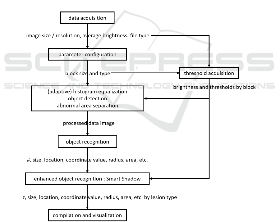
3 PROPOSED PROCEDURE
The proposed procedure is composed of five stages:
Preprocessing, Parameter Configuration, Abnormal
Area Separation, Lesion Recognition, and
Categorization and Visualization. The flowchart for
the overall procedure is shown in Figure 2, followed
by its pseudocode shown in Figure 3.
3.1 Preprocessing
To analyse skin and its lesions, an image is taken by
skin diagnosis device. As the device does not touch
the skin and is exposed to external light source
(sunlight, indoor lamp, etc.), various factors such as
direction and amount of light, vignetting and lens
distortion should be considered. So, color
temperature and lens distortion calibration,
vignetting and chromatic abberation correction and
other necessary processing should be applied on the
stage of imaging the skin.
After the correction and calibration of the image,
basic properties such as the image size, resolution
and the image file type are acquired for further
analysis.
3.2 Parameter Configuration
Detailed and practical properties such as the average
brightness for entire image and the image histogram
are obtained in this stage.
In addition, the image is divided into number of
image blocks. For each image block, average
brightness is obtained so that the threshold for each
block is configured using the average brightness.
The threshold for each block is used to separate the
lesion part from the skin part. This adaptive
threshold configuration enables the separation of the
skin and its lesions effectively.
SIGMAP 2019 - 16th International Conference on Signal Processing and Multimedia Applications
338
3.3 Abnormal Area Separation
Using the parameters and properties acquired from
Section 3.1 and 3.2, the lesion part is separated from
the skin part.
By the threshold value on each designated image
block, each pixel is classified upon the binarization
method (black and white manner).
3.4 Lesion Recognition
Among the number of image processing and object
recognition technologies described in Section 2.3,
CHT is one of the most appropriate algorithms to
detect and recognize the skin lesions.
In general, lesions are dark and circular in shape
compared to the skin, so it is expected that CHT is
possible to detect and recognize the skin lesions
effectively.
Furthermore, as there are lesions in a concave
shape such as pores and a convex shape such as
moles, the location of dark and bright part of those
lesions is different according to the direction of light.
Therefore, it is possible to distinguish lesions by the
direction of the shadow. In this stage, the Smart
Shadow analysis is performed; the discrimination of
the lesion by its shadow.
3.5 Categorization and Visualization
After obtaining the concave and convex lesions
according to the shadow analysis, each feature is
analysed and marked in the final stage.
Since the information obtained by the CHT
includes the position (coordinates) and size of each
lesion, the detected lesions can be displayed in an
image for visual understanding, and statistical
features such as area can also be analysed.
Figure 2: Flowchart for the proposed procedure.
Effective Image Processing Procedure for Skin Lesion Recognition in Contactless Skin Diagnosis Devices
339

INPUT : skin_Image
OUTPUT : p_center and p_radius according to lesions
/* parameter definition & configuration */
skin_Image // skin image to analyze
skin_h, skin_w // size of skin_Image
n_blocks // number of blocks (horizontal and vertical)
th_lesion // threshold value to classify lesions
p_comp // compensation parameter
p_center // array of p_center coordinate values of lesions
p_radius // array of radii of lesions
LESION // value for separating lesions
n_blocks_h=skin_h/n_blocks
n_blocks_w=skin_w/n_blocks
/* th_lesion acquisition */
for k=1 to n_blocks
s=(k-1)*n_blocks_h
for m=1 to n_blocks
n=(m-1)*n_blocks_w
g=1
for ni=1+s to n_blocks_h+s
e=1
for nj=1+n to n_blocks_w+n
tmp_Image(g,e)=skin_Image(ni,nj,1)
// the R value
e=e+1
g=g+1
th_lesion(k,m)=mean(mean(tmp_Image))-p_comp
/* lesion (abnormal area) separation */
for k=1 to n_blocks
s=(k-1)*n_blocks_h
for m=1 to n_blocks
n=(m-1)*n_blocks_w
for ni=1+s to n_blocks_h+s
for nj=1+n to n_blocks_w+n
if (skin_Image(ni,nj,1)<=th_lesion(k,m))
proc_Image(ni,nj,1)=LESION
proc_Image(ni,nj,2)=LESION
proc_Image(ni,nj,3)=LESION
/* object detection & recognition */
// to find abnormal area (dark area regarded as lesions) and localize
// put the center points and radii into arrays of p_center and p_radius
// use imfindcircles in MATLAB (Circular Hough Transform is used)
/* enhanced object recognition: Smart Shadow */
for i=1 to size(p_center)
if (a(p_center(i,2)+p_radius(i)/4,p_center(i,1),1)
>=a(p_center(i,2)-p_radius(i)/4, p_center(i,1),1))
// assumed the light direction goes downwards
p_center(j,:)=p_center(i,:)
p_radius(j)=p_radius(i)
j=j+1
Figure 3: Pseudocode of the proposed procedure.
SIGMAP 2019 - 16th International Conference on Signal Processing and Multimedia Applications
340
4 EXPERIMENTAL RESULTS
The procedure proposed in Section 3 has been
implemented using Mathworks MATLAB R2016a
on Microsoft Windows 7. After a number of close
concern of various skin lesion images taken by skin
diagnosis devices, the skin images are artificially
produced using Adobe Photoshop CC 2018.
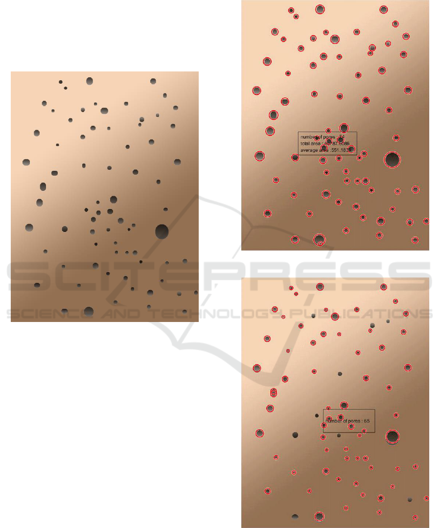
Figure 4: The original skin.
Figure 4 shows the original skin image, regarded
as taken from a skin diagnosis device. It is regarded
as previously processed to address the issues of
color temperature difference, lens distortion,
vignetting and chromatic abberation. It is set that the
light source is at the upper left (the brightness
gradually decreases from upper left to lower right)
with the light brown skin.
Figure 5 shows the result of separating abnormal
area which are regarded as skin lesions. Red circles
denote the abnormal area. After the analysis of
recognized lesions, the number of lesions is 74 with
the total area of 40787.6 (pixels
2
) and the average
area per lesion of 551.2 (pixels
2
).
After the enhanced object recognition using the
shadow analysis called Smart Shadow, possible
pores (concave objects) are shown in Figure 6 and
possible moles (convex objects) are shown in Figure
7. After the analysis, the number of possible pores is
65 and the number of possible moles is 8. The
overlapped parts in Figure 5 and Figure 6 are
identified as errors, mainly due to the CHT
parameter settings.
Figure 5: The result of recognizing skin lesions.
Figure 6: The result of recognizing possible pores.
Effective Image Processing Procedure for Skin Lesion Recognition in Contactless Skin Diagnosis Devices
341
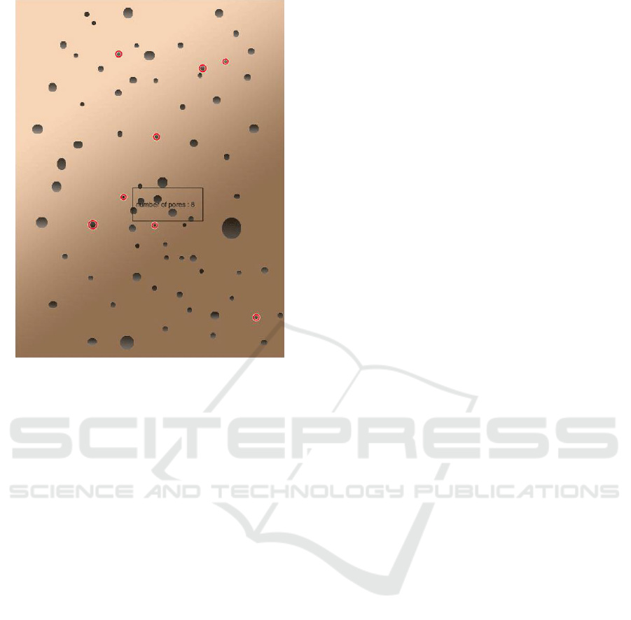
Figure 7: The result of recognizing possible moles.
5 CONCLUSIONS
An effective image analysis procedure for
discriminating and recognizing various skin lesions
under contactless skin diagnosis environment is
proposed. Issues such as uneven distribution of light
are properly addressed, and various skin lesions are
effectively discriminated according to their
characteristics, using the image processing technique
and the shadow analysis.
To generalize and objectify the proposed
procedure, additional analysis and comparison of
detailed skin lesion would be performed under
additional light source including the variety of its
strength, angle, wavelength, etc. Further researches
after this work include detailed lesion discrimination
upon appropriate decision-making frameworks such
as deep learning, and Bid Data analysis over various
kinds of skin image.
ACKNOWLEDGEMENTS
This work was supported by the National Research
Foundation of Korea (NRF) grant funded by the
Korea government (MSIT) (No.
2018R1C1B5043326).
REFERENCES
Abbadi, N. K. A., Dahir, N. S., Al-Dhalimi, M. A., and
Restom, H., 2010. Psoriasis Detection Using Skin
Color and Texture Features. In Journal of Computer
Science, 6(6):648-652.
Ahn, Y. K., 2019. Genie Skin. In AMC Co., Ltd.,
http://www.amc11.com
Funk, N., and Yang, Y.-H., 2007. Using a Raster Display
for Photometric Video. In Proceedings of Canadian
Conference of Computer and Robot Vision (CRV
2007), 201-207, Montreal, Canada.
Gonzalez, R. C., and Woods, R. E., 2018. Digital Image
Processing, 4
th
Ed. By Pearson Education.
Hu, Z., and Yu, C. S., 2013. Functional Research and
Development of Skin Barrier. In Chinese Journal of
Clinicians, 7(7):3101–3103.
Kim, J., and Kim, D., 2014. Matching Points Filtering
Applied Panorama Image Processing Using SURF and
RANSAC Algorithm. In Journal of the Institute of
Electronics and Information Engineers, 51(4):820-835.
Market Research Report, 2018. Dermatology Devices
Market Size, Share & Trends Analysis Report By End
Use (Clinics, Hospitals), By Product & Application
(Diagnostic, Treatment), By Region, And Segment
Forecasts, 2018 – 2025. In Grand View Research,
2018.
Menaka, R., and Rohini, S., 2014. Efficient Detection of
Ischemic Stroke from MRI Images Using Wavelet
Transform. In International Journal of Computer
Science and Information Technology Research,
2(3):446-454.
Nillius, P., and Eklundh, J. O., 2001. Automatic
Estimation of the Projected Light Source Direction. In
Proceedings of the 2001 IEEE Computer Society
Conference on Computer Vision and Pattern
Recognition (CVPR 2001).
Vegt, S. E., 2015. A Fast and Robust Algorithm for the
Detection of Circular Pieces in a Cyber Physical
System. In ES Reports, Eindhoven University of
Technology, 1-5.
Vezhnevets, V., Sazonov, V., and Andreeva, A., 2003. A
Survey on Pixel-Based Skin Color Detection
Techniques. In Proceedings of Graphicon 2003, 85–
92, Moscow, Russia.
Wei, L., Gan, Q., and Ji, T., 2018. Skin Disease
Recognition Method Based on Image Color and
Texture Features. In Computational and Mathematical
Methods in Medicine, 2018(7):1-10.
Yang, J., Sun, X., Liang, J, and Rosin, P. L., 2018.
Clinical Skin Lesion Diagnosis Using Representations
Inspired by Dermatologist Criteria. In The IEEE
Conference on Computer Vision and Pattern
Recognition (CVPR), 1258-1266.
Zaqout, I. S., 2016. Diagnosis of Skin Lesions Based on
Dermoscopic Images Using Image Processing
Techniques. in International Journal of Signal
Processing, Image Processing and Pattern
Recognition, 9(9):189-204.
SIGMAP 2019 - 16th International Conference on Signal Processing and Multimedia Applications
342
