
SPECTRAL AND CROSS-SPECTRAL ANALYSIS OF
CONDUCTANCE CATHETER SIGNALS
New Indexes for Quantification of Mechanical Dyssinchrony
Sergio Valsecchi
Medtronic Italia, Rome, Italy
Luigi Padeletti
University of Florence, Florence, Italy
Giovanni Battista Perego
Istituto Auxologico Italiano, Ospedale S Luca, Milan, Italy
Federica Censi, Pietro Bartolini
Dept Technologies and Health - Italian National Institute of Health, Rome, Italy
Jan J. Schreuder
Dept of Cardiac Surgery, San Raffaele Hospital, Milan, Italy
Keywords: Conductance catheter, spectral analysis, coherence function, heart failure, mechanical ventricular
dyssynchrony.
Abstract: We hereby present novel index to quantify ventricular mechanical dyssynchrony by using
spectral and cross-spectral analysis of conductance catheter volume signals. Conductance
catheter is a volume measurement technique based on conductance measurement: the
intraventricular volume, i.e. the time-varying volume of blood contained within the
heart cavity, is
estimated by measuring the electrical conductance of the blood employing a multi-pole catheter. Five
segmental volume signals (SV
i
, i=1,…5) can be acquired; total volume (TV) is estimated as the
instantaneous sum of the segmental volumes. We implemented classical time-domain dyssynchrony indexes
already utilized in conductance catheter signals analysis, and new frequency-domain indexes. Study
population consisted of 15 heart failure (HF) patients with left bundle branch block and 12 patients with
preserved left ventricular (LV) function. We found that spectral measures seem to out-perform classical
time-domain parameters in differentiating atrial HF patients from no-HF group. These findings encourage
the use of spectral analysis
to obtain crucial quantitative information from conductance catheter
signals.
1 INTRODUCTION
In a normal heart, mechanical activation of the
ventricles occurs in a coordinated manner and
depends on the rapid spread of electric signals via
specialized fibers (His-Purkinje system) which
branch out throughout the right ventricular (RV) and
left ventricle (LV) endocardium (Uhley, 1960).
When the activation is slowed-down or blocked,
ventricle activation and contraction become
dyssynchronous. Ventricular mechanical
dyssynchrony is most commonly identified clinically
by a prolonged QRS duration with left bundle-
branch block (LBBB) morphology on surface
437
Valsecchi S., Padeletti L., Battista Perego G., Censi F., Bartolini P. and J. Schreuder J. (2008).
SPECTRAL AND CROSS-SPECTRAL ANALYSIS OF CONDUCTANCE CATHETER SIGNALS - New Indexes for Quantification of Mechanical
Dyssinchrony.
In Proceedings of the First International Conference on Bio-inspired Systems and Signal Processing, pages 437-444
DOI: 10.5220/0001060104370444
Copyright
c
SciTePress
electrocardiogram but can also be detected by
echocardiographic imaging of contraction timing.
Ventricular mechanical dyssynchrony plays a
regulating role already in normal physiology
(Brutsaert, 1987) but is especially important in
pathological conditions, such as hypertrophy (Villari
et al., 1996), ischemia (Heyndrickx and Paulus,
1990), infarction (Gepstein et al., 1998), or heart
failure (HF) (Nelson, 2000). Dyssynchrony
exacerbates heart failure (HF) in a variety of ways,
generating cardiac inefficiency as well as pathologic
changes at the biologic tissue, cellular, and
molecular levels. Currently, the conductance
catheter method has been extensively used to assess
global systolic and diastolic ventricular function.
More recently the ability of this instrument to pick-
up multiple segmental volume signals has been used
to quantify mechanical ventricular dyssynchrony.
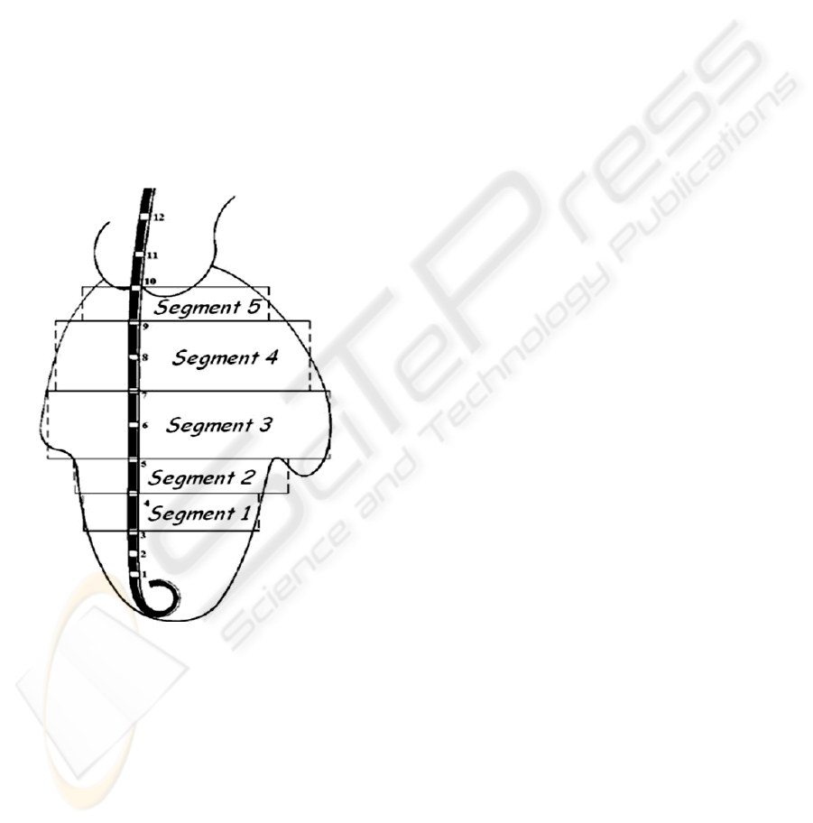
Figure 1: The conductance catheter positioning inside the
left ventricle.
Conductance catheter was first introduced in
1981, by Baan and co-workers as a new volume
measurement technique based on conductance
measurement (Ban et al., 1981, Ban et al., 1984).
Intraventricular volume, i.e. the time-varying
volume of blood contained within the heart cavity, is
estimated by measuring the electrical conductance of
the blood employing a multi-pole catheter
(conductance catether, Figure 1). The conductance
catheter has 12 electrodes and should be positioned
along the long axis of the LV in such a way that the
electrode at the tip is situated within the apex and
the proximal one just above the aortic valve. A weak
alternating current (0.4 mA peak-to-peak, 20 kHz) is
induced between the two most distal and two most
proximal electrodes, in order to set up an electrical
field within the ventricular cavity. Other 6 electrodes
are used pair wise to measure segmental
conductance signals. Two electrodes are used to
record the intracardial ECG. A micromanometer
measures real-time LV pressure. The induced
voltage is then measured with six electrodes in
between, yielding 5 segmental voltages. Since the
conductance of the blood itself is constant
(neglecting long term changes in haematocrite) the
measured voltage will be proportional to blood
resistivity, and thus inversely proportional to the
conductance or amount of blood between the
measuring (voltage) electrodes. This method has
several advantages over other methods which
determine intra-ventricular volumes. The results are
obtained immediately, i.e. on-line, and precise
geometric assumptions regarding the ventricle or
labor-intensive analyses are not required. Recently,
Steendijk et al., first introduced time-domain
quantitative indexes of dyssynchrony based on
volume signals acquired with the conductance
catheter (Steendijk et al., 2004). Spectral analysis of
conductance catheter signals has not been tempted
yet. Frequency domain analysis has been extensively
used to characterize a number of physiological
signals, with promising results in terms of both
classification schemes (Schumann et al., 2002, 1
Zywietz et al., 2004, Severi et al., 1997) and
understanding of physiological mechanisms (Asyali
et al., 2007, Cerutti et al., 1988, Montano et al.,
2001). During ventricular dysfunction, segmental
ventricular volumes experience abnormal changes
which could result in unexpected spectral
components. Also, the asynchrony and
incoordination between ventricular segments, that
seem to be quintessential to ventricular dysfunction,
could be promisingly explored by cross-spectral
analysis. The coherence spectrum is a frequency
domain measure that may be used to make a
quantitative comparison between activity of two
heart regions. In the present study, coherence spectra
have been used to quantify the relation between
spectral components of ventricular volumes from
different regions. The coherence spectrum would
provide a measure of the synchrony and
coordination between ventricular sites, and thus be
indicative of the organization of electrical activity.
Such a measure would be a useful tool in the
BIOSIGNALS 2008 - International Conference on Bio-inspired Systems and Signal Processing
438

characterization and detection of synchronous
contraction. Coherence measurements may provide a
means to quantify the terms "synchronous" and
"dissynchronous" as applied to ventricular
contraction.
Aim of this paper is to characterize the
conductance catheter signals in the frequency
domain and to propose new indexes for ventricular
mechanical dyssynchrony quantification.
2 METHODS AND MATERIALS
2.1 Study Population
The study population consisted of 27 consecutive
patients with indications for electrophysiologic study
or device implantation: 15 HF patients with left
bundle branch block and 12 patients with preserved
LV function. Table 1 shows the clinical
characteristics of the study population. Age, sex and
QRS duration were similar between groups. Subjects
with a previously implanted device, valvular
insufficiency or stenosis were excluded from
analysis.
Table 1: Clinical characteristics of the study population.
non-HF group
(n=12)
HF group
(n=15)
Male gender, n 7 11
Age, years
67±14 68±6
Ischemic
Cardiomyopathy, n
- 7
NYHA Class -
3.1±0.5
Ejection Fraction, %
57±9 26±6*
QRS duration, ms
88±21 167±24*
p-values: * < 0.05
2.2 Experimental Protocols
A conductance catheter was placed in the LV via the
femoral artery, and a temporary pacing lead was
positioned in the right atrium. The conductance
catheter enables online measurement of 5 segmental
volume (SV
i
, i=1,…,5) slices perpendicular to the
LV long axis. We used 7-Fr combined pressure-
conductance catheters with 1-cm interelectrode
spacing (CD Leycom; Zoetermeer, The
Netherlands). The catheter was connected to a
Cardiac Function Lab (CD Leycom) for online
display and acquisition (sample frequency 250 Hz)
of segmental and LV total volumes (TV), LV
pressure, and ECG. TV was obtained as the
instantaneous sum of the segmental volumes.
Two stimulation protocols have been used, i.e.
during spontaneous ventricular activation and atrial
pacing. For this protocol, hemodynamic status was
evaluated using multiple parameters. Indices of LV
pressure, volume, and function were calculated and
averaged over 8 to 10 beats at end expiration from
the raw LV pressure and conductance volume data.
Sequences of 30 s, i.e. 40-50 consecutive non-
arrhythmic cardiac cycles at fixed heart rate induced
by atrial pacing (at 10 bpm above the sinus rate) and
steady-state conditions, were selected for off-line
analysis using custom-designed software.
2.3 Classical Dyssynchrony Parameters
Estimation
From the conductance catheter signals, we estimated
the following classical time-domain parameters:
mechanical segmental dyssynchrony (DYS), Internal
flow fraction (IFF), Mechanical Dispersion (DISP),
Cycle Efficiency (CE) and Time exceeding aortic
closure (TExAC). See appendix for more details.
2.4 Spectral and Cross-Spectral
Analysis
First we analysed the segmental and total volume
signals in the frequency domain. For the spectral
analysis, the periodogram of the signals was
estimated. To reduce spectral leakage a Hamming
window was applied after removal of the mean
value. The length of segments was 1000 samples and
a segment-overlap of 30% was used. Then we
divided the signal bandwidth in 4 frequency bands
(0-1 Hz, 1-5 Hz, 5-20 Hz and >20 Hz), and we
estimated the percentage powers (PP) and the peak
frequencies (PF) in each bands (PP
0-1Hz
, PP
1-5Hz
, PP
5-
20Hz
, PP
>20Hz
and PF
0-1Hz
, PF
1-5Hz
, PF
5-20Hz
, PF
>20Hz
,
respectively).
The continually changing temporal or phase
relationship between two volume signals has been
quantified in the frequency domain by magnitude-
squared coherence (Ropella et al., 1989).
Magnitude-squared coherence (C(f)) between two
recordings is defined as
)()(
)(
)(
2
fSfS
fS
fC
yyxx
xy
=
Where x(t) and y(t) are two simultaneous recordings,
Sxy is the cross power spectrum between signals x
and y, and Sxx and Syy are the individual power
spectra for signals x and y, respectively. C(f) is a
measure of the linear relation between signals as a
SPECTRAL AND CROSS-SPECTRAL ANALYSIS OF CONDUCTANCE CATHETER SIGNALS - New Indexes for
Quantification of Mechanical Dyssinchrony
439

function of frequency, f, and is a real quantity with
value between zero and one. In other terms, C(f)
measures the constancy of the time delay (phase) at
a specific frequency between signals x and y. Two
linearly related signals (in the absence of noise) will
have a C(f) function equal to one at all frequencies
present in both signals, while two random,
uncorrelated signals will have a C(f) equal to zero at
all frequencies. Any linear operation (multiplication
by a constant or addition of a constant) on one or
both of the signals will not alter the C(f) between x
and y. However, additive, uncorrelated noise and
system nonlinearities will reduce C(f) for two
similar signals. C(f) may be estimated for sampled
data using a method of overlapped and averaged
FFT spectral estimates (Carter et al., 1973).
Basically, estimates of Sxx, Syy and Sxy are
determined using a periodogram technique (2048-
samples long window, overlap 512 samples), and
their estimates are then used in the definition of C(f).
The C(f) functions between each segmental volume
SVi and the TV have been estimated. A Total
Coherence function has been defined over the band
0-125 Hz by averaging the 5 C(f) functions. From
the Total Coherence function, 5 new frequency
domain indexes have been extracted:
- mean value of the Total Coherence over the band
0-125 Hz (Coh
Tot
)
- mean value of the Total Coherence from 0 to 1 Hz
(Coh
0-1Hz
),
- mean value of the Total Coherence from 1 to 5 Hz
(Coh
1-5Hz
),
- mean value of the Total Coherence from 5 to 20 Hz
(Coh
5-20Hz
),
- mean value of the Total Coherence from 20 to 125
Hz (Coh
>20Hz
).
2.5 Statistical Analysis
All data are presented as means±SD. Differences
between distributions were compared by a t-test for
Figure 2: Examples of SV and TV time series and power spectra, for a no-HF patinet and a HF one. Coherence function
between SV
1
and TV are showed, as well as the total coherence function.
No-HF patient
HF patient
Power spectrum
Power spectrum
Power spectrum
Power spectrum
Coherence function Sv
1
-TV
Coherence function Sv
1
-TV
Total Coherence
Total Coherence
180
200
150
170
20
40
20
40
[ml]
[ml]
[ml]
[ml]
Total volume
Segment-1 volume
Total volume
Segment-1 volume
0 [s] 10
0 [s] 10
0 [Hz] 20
0 [Hz] 20
0 [Hz] 20
0 [Hz] 20
0 [Hz] 125 0 [Hz] 125
0 [Hz] 125 0 [Hz] 125
0
1
0
1
0
1
0
1
0
300
[ml
2
/H
0
300
[ml
2
/H
0
3x10
4
[ml
2
/H
0
3x10
4
[ml
2
/H
BIOSIGNALS 2008 - International Conference on Bio-inspired Systems and Signal Processing
440

Gaussian variables, and by Mann-Whitney
nonparametric test for nongaussian variables.
Statistical correlations between variables were tested
by least-squares linear regression. A P value < 0.01
was considered significant. We performed receiver-
operating characteristic (ROC) curve analysis to test
the diagnostic performance of the indexes to
discriminate the patient groups. Sensitivities and
specificities at the optimal cut-off point were
determined.
3 RESULTS
Example of the power spectrum of a TV signal are
showed in figure 2, for a HF patient and a no-HF
one. The coherence function between one SV and
the TV and the Total coherence are also showed.
The characteristics of the power spectrum of TV
signals are reported in Table 2 (similar results were
obtained for SVi signals, but were not reported). The
majority of the signal power is in the band from 1 to
5 Hz (programmed heart rate during acquisition
from 70 to 100 bpm). The components above 20Hz
are associated to less than 1% of the total signal
power. The frequency peak in the 0 – 1 Hz band
matches with the respiratory rate and the power in
this band seems higher in HF group. Table 3
summarizes the results of the comparison between
groups for all indexes considered in the analysis,
represented as mean ± standard deviation. Overall, 3
parameters permitted to discriminate the two groups
(p<0.01): Coh1-5, Coh5-20 and CE. Table 4 shows
the results of the ROC curve analysis. Sensitivity
and specificity for Coh1-5 are 0.67 and 0.92, those
obtained for Coh5-20 are 1.00 and 0.92 and those
relatve to CE are 0.80 and 0.83, respectively. In
Figure 3 the ROC curves are shown.
Table 2: Characteristics of the power spectrum of TV
signal.
no-HF HF p-values
PP
0-1Hz
4.34±6.26 9.12±12.00 0.071
PF
0-1Hz
0.41±0.17 0.45±0.12 0.488
PP
1-5Hz
93.24±6.45 88.44±11.85 0.194
PF
1-5Hz
1.52±0.20 1.44±0.13 0.301
PP
5-20Hz
2.16±1.84 2.21±1.34 0.946
PF
5-20Hz
8.37±0.88 7.63±0.80 0.036
PP
>20Hz
0.25±0.25 0.23±0.15 0.814
PF
>20Hz
37.59±5.12 44.14±7.59 0.013
4 DISCUSSION
Quantification of nonuniform mechanical function
and dyssynchrony may lead to a more complete
diagnosis of ventricular dysfunction (Schreuder wet
al., 1997, Schreuder et al., 2000). Moreover, it may
guide therapy, because patients with extensive
dyssynchrony are likely to benefit from
resynchronization therapy (Leclercq et al., 2002).
The visualization of mechanical dyssynchrony
provided by methods based on magnetic resonance
imaging and echocardiography, although further
emphasize the important role of mechanical
dyssynchrony in cardiac dysfunction, requires
laborious procedures and require substantial operator
interaction and expertise.
Table 3: Indexes of mechanical dyssynchrony in no-HF
and HF groups.
no-HF HF p-values
DYS, % 26.0±7.2 32.6±3.9 0.012
IFF, % 25.8±18.8 40.8±13.6 0.033
DISP, ms 23.4±16.4 35.6±13.2 0.068
CohTot 0.44±0.07 0.37±0.10 0.016
Coh0-1 0.63±0.19 0.51±0.18 0.099
Coh1-5 0.69±0.10 0.57±0.10 0.004*
Coh5-20 0.47±0.07 0.32±0.04 0.000*
Coh>20 0.43±0.08 0.37±0.12 0.041
CE 0.78±0.12 0.58±0.16 0.000*
TExAC, ms 6.9±8.8 15.7±10.5 0.016
*p<0.01
Figure 3: ROC curve analysis.
SPECTRAL AND CROSS-SPECTRAL ANALYSIS OF CONDUCTANCE CATHETER SIGNALS - New Indexes for
Quantification of Mechanical Dyssinchrony
441

Table 4: ROC curve analysis of the tested variables.
Area Under Curve
(95% CI)
p-value Cut-off
Coh1-5
0.81
(0.65-0.98)
0.006 0.57
Coh5-20
0.98
(0.94-1.02)
0.000 0.40
CE
0.88
(0.75-1.00)
0.001 0.70
Recently, novel indexes were introduce to quantify
dyssynchrony based on volume signals acquired by
the conductance catheter during cardiac
catheterization (9). Such indexes were based on a
time-domain approach and provided additional, new,
and quantitative information on temporal and spatial
aspects of mechanical dyssynchrony.
To our knowledge, conductance catheter volume
signals have never been studied in the frequency
domain. Since dyssynchrony refers to the
organization of the mechanical contraction of the
ventricle, it is natural to investigate such a
phenomenon by spectral and cross-spectral analysis
of ventricular segmental movements. The frequency-
domain analysis can indeed discover particular
aspects of interaction between volume signals
beyond the temporal relationships.
Present analysis permitted to describe some
characteristics of the conductance-volume signals.
The frequency analysis evidenced the absence of
relevant components above 20 Hz: this result
corroborates the validation of segmental signals
acquisition obtained by comparison with cine-
computerized tomography (16), whose sampling rate
has approximately the same value. The amplitude of
the components in the range 0-1 Hz, attributable to
the respiratory artefact, resulted markedly higher in
HF patients, this may be due to the higher
(mechanical) cardio-pulmonary interaction or to an
altered vasovagal activity.
More interesting results have been obtained by
cross-spectral analysis. The spectral coherence
function provides a quantitative measure of that
temporal synchrony and coordination between
activities of ventricular regions. During synchronous
mechanical contraction, multiple sites are activated
in an coordinated manner, and the phase relation
between activity from two sites is relatively
unchanging, resulting in a high (close to 1)
coherence. When ventricular contraction is
dyssynchronous, the activity observed at one region
is likely to be unrelated to the activity observed at
other distant regions. Thus the coherence between
two such sites would be very low at all frequencies
due to a continually changing phase relation.
In the present study, the spectral coherence was
confirmed to be significantly greater for ventricular
contraction of no-HF patients than for HF ones. We
found that the most significant parameters in the
discrimination between HF and no-HF group were
Coh1-5 and Coh5-20, with the latter reaching a
sensitivity of 1 and a specificity of 0.92. Spectral
measures seem to out-perform classical time-domain
parameters (9) in differentiating atrial HF patients
from no-HF group.
Since no previous studies have been performed
on a similar topic, the frequency bands have been
chosen on empirical basis. The choice of the optimal
frequency bands in term of discriminating power
would require larger population and/or modelling of
ventricular contraction.
In conclusion, this paper encourages the use of
spectral analysis to obtain crucial quantitative
information from conductance catheter signals.
REFERENCES
Uhley H: Peripheral distribution of the canine A-V
conduction system; observations on gross
morphology. Am J Cardiol 5:688- 691, 1960.
Brutsaert DL. Nonuniformity: a physiologic modulator of
contraction and relaxation of the normal heart. J Am
Coll Cardiol 9: 341–348, 1987.
Villari B, Vassalli G, Betocchi S, Briguori C, Chiariello
M, and Hess OM. Normalization of left ventricular
nonuniformity late after valve replacement for aortic
stenosis. Am J Cardiol 78: 66–71, 1996.
Heyndrickx GR and Paulus WJ. Effect of asynchrony on
left ventricular relaxation. Circulation 81: 41–47, 1990.
Gepstein L, Goldin A, Lessick J, Hayam G, Shpun S,
Schwartz Y, Hakim G, Shofty R, Turgeman A,
Kirshenbaum D, and Ben-Haim S. A
Electromechanical characterization of chronic
myocardial infarction in the canine coronary occlusion
model. Circulation 98: 2055–2064, 1998.
Nelson GS, Curry CW, Wyman BT, Kramer A, Declerck
J, Talbot M, Douglas MR, Berger RD, McVeigh ER,
and Kass DA. Predictors of systolic augmentation
from left ventricular preexcitation in patients with
dilated cardiomyopathy and intraventricular
conduction delay. Circulation 101: 2703–2709, 2000.
Baan J, Jong TTA, Kerkhof PLM, et al. Continuous stroke
volume and cardiac output from intraventricular
dimensions obtained with impedance catheter.
Cardivascular Research 1981;15:328-334.
Baan J, van der Velde ET, de Bruin HG, et al. Continuous
measurement of left ventricular volume in animals and
humans by conductance catheter. Circulation
1984;70:812-823.
BIOSIGNALS 2008 - International Conference on Bio-inspired Systems and Signal Processing
442

Steendijk P, Tulner SAF, Schreuder JJ, et al.
Quantification of left ventricular mechanical
dyssynchrony by conductance catheter in heart failure
patients. Am J Physiol 2004;286:H723-H730
Schumann A, Wessel N, Schirdewan A, Osterziel KJ,
Voss A. Potential of feature selection methods in heart
rate variability analysis for the classification of
different cardiovascular diseases. Stat Med. 2002 Aug
15;21(15):2225-42.
Zywietz CW, Von Einem V, Widiger B, Joseph G. ECG
analysis for sleep apnea detection. Methods Inf Med.
2004; 43(1):56-9.
Severi S, Cavalcanti S, Avanzolini G. Heart rate
variability spectral indices for haemodynamic
classification of haemodialysis patients. Physiol Meas.
1997 Nov; 18(4):339-53.
Asyali MH, Berry RB, Khoo MC, Altinok A. Determining
a continuous marker for sleep depth. Comput Biol
Med. 2007 Apr 12;
Cerutti S, Alberti M, Baselli G, Rimoldi O, Malliani A,
Merri M, Pagani M. Automatic assessment of the
interaction between respiration and heart rate
variability signal. Med Prog Technol. 1988; 14(1):7-19.
Montano N, Porta A, Malliani A. Evidence for central
organization of cardiovascular rhythms. Ann N Y
Acad Sci. 2001 Jun;940:299-306. Review.
Ropella KM, Sahakian AV, Baerman JM, Swiryn S. The
coherence spectrum. A quantitative discriminator of
fibrillatory and nonfibrillatory cardiac rhythms.
Circulation 1989;80:112-9.
Carter C, Knapp CH, Nuttall B. Estimation of the
magnitudesquared coherence function via overlapped
fast Fourier Transform processing. IEEE Trans Audio
Electroacoustics. 1973;21:337-44.
Schreuder JJ, Steendijk P, Van der Veen FH, et al. Acute
and short-term effects of partial left ventriculectomy in
dilated cardiomyopathy: assessment by pressure-
volume loops. J Am Coll Cardiol 2000;36:2104-2114.
Schreuder JJ, van der Veen FH, van der Velde ET, et al.
Left ventricular pressure-volume relationships before
and after cardiomyoplasty in patients with heart
failure. Circulation. 1997;96:2978-2986.
Leclercq C, Kass DA. Retiming the failing heart:
principles and current clinical status of cardiac
resynchronization. J Am Coll Cardiol 39: 194–201, 2002.
APPENDIX
Mechanical dyssynchrony. At each time point, a
segmental signal is defined as dyssynchronous if its
change (i.e., dSV/dt) is opposite to the simultaneous
change in the total LV volume (dTV/dt). Segmental
dyssynchrony is quantified by calculating the
percentage of time within the cardiac cycle that a
segment is dyssynchronous. Overall LV
dyssynchrony (DYS) is calculated as the mean of the
segmental dyssynchronies. DYS may be calculated
within each specified time interval, i.e. during
systole and diastole, with systole defined as the
period between the moments of dP/dtmax and
dP/dtmin.
Internal flow. Nonuniform contraction and filling
is associated with ineffective shifting of blood
volume within the LV. This internal flow (IF) is
quantified by calculating the sum of the absolute
volume changes of all segments and subtracting the
absolute total volume change:
[
]
2//)(/)()(
∑
−= dttdTVdttdSVitIF
Note that dTV(t)/dt represents the effective flow into
or out of the LV. Thus IF measures the segment-to-
segment blood volume shifts, which do not result in
effective filling or ejection. Division by two takes
into account that any non-effective segmental
volume change is balanced by an equal but opposite
volume change in the remaining segments. IF
fraction (IFF) is calculated by integrating IF(t) over
the full cardiac cycle and dividing by the integrated
absolute effective flow.
Mechanical dispersion. In the HF patients, a
substantial dispersion is present in the onset of
contraction between the segments. This dispersion is
assessed by segmental lag times which are
determined by calculating the cross correlations
between TV(t) and SV(t) for all systolic time points
(i.e., between dP/dtmax and dP/dtmin). For each
segment the lag which produces the highest linear
correlation is determined. Mechanical dispersion
(DISP) is defined as 2 standard deviation of the
segmental lag times. Recently, new parameters have
been introduced to quantify LV dyssynchrony with
echocardiographic techniques. These indices can be
directly applied to conductance method.
Cycle Efficiency. Calculated as previously described
by the formula: CE=SW/[ LVP* LV volume], with
SW = stroke work, LVP = end-systolic – end-
diastolic LV pressure. This index quantifies
distortions in the shape of the pressure-volume
diagram. The calculation assumes that the optimal
contraction would have CE value near 1.0,
corresponding to a rectangular pressure volume
diagram. Decreases in cycle efficiency may be
caused by multiple factors including isovolumic
volume shifts as well as changes in afterload and
ventricular stiffness. Similarly, regional cycle
efficiency can be calculated from the most basal to
the most apical segmental volume signal plotted
against LV pressure. Differences in regional cycle
efficiency during isovolumic filling or emptying
may indicate inefficient patterns contraction or
relaxation due to dyssynchrony.
SPECTRAL AND CROSS-SPECTRAL ANALYSIS OF CONDUCTANCE CATHETER SIGNALS - New Indexes for
Quantification of Mechanical Dyssinchrony
443

Time exceeding aortic closure. In order to measure
diastolic dyssynchrony and specifically to quantify
LV contraction in diastolic phase, a new index was
proposed, quantitatively reflecting the whole
temporal amount spent by 12 LV segments in
contracting after aortic valve closure. Using strain
imaging that reflects myocardial deformation, the
time of strain tracing exceeding aortic valve closure
(ExcT) was measured in each segment as the
interval between the marker of aortic closure and the
nadir of the strain tracing. ExcT was considered 0
when the nadir of strain curve did not exceed aortic
valve closure. The overall time of strain exceeding
aortic valve closure (oExcT) was computed as the
sum of the 12 segmental ExcTs. The index may be
implemented in conductance method by considering
each segment presenting a systolic phase (negative
dSVi/dt) persisting during the phase of global
diastole (positive dTV/dt). oExcT is estimated as the
sum of these delays for all segments.
BIOSIGNALS 2008 - International Conference on Bio-inspired Systems and Signal Processing
444
