
Effects of Electrical Stimulation of the Calf Muscles
on Jumping Performance
Nagaoka Daichi
1
, Ogiso Kazuyuki
1
, Takenaka Mutsumi
1
and Tokui Masato
2
1
Faculty of education Kogakkan University, Ise, Mie, Japan
2
Department of Sport Science, Kyusyu Kyoritsu University, 1-8 Jiyugaoka, Yahatanishi-ku, Kitakyusyu, Fukuoka, Japan
1 OBJECTIVES
The calf muscles contract almost isometrically while
the Achilles tendon stretches and shortens during the
contact period when walking (Fukunaga et al.,
2001). This interaction between them makes it
possible for the muscles to exert a larger force and
for the tendinous tissue to function as a spring so
that walking can be performed more efficiently. In
addition, jumping requires an even larger force than
walking, and it is likely then that the functions of the
tendinous tissue influence performance
considerably. However, it is difficult to control the
functions of the tendinous tissue during jumping
because it is not innervated by afferent nerves. In
this study, therefore, to investigate the effects of
tendinous tissue on jumping performance, we
induced lengthening and shortening of the Achilles
tendon by forcibly contracting the calf muscles by
electrical stimulation.
2 METHODS
2.1 Subjects
Fifteen healthy men participated in this study (age,
21.1 ± 1.3 years; height, 173.5 ± 7.0 cm; weight,
69.0 ± 10.5 kg). All subjects were in good health,
with no orthopedic or neuromuscular abnormalities.
Subjects were fully informed of the nature and
possible consequences of the study before providing
written informed consent. The experiments were
conducted in accordance with the Declaration of
Helsinki. Approval was obtained from the Ethics
Committee of Kogakkan University.
2.2 Protocol
Subjects were instructed to perform 10 consecutive
two-legged jumps at maximum effort (100% jump)
and at 50% of the maximal jump height (50% jump).
Jumps were performed on a jump-measuring mat
(PH-1260, DKH, Tokyo, Japan) to measure jump
height, ground contact, and flight time. Subjects
were instructed to place both hands on their waist
and reduce ground contact time as much as possible.
Both normal and electrically stimulated jumps were
performed.
An electrical stimulus was applied over the calf
muscle during the jump at a frequency of 20 Hz
(ES20) or 60 Hz (ES60). Six sets of jumps were
performed with intervals of at least 5 min. Electrical
stimulation intensity was set to 20% of the
maximum ankle plantar-flexion torque, using an
electromyography / evoked potential measuring
system (MEB-2306, NIHON KODEN, Tokyo,
Japan). Two anodes and one cathode were placed on
the proximal and distal ends of the triceps surae
muscle, respectively. Reference marks were placed
on the right caput of the ossis metatarsalis V, ankle
joint, knee joint, greater trochanter, acromion,
tragus, and on the top of the subject’s head.
Jumping movements were filmed in the sagittal
plane with a high-speed camera (300 fps; EXLIM-
F1, CASIO, Tokyo, Japan) with 2 reference marks
placed on the ground at an interval of 2 m. The
subjects were questioned about their jump
performance and asked to rate the force required for
the jump and the ease of control on a 5-point scale
(5: very light or easy; 4: light or easy; 3: normal; 2:
heavy or difficult; 1: very heavy or difficult). In
addition, they were questioned about the extent (1:
none to 5: severe) and location of muscle soreness
each day for 6 days after the experiment.
2.3 Data Processing
The reference points in each frame were
automatically digitized (DARTFISH SOFTWARE,
DARTFISH, Fribourg, Switzerland), smoothed, and
converted to real coordinates. The ankle, knee, and
hip joint angles were computed during the ground
contact phase. Distances between the reference mark
Daichi N., Kazuyuki O., Mutsumi T. and Masato T..
Effects of Electrical Stimulation of the Calf Muscles on Jumping Performance.
Copyright
c
2014 SCITEPRESS (Science and Technology Publications, Lda.)
on the ground in front of the subject and the right
caput of the ossis metatarsalis V (landing point)
were also computed to evaluate the stability of the
jump. The rebound jump index was computed using
the ground contact and flight times obtained from
the jump-measuring mat.
2.4 Statistics
Data are presented as the means ± SD. One-way
analysis of variance was used to analyze the
differences in jumping performance, movement, and
5-grade evaluations of jumping performance and
muscle soreness. Fisher’s post hoc comparison was
performed when significance was found. The
probability level accepted for statistical significance
was p<0.05.
3 RESULTS
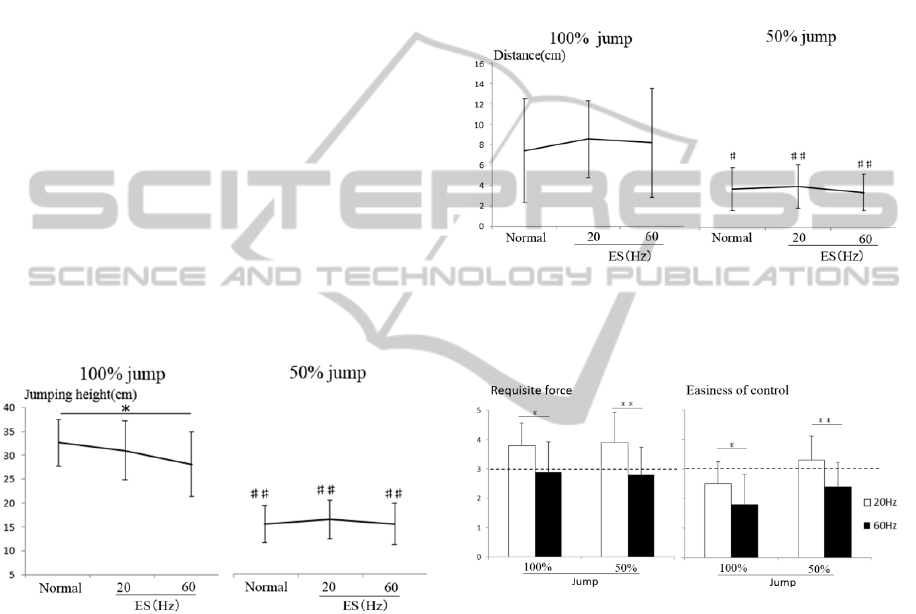
Jump height was significantly lower in the 100%
jump with ES60 than in the 100% normal jump. No
significant differences in jump height were observed
between the other conditions (Fig. 1).
Figure 1: Jump height. * and ## denote significant
differences (p<0.05 and p<0.01, respectively) among
jump conditions and between 100% and 50% jumps,
respectively.
Stability of the jump increased significantly in
the 50% jump compared with the 100% jump.
Electrical stimulation had no effect on stability (Fig.
2). The results of the self-evaluations were as
follows: jump performance was rated significantly
lower for 60ES than for 20ES in both the 100% and
50% jumps; force required was rated as high at 20
Hz and almost the same at 60 Hz compared with the
normal jump; whereas ease of control was rated
almost the same at 20 Hz but lower at 60 Hz
compared with the normal jump (Fig. 3).
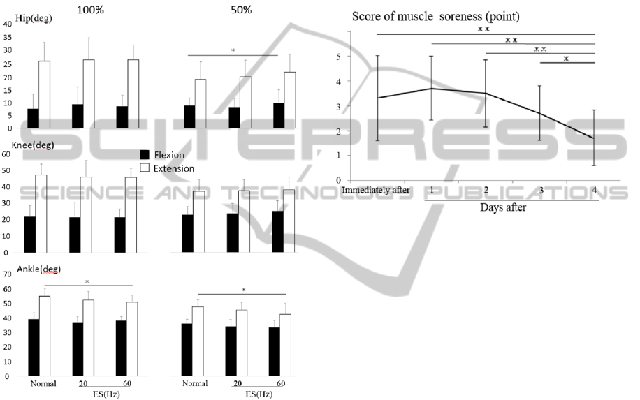
No obvious differences were observed in angular
displacement of the lower joints during jumps
between the normal, ES20, and ES60 jumps.
Significant differences were observed only in hip
flexion in the 50% jump and in ankle plantar-flexion
in both the 100% and 50% jumps between the
normal and ES60 jumps (Fig. 4).
After the experiment, all subjects reported severe
muscle soreness at the myotendinous junction of the
gastrocnemius muscle. However, it gradually
decreased day by day (Fig. 5).
Figure 2: Distances between the reference mark and
landing point during jumps. # and ## denote significant
differences (p<0.05 and p<0.01, respectively) between
100% and 50% jumps.
Figure 3: Results of self-evaluations on jump performance
by assessing the force required for the jump and ease of
control. * and ** denote significant differences (p<0.05
and p<0.01, respectively) between ES20 and ES60.
4 DISCUSSION
In this study, the intensity of the electrical
stimulation applied to the calf muscle during
jumping was adjusted so that that the calf muscle
could generate 20% of the maximum torque at
frequencies of 20 Hz and 60 Hz. However, electrical
stimulation at 60 Hz did not increase jump height;
instead it significantly decreased it when the jump
was performed at full effort. This may be related to
the difficulty in controlling jumps stimulated at 60
Hz, as revealed by the results of the self-evaluation
on ease of control. This implies that jump height was
sacrificed to stabilize the landing point and may be
supported by the significant decreases in ankle
plantar-flexion during the late contact phase of
jumps stimulated at 60 Hz. On the other hand, for
the electrical stimulation at 20 Hz, subjects reported
performing the jump with less effort, although jump
height remained unchanged. This suggests
appropriate use of the tendinous tissue elasticity
during these jumps.
Figure 4: Angular displacement of flexion and extension
of each joint during the jumps. * denotes significant
differences (p<0.05) among jump conditions.
Subjects reported severe muscle soreness after
the exercise. Since they performed only 6 sets of 10
consecutive two-legged jumps with sufficient
intervals, the pain was likely due to the electrical
stimulation applied to the calf muscle. Muscle
soreness was concentrated at or near the
myotendinous junction of the gastrocnemius muscle.
Muscle strain injury that is caused by combining a
large force with substantial stretch (Garrett, 1990)
has been reported to occur at or near the
myotendinous junction (Tidball et al., 1993), and a
clinical report revealed that most muscle strain
injuries occur at or near the myotendinous junction
during high-intensity or explosive voluntary
movements such as sprint and quick turn (Okuwaki,
2009). Therefore, forced contraction of the calf
muscle by electrical stimulation might induce a large
force with substantial stretch at or near the
myotendinous junction during jumping. However, it
is not clear which experimental condition in the
jump exercise induced the muscle soreness reported
in this study. Further studies are needed to perform
jumps with electrical stimulation applied to the
muscle to ensure safe and appropriate training of the
myotendinous units.
Figure 5: Change in muscle soreness. * and ** denote
significant differences (p<0.05 and p<0.01, respectively)
among days after the experiment.
REFERENCES
Fukunaga, T., Kubo, K., Kawakami, Y., Fukashiro, S.,
Kanehisa, H., Maganaris, C.N., 2001. In vivo
behaviour of human muscle tendon during walking.
Proc. R. Soc. Lond. B 268: 229-233.
Garrett, W.E., Jr., 1990. Muscle strain injuries: clinical
and basic aspects. Med Sci Sports Exerc 22: 436-443.
Okuwaki, T., 2009. Muscle strains of top-level athletes in
Japan. J Japan Soc Clin Sports Med 17: 497-505.
Tidball, J.G., Salem, G., Zernicke, R., 1993, Site and
mechanical conditions for failure of skeletal muscle in
experimental strain injuries. J Appl Physiol 74: 1280-
1286.
Zuurbier, C.J., Everard, A.J., van der Wees. P., Huijing,
P.A., 1994. Length-force characteristics of the
aponeurosis in the passive and active muscle condition
and in the isolated condition. J Biomech 27: 445–453.
