Biomedical Device for Early Breast Cancer Detection: Device
Performance Improving by Plasmonic-Photonic Mask
Sanem Meral, Ezel Yalcinkaya, Metin Eroglu, Ahmad Salmanogli and H. Selcuk Gecim
Faculty of Engineering, Cankaya University Electrical and Electronics Engineering Department, Ankara, Turkey
salmanogli@cankaya.edu.tr, gecim@cankaya.edu.tr
Keywords: Biomedical Imaging Device, In-vivo Imaging, NIR, Plasmonic Resonance, Plasmonic Mask.
Abstract: In this article, a new device to detect breast cancer at an early stage, is presented. The main advantages of the
device are its easy operational procedure, portability, high accuracy due to usage of plasmonic-photonic mask
and the low cost. In fact, the novelty of the device presented is to apply the new mask called plasmonic-
photonic mask for precise analysis of the captured images. In the early stage of the work, a phantom model is
employed and the operation of the system is realized. It is shown that the image processing toolbox is safely
matched with the device. It should be noted that for the in-vivo imaging, the device should be completed and
equipped with a high accuracy charge coupled device (CCD) and laser.
1 INTRODUCTION
The early and comparatively easier detection of
cancer is a valuable task in medicine. Recently,
detecting cancerous tumors at an earlier stage have
been a major problem in medical imaging. There are
some classical imaging devices such as magnetic
resonance imaging (MRI) (Grover et al., 2015),
computed tomography (CT) (Goldman, 2007),
positron emission tomography/computed
tomography (PET/CT) (Basu et al., 2014), X-Ray
(Schueler, 1998), mammography (Moseley, 2016)
etc. Such imaging devices have initiated a new area
of image processing in the medical industry (Yasrib
and Suhaimi, 2003). Using image processing
techniques (Gonzalez, 1993) lead to analyze the
captured images in more details based on the
advanced algorithms (Joo et al., 2004). Also, there are
some devices in biomedical imaging which utilizes
the fluorescence to enhance the imaging performance
(Moon et al., 2003).
Early breast cancer detection using plasmonic-
photonic mask is an innovational imaging technique.
The device presented aims to apply a mask called
plasmonic-photonic mask for precise analysis of
captured images. As it is a promising imaging system
to be an alternative to mammography, trials are still
executed on a phantom model. A superiority of the
device presented is its portability. It is beneficial in
both lab environment and transportation purposes.
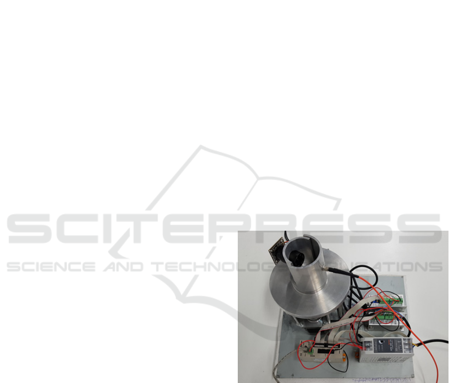
Visuals of the device can be seen in Fig. 1.
Figure 1: Photograph of the investigated system.
A short introduction of the imaging system
introduced in this work is as follows; since resolution
in medical imaging is crucially important to detect the
small tumors (DeSchepper et al., 1997), in this study,
precise detection is aimed by the usage of plasmonic-
photonic mask. This system operates at a specific
near-infrared (NIR) wavelength, 808 nm. The system
is operated at 808 nm wavelength which results in the
penetration depth facilitated imaging soft tissues. The
absorption ratio of the tissue at that wavelength is
reported to be quite small i.e. about 4-5 mm (Cletus
Meral, S., Yalcinkaya, E., Eroglu, M., Salmanogli, A. and Gecim, H.
Biomedical Device for Early Breast Cancer Detection: Device Performance Improving by Plasmonic-Photonic Mask.
DOI: 10.5220/0007679301610166
In Proceedings of the 12th International Joint Conference on Biomedical Engineering Systems and Technologies (BIOSTEC 2019), pages 161-166
ISBN: 978-989-758-353-7
Copyright
c
2019 by SCITEPRESS – Science and Technology Publications, Lda. All rights reserved
161
et al., 2009). Thus, the penetration depth in soft
tissues (Clement et al., 2005) – (Ash et al., 2017), at
the NIR wavelengths, 808 nm, is preferred. Since the
main purpose of this device is to detect the small
tumors, this study is claimed to offer comparatively
higher operational accuracy as compared to the
classical mammography. This is because the system
realized in this study includes additional NIR laser to
scan the area of examination. Also using the
plasmonic-photonic mask helps to improve the
performance of the system (Salmanogli and Salimi,
2017) – (Salmanogli and Farhadnia, 2016).
Interaction of the light with metal nanoparticle (NP)
produces the mode called plasmonic mode
(Salmanogli and Gecim, 2018) – (Salmanogli et al.,
2018). Then the plasmonic mode is effectively
coupled to the far-field by utilizing the photonic
mode. In other words, using photonic structure leads
to merging two presented modes (Salmanogli and
Salimi, 2017). The mentioned phenomenon is
employed in image data acquisition.
It is claimed that performance of this device can
enhance the mammography in some critical
applications requiring high accuracy, together with
mobility of the system, low risk, and low cost.
Finally, it should be noted that as an important point
the plasmonic-photonic mask used in this system can
strongly improve the imaging system accuracy and
resolution (Salmanogli and Salimi, 2017).
2 SYSTEM DEFINITION
In this section, the system employed in this study is
explained in detail. The correlation between the
device’s inner and outer elements is established
according to the operational principles of the
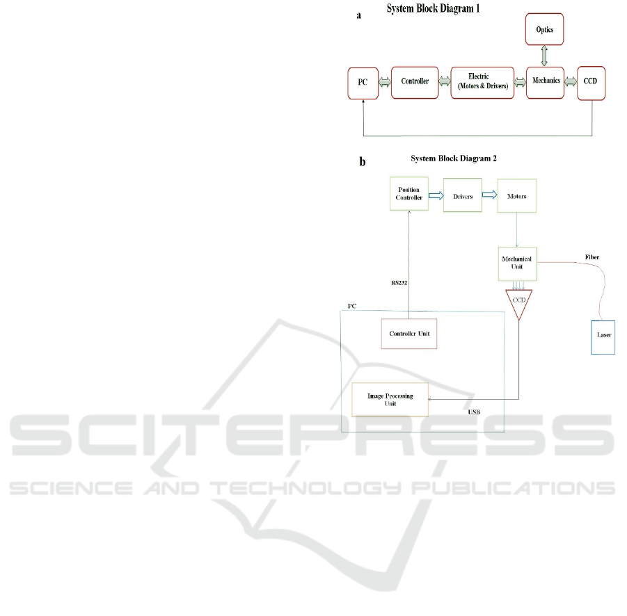
system. The layout of the system is illustrated in Fig.
2. It can be clearly seen that the mechanical parts are
combined with optics and charge coupled device
(CCD). The CCD is connected to the computer for
further image processing. In this system a NIR laser
is coupled to mechanical part through a single mode
fiber. After the interaction between light and matter,
photons are collected by the CCD camera. The
image captured is transferred to the image
processing unit in computer. The controller unit in
the computer is used to control the position of the
plates through RS232.
Drivers in the system are controlled by the
position controller, to manage the rotation of the
motors.
Figure 2: Operational principle of the system (a) Layout of
the system, (b) Inner structure of the system.
The motors are directly linked to the plates. The
electro-mechanical pieces of the system are
connected to the computer. Mechanical unit of the
system is shown in Fig. 3. The camera is positioned
on Plate 1, whereas the optical fiber comes through
Plate 2. Thus, optics and CCD are directly connected
to the mechanical unit. For a better understanding, the
top view of the system is shown in Fig. 3. The
mechanical unit is composed of two moving plates
with different radius values. Radiuses of the plates 1
and 2, are marked as r1 and r2. Plate 1 has a bigger
radius as r
1
. Also side view of the system is illustrated
in Fig. 3.
BIOIMAGING 2019 - 6th International Conference on Bioimaging
162
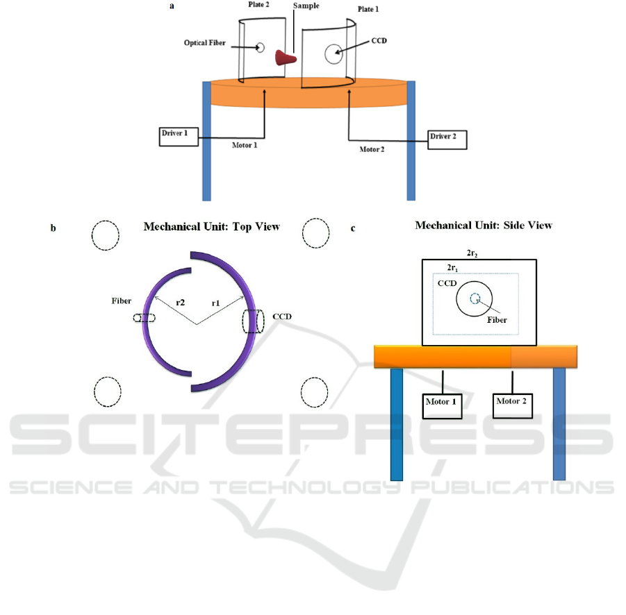
Figure 3: (a) Mechanical unit of the system, (b) Top view of the system, (c) Side view of the system.
The operational principle of the optics and CCD
is introduced in Fig. 4. Laser beam interact with the
sample mounted. Then scattered beam is multiplied
and collected by the camera. Intensified scattered
beam can clearly be observed. Fig. 4. By interaction
of the scattered photons with the small NPs such as
Au NPs, the surface plasmonic modes are created.
This is due to the incident wave sent, being aligned in
phase with the electrons on the near-field of
nanoparticles. Then the produced near-field
plasmonic modes are coupled to the photonic mode.
The consequent mode is transferred to the far-field
called focal point in this study. While the bandwidth
of the scattered photons decrease, their intensity
increases. Thus low intensity photon beam sent,
transforms into high intensity photon beam.
Consequently the final image is re-constructed using
the plasmonic-photonic effect. In fact, using
plasmonic-photonic effect, leads to improvement of
the imaging system accuracy. It is claimed that in the
study, the presented system can be used to detect the
small breast cancer tumors. Also, it has been reported
(Salmanogli et al., 2017) that using low power laser
to scan the breast, results in the detection of a very
small amount of the scattered photons. Therefore the
plasmonic-photonic mask is offered.
The plasmonic-photonic mask used in the system
is an array of gold nanoparticles. The inter-distance
of nanoparticles are the same as the operation
wavelength, which 808 nm. This wavelength is
chosen for maximum amplification (Salmanogli et
al., 2017). Top view and side view of the plasmonic-
photonic mask is shown in Fig. 5. So from that point
of view, the contribution of the study is to be able to
produce high intensity photon beam for imaging
without using high power laser beam which may
harm human body. That can also be explained as the
captured image is always constructed by low intensity
photon beam. The major advantage offered by the
study is utilizing a plasmonic-photonic mask
producing high intensity photon beam needed.
Finally, the constructed image quality is improved.
Biomedical Device for Early Breast Cancer Detection: Device Performance Improving by Plasmonic-Photonic Mask
163
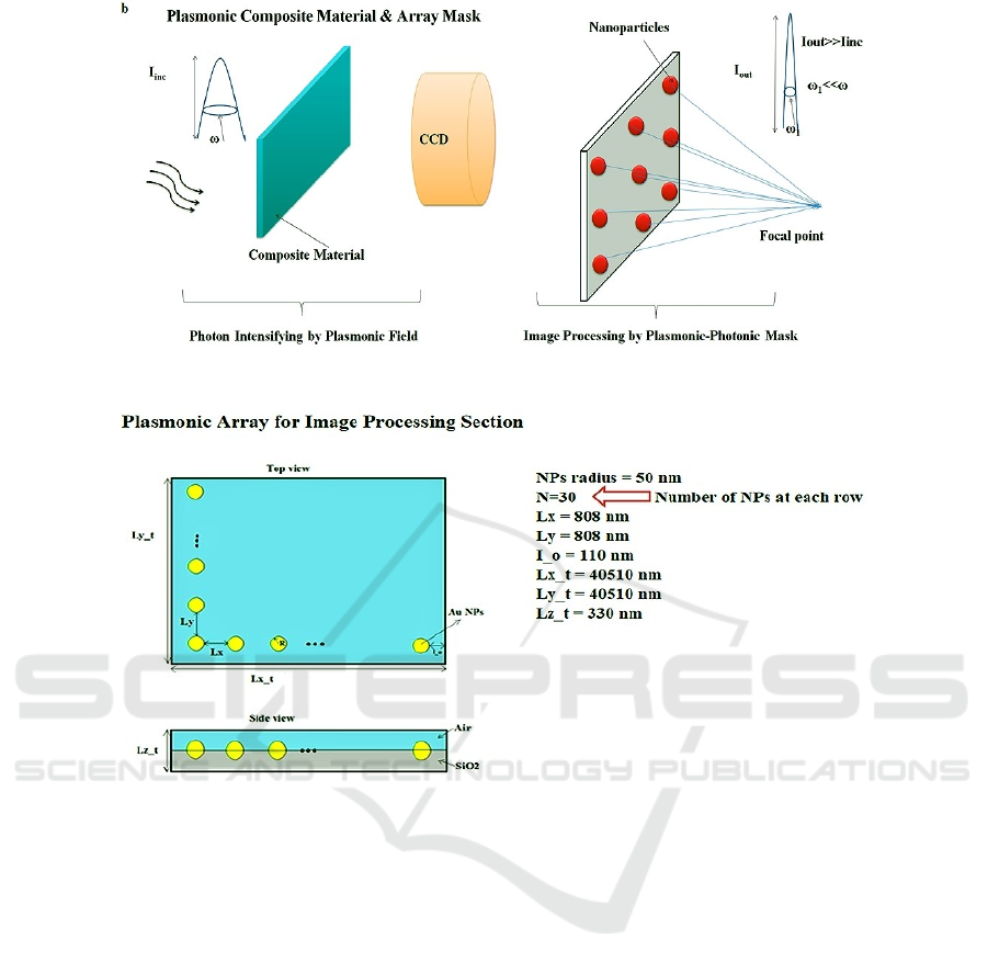
Figure 4: (a) Optics and CCD working principle, (b) Plasmonic-photonic mask.
Figure 5: Plasmonic array mask, regular arranging of plasmonic NPs; top view and side view.
3 RESULT AND DISCUSSION
In this study operational results are shortly discussed.
To obtain preliminary results, a phantom model
(Salmanogli et al., 2017) is employed in the system
and data related to imaging are collected. After number
of operational runs, some images were selected from
the collected data in order to investigate the effect of
plasmonic photonic mask. In Fig. 6 it can be clearly
seen that the plasmonic mask can effectively help to
clarify the blurred image. One has to realize that the
blurred images occur as a result of low intensity
scattered photons from the breast. As additional
information some results obtained with the plasmonic
mask on the captured images is shown in Fig. 6.
As a matter of fact, the comparison between the
states are done and illustrated in Fig. 6b and d. In Fig.
6, we considered two original figures (Fig. 6a and Fig.
6c). Fig. 6a is recorded with high intensity input, while
the picture illustrated in Fig. 6c is recorded with low
intensity laser. The main objective is to compare the
plasmonic-photonic mask effect on the images
considered. In other words, it is shown that using
plasmonic-photonic mask improve the images that has
been recorded with low intensity incoming photons.
These cases are performed due to scattered photons
from the area like the breast being dramatically
attenuated. Therefore, the original images obtained
without the plasmonic-photonic mask is unclear to the
observant.
Before studying the processed image by the
plasmonic-photonic mask, a short description of the
mask is explained. The Au NPs radius is 50 nm, and
the inter-distance between the Au NPs is around 808
nm. The experimental setup revealed that due to the
photonic effect, the structure’s plasmonic peak has a
large red-shift and is about 808 nm whereas the Au
NPs plasmonic peak is 532 nm. In this work, a
plasmonic resonance at 808 nm which is the same with
the incidence wavelength is needed. By applying this
BIOIMAGING 2019 - 6th International Conference on Bioimaging
164
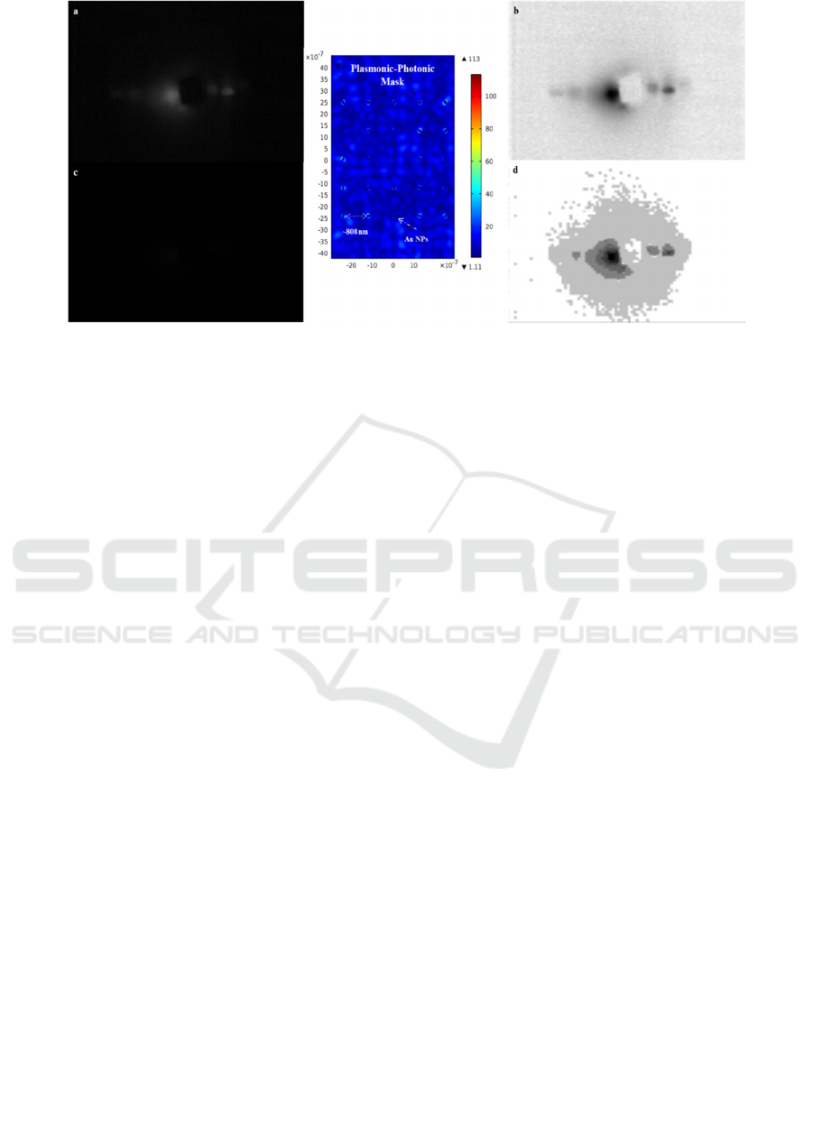
Figure 6: Image segmentation and edge detection through the gradient image processing by plasmonic mask (a) Original
image, (b) Image after applying plasmonic mask, (c) Original image, (d) Image after applying plasmonic mask, (e) The middle
image is the designed plasmonic-photonic mask, the color bar in the right side illustrates the normalized NPs localized
plasmonic field (Salmanogli et al., 2017).
mask on Fig. 6a, the discontinuity of the intensity at
different interfaces is clearly seen in Fig. 6b. On the
other hand, we need to detect discontinuity by this
mask. For this reason, the designed plasmonic-
photonic mask is applied on the poor image shown in
Fig. 6c. Low intensity incoming wave is enhanced by
the high intensity generated around the near-field of
the nanoparticles. Afterwards; amplified signals are
detected and captured by the traditional CCD. To put
it in other words, the image is constructed by the
intensified plasmonic field rather than the traditional
incident wave which is scattered from the area.
After applying the plasmonic-photonic mask, the
boundaries due to augmentation of the gradient image
can be easily detected. The result is depicted in Fig.
6d.
The results indicate that utilizing the plasmonic-
photonic mask clearly improves the imaging system
performance. Please note that this is a short paper
about the general features of the studied system.
However, one can find more details in cited
references. All of the mentioned cases can be found
in (Salmanogli and Salimi, 2017) – (Salmanogli and
Farhadnia, 2016) – (Salmanogli and Gecim, 2018) –
(Salmanogli et al., 2018) – (Salmanogli et al., 2017).
4 CONCLUSION
In this study a new biomedical imaging system is
presented. This system is assumed to operate for early
detection of the breast cancer. In this work, the block
diagram of the system is illustrated. Necessary
matching between the system operation and image
processing toolbox is successfully established.
Examples of some results obtained from this study
were shown in Fig. 6. Finally, it can be concluded that
using plasmonic-photonic mask clearly results in
improvement on the blurred images.
ACKNOWLEDGEMENTS
This work is supported by Cankaya University,
Ankara, Turkey.
REFERENCES
Grover, V., Tognarelli, J., Crossey, M., Cox, I., Taylor-
Robinson, S. and McPhail, M., 2015. Magnetic
Resonance Imaging: Principles and Techniques:
Lessons for Clinicians. Journal of Clinical and
Experimental Hepatology, 5(3), pp.246-255.
Goldman, L., 2007. Principles of CT and CT Technology.
Journal of Nuclear Medicine Technology, 35(3),
pp.115-128.
Basu, S., Hess, S., Nielsen Braad, P., Olsen, B., Inglev, S.
and Høilund-Carlsen, P., 2014. The Basic Principles of
FDG-PET/CT Imaging. PET Clinics, 9(4), pp.355-370.
Schueler, B., 1998. Clinical applications of basic x-ray
physics principles. RadioGraphics, 18(3), pp.731-744.
Moseley, T., 2016. Digital Mammography and Digital
Breast Tomosynthesis. Clinical Obstetrics and
Gynecology, 59(2), pp.362-379.
Yasrib, A. and Suhaimi, M., 2003. Image Processing in
Medical Applications. Journal of Information
Technology Impact, 3(2), pp.63-68.
Biomedical Device for Early Breast Cancer Detection: Device Performance Improving by Plasmonic-Photonic Mask
165

Gonzalez, D., 1993. Digital Image Processing. Addison
Wesley.
Joo, S., Yang, Y., Moon, W. and Kim, H., 2004. Computer-
Aided Diagnosis of Solid Breast Nodules: Use of an
Artificial Neural Network Based on Multiple
Sonographic Features. IEEE Transactions on Medical
Imaging, 23(10), pp.1292-1300.
Moon, W., Lin, Y., O'Loughlin, T., Tang, Y., Kim, D.,
Weissleder, R. and Tung, C., 2003. Enhanced Tumor
Detection Using a Folate Receptor-Targeted Near-
Infrared Fluorochrome Conjugate. Bioconjugate
Chemistry, 14(3), pp.539-545.
DeSchepper, A., Parizel, P., Ramon, F., DeBeuckeleer,
L. and Vandevenne, J., 1997. Imaging of Soft Tissue
Tumors. Springer-Verlag Berlin Heidelberg.
Cletus, B., Künnemeyer, R., Martinsen, P., McGlone, A.
and Jordan, R., 2009. Characterizing liquid turbid
media by frequency-domain photon-migration
spectroscopy. Journal of Biomedical Optics, 14(2), p.
24-41.
Clement, M., Daniel, G. and Trelles, M., 2005. Optimising
the design of a broadband light source for the
treatment of skin. Journal of Cosmetic and Laser
Therapy, 7(3-4), pp.177-189.
Ash, C., Dubec, M., Donne, K. and Bashford, T., 2017.
Effect of wavelength and beam width on penetration in
light-tissue interaction using computational methods.
Lasers in Medical Science, 32(8), pp.1909-1918.
Salmanogli A.,Salimi, K., 2017. Lattice plasmon effect on
imaging resolution: Point-spread function enhancing.
Sensors and Actuators A: Physical, 267, pp.21-29.
Salmanogli A., Farhadnia, F., 2016. Portable in-vitro & in-
vivo imaging system in the low photon condition based
on the plasmonic-photonic virtual mask image
processing system, IR PATENT: G06T; A61B; G06K.
Salmanogli A., Gecim, H., 2018. Quantum eye: Lattice
plasmon effect on quantum fluctuations and photon
detection. Annals of Physics, 394, pp.162-178.
Salmanogli, A., Gecim, H. and Piskin, E., 2018. Plasmonic
System as a Compound Eye: Image Point-Spread
Function Enhancing by Entanglement. IEEE Sensors
Journal, 18(14), pp.5723-5731.
Salmanogli, A., Salimi, K., Farhadnia, F. and Usta, D.,
2017. Sensitive plasmonic-photonic nanosensor as a
morphologic mask. Optical Materials, 70, pp.73-82.
BIOIMAGING 2019 - 6th International Conference on Bioimaging
166
