
E-LEARNING TOOLS FOR WOUND IMAGE UNDERSTANDING
Augustin Prodan, Mădălina Rusu and Remus Câmpean
Department of Mathematics and Informatics, Iuliu Haţieganu University, Cluj-Napoca, Romania
Rodica Prodan
MedFam Group, Str. Constanţa 1, Cluj-Napoca, Romania
Keywords: Web-based Education, e-Learning Scenario, Java and XML Technologies, Artificial Intelligence, Intelligent
Tutoring Systems, Wound Image Understanding, Wound Healing Simulation, Virtual Learning
Communities.
Abstract: This paper presents an e-learning framework for analyzing, processing and understanding wound images, to
be used in teaching, learning and research activities. We intend to promote e-learning technologies in
medical, pharmaceutical and health care domains. Our approach to e-learning is so called blended learning,
which combines traditional face-to-face and Web-based on-line learning, with focus on principles of active
learning. Using Java and XML technologies, we build models for various categories of wounds, due to
various aetiologies. Based on colour and texture analysis, we identify the main barriers to wound healing,
such as tissue non-viable, infection, inflammation, moisture imbalance, or edge non-advancing. This
framework provides the infrastructure for preparing e-learning scenarios based on practice and real world
experiences. We make experiments for wound healing simulation using various treatments and compare the
results with experimental observations. Our experiments are supported by XML based databases containing
knowledge extracted from previous wound healing experiences and from medical experts knowledge. Also,
we rely on new paradigms of the Artificial Intelligence for creating e-learning scenarios to be used in a
context of active learning, for wound image understanding. To implement the e-learning tools, we use Java
technologies for dynamic processes and XML technologies for dynamic content.
1 INTRODUCTION
Medical images are valuable in didactic activities for
students in medicine and pharmacy. Digital pictures
are in great demand, because digital technologies
provide unlimited resources for medical and
pharmaceutical education. Computerized image
processing contains methods for non-invasive
wound evaluation, allowing an accurate diagnosis in
a large category of patients with damaged and
wounded skin. Traditional non-invasive technologies
are limited frequently to subjective visual
evaluations. Colour and texture information provide
the infrastructure for a structured approach to non-
invasive wound assessment. We use the RGB (Red-
Green-Blue) colour space to define a set of image
features for every category of wounds. To identify a
wound in an image, we implement specific methods
based on some generic criteria, such as normal skin
and wounded skin. For some applications we use as
main colours Red, Yellow and Black to assess the
gravity of a wound. Generally, wounds have a non-
uniform mixture of yellow slough, red granulation
tissue and black necrotic tissue. Relying on a high
quality of image acquisition, we can analyse a
succession in time of more images for the same
wound and assess changes in wound healing, i.e. the
recovery or worse evolution.
We intend to develop e-learning tools for
students and residents in medicine, pharmacy and
health care, to be used in both didactic and research
activities. Our aim is to create and implement in
Java an automatic method which can be used as a
reference standard for colour and texture wound
analysis. The purpose is to create e-learning
scenarios for wound image understanding and
wound healing simulation, by applying this method
to large amounts of wound image data stored in
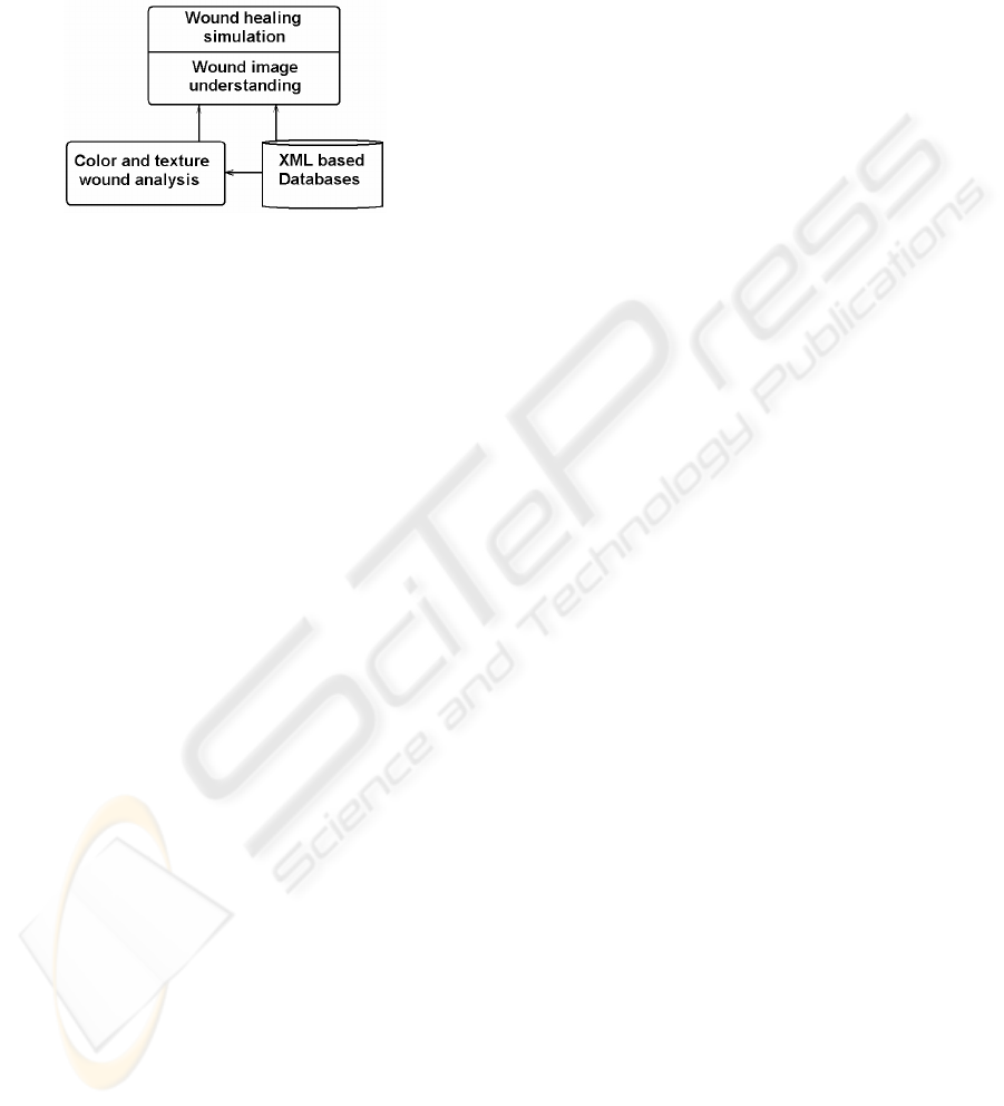
XML based knowledge bases (Figure 1). Our
388
Prodan A., Rusu M., Câmpean R. and Prodan R. (2008).
E-LEARNING TOOLS FOR WOUND IMAGE UNDERSTANDING.
In Proceedings of the Fourth International Conference on Web Information Systems and Technologies, pages 388-393
DOI: 10.5220/0001523303880393
Copyright
c
SciTePress
objective is to develop appropriate skills in wound
management for a learner that traverses such an
e-earning scenario. The e-learning scenarios are
practice driven and relevant to professional practice,
being used by students in medicine and pharmacy, at
graduate, postgraduate and residency levels.
Figure 1: The general method applied for wound image
understanding and wound healing simulation.
Wound image understanding is a difficult
knowledge-based process and we have to use the
new paradigms of the Artificial Intelligence (e.g.
Bayesian Inference, Case Based Reasoning and
Intelligent Agents) to manage it. Relying on large
amounts of wound image data collected from
medical and health care environments, we intend to
create XML and CBR (Case Based Reasoning)
knowledge bases, working in a continuous
collaboration with physicians and wound care
experts from our university and from health care and
medical units. We have continuous access to actual
medical records, to monitor the wound evolution and
to verify both the accuracy and the consistency of
our system.
The originality of our work consists in relying on
new paradigms of artificial intelligence for creating
intelligent and practical e-learning tools to be used
in a context of blended and active learning. To
implement these e-learning tools, we use Java
technologies for dynamic processes and XML
technologies for dynamic content. We create XML
based databases containing knowledge extracted
from previous wound healing experiences and from
medical experts knowledge. The methods presented
in this paper should be useful as an adjunct to
traditional teaching and learning resources. In a
context of blended learning, the teachers and
learners may combine the colour and texture based
parameters with traditional parameters, such as
smell, venous and arterial status, patient history, etc.
2 IMAGE PROCESSING
The infrastructure of our system is based on a
collection of Java class libraries, containing methods
for processing images specific to various categories
of wounds (Prodan et al., 2006). We implemented
general methods that create many common special
effects and use them in analysing the wounds.
A digital image consists of a two dimensional
array of pixels P
mn
with m rows and n columns.
Using Java language, this image is represented in
internal memory as a three dimensional array P
mn4
,
each pixel being described in a specific RGB format
by four unsigned 8-bit integers (Prodan and Prodan,
1997). The first three integers represent the base
colour components (Red, Green and Blue), and the
fourth integer, referred to as α (alpha) represents the
transparency. A specific colour is obtained by
mixing different amounts of basic colours (red,
green and blue) with a specific transparency. The
standard Core Java Technologies provides methods
for processing digital images, such as blur, sharpen,
brighten or tone down an image. We will create the
Java framework by implementing the image
processing algorithms into one of the following two
layers: (1) for low-level implementations, allowing
to operate directly on pixels; and (2) for high-level
implementations, based on standard Java libraries
such as JAI (Java Advanced Imaging) API.
For a given wound, we must find out some
quantitative and qualitative attributes for assessing
the healing state. As quantitative attributes we
measure its surface area and its volume (evaluating
depth). The original image is processed with the
purpose to emphasize the distinction between wound
and non-wound area. We use some general methods
to enhance the image, because we must exaggerate
the distinction between wound and non-wound. As
an example, for individuals with fair skin, we lighten
the images and then view them using shades of
green with the red and blue minimized. This way
more clearly exhibit the borders of the wound than
in the original image. Removal of the red and blue
leaves the wound black and the rest of the image
green. For images of individuals with dark skin, both
the red and green are accentuated while the blue is
minimized. This procedure also leaves the non-
wound area green, but colours the wound red. In
either case, the wound can easily be distinguished
from the non-wound without difficulty. We
implement e-tools that will enable to assess the
current state of the wound and to gain insight into
the wound evolution, by comparing the series of
wound data collected over time. Based on this
E-LEARNING TOOLS FOR WOUND IMAGE UNDERSTANDING
389
knowledge we can design an e-tool for simulating
the process of wound healing. The colour image
processing is the most acceptable automatic method
of objectively and reproducibly analysing skin
wounds and lesions.
3 IDENTIFYING THE WOUND
The first task we face with in our system is to
identify the wound in a digital image. Indeed, before
analysing a wound image, it is necessary to identify
it. For this purpose, we implemented specific
methods based on some generic criteria, such as
normal skin and wounded skin. As a general
approach, to identify the wounds it is necessary to
traverse two phases: a pre-process phase and an
identification phase. In pre-process phase, the
original image is transformed with the purpose to
emphasize the distinction between wound and non-
wound area. As specified in previous section, we use
some general methods to enhance the image,
because we must exaggerate the distinction between
wound and non-wound. In the identification phase,
the image is divided into little boxes, then start
analyzing each box for colour profile, determining
the percentage of main colours. It is examined the
difference in the colour profile of each box to the
colour profile of a box covering healthy skin, taken
from outside the wound area. The distribution
obtained from a box with healthy skin can be used as
a benchmark. Other distributions are then compared
in statistical terms with this baseline distribution and
decisions are made on determining the edge. Wound
area and different colour percentages follow from
this as well. The degree of deviations from this
benchmark distribution can then be used to classify
wounds. Assuming normality, the first two moments
(the mean and the standard deviation) estimated
from a sample will determine the colour and texture
distributions. The edge identification has an element
of subjectivity which is left to the medic or wound
specialist to set. Say for example, that wound edge
starts if the colour profile changes 40%, 70% or
90%, depending on how sensitive we want the
detector to be.
We implement the process of wound
identification in user oriented applications, endowed
with friendly GUI (Graphical User Interface), to be
used in didactic and research activities. When an
application is launched, it makes the following
general actions: (a) Reads the digital image in main
memory; (b) Converts pixel data of the digital image
into a three-dimensional array that is better suited
for processing; (c) Make a working copy of the
three-dimensional array, in order to avoid having to
make changes to the original array of pixel data; and
(d) Display on the same frame both the original
image and the modified image that contains the
output results.
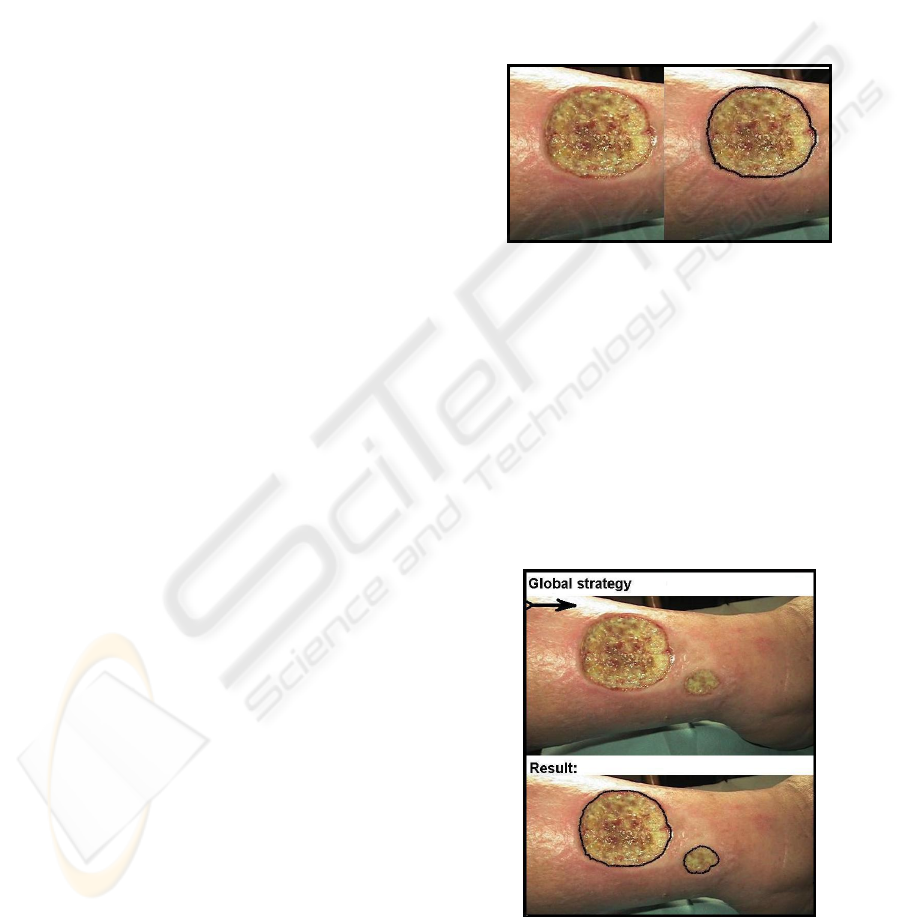
Figure 2 shows the output result displayed by
such an application launched to identify a wound.
The left hand side contains the original image, while
the right hand side contains the clone image,
processed and marked with the contour of the
wound.
Figure 2: The output result of identifying a wound.
Our system contains two general strategies for the
process of identifying the wounds: a global strategy
and a wound by wound strategy. The user may
choose one of the two general strategies, or may
combine them using a friendly graphical user
interface. When apply the global strategy, the whole
image is traversed from top-left corner towards
bottom-right corner, applying specific methods for
edge-detection and wound identification. The output
result presents all the wounds identified inside the
current image (see Figure 3).
Figure 3: Applying the global strategy.
When apply the wound by wound strategy, each
wound is identified in a separate process, based on a
WEBIST 2008 - International Conference on Web Information Systems and Technologies
390
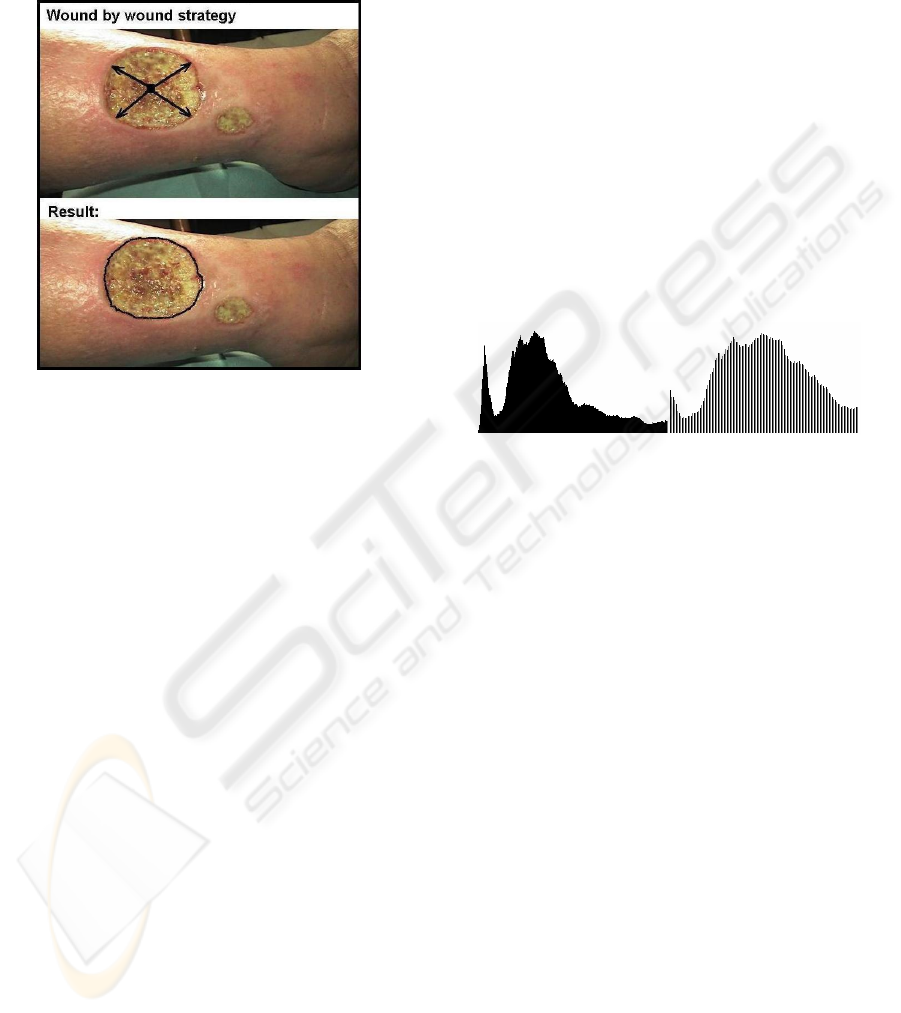
representative area belonging to it. In this case, only
the selected wound is traversed, starting with
representative area and going towards the four main
points: top-left, top-right, bottom-left and bottom-
right (see Figure 4).
Figure 4: Applying the wound by wound strategy.
4 CLASSIFICATION METHODS
Our work is based on a continuous collaboration
with physicians and wound care experts, because it
is necessary to make a rigorous classification for
various categories of wounds. We collected large
amounts of wound image data and we calculate
statistical parameters as mean, median, standard
deviation, confidence interval, skewness and
kurtosis for them. These historical data are included
in XML based databases, to be used as inputs to
classification algorithms. The general purpose is to
make distinction between infected and non-infected,
inflamed and non-inflamed wounds. Based on colour
analysis, we build a statistically significant
differentiation of mild, moderate and severe wounds.
Our system analyses the differences in calibrated
hue between injured and non-injured skin, obtaining
a repeatable differentiation of wound severity for
various time intervals. As an example, burn wounds
are characterized according to their depth as: (a)
superficial – with bright red colour and the presence
of blisters (usually with brown colour); (b) deep –
with red-whitish colour and with dark dots; (c) full
thickness – with creamy or dark brown colour.
The system contains classification methods for
classifying wound images into different groups
based on colour and texture information. We
investigated the suitability of statistical parameters
for providing useful inputs to the classification
algorithms.
Assuming normality, the first two moments
(mean and standard deviation) characterize very well
the colour distribution. The mean represents the
centre point of the distribution, separating the values
into two equally probable subsets. Standard
deviation represents the dynamics of the values, how
wide around the mean the colours of the wound
image are distributed. The first two moments (mean
and standard deviation) are used to modify the
contrast and the brightness of an image. The contrast
is determined by the width of the distribution, while
the brightness is determined by the location of the
grouping colour values (Baldwin, 2005). We
implemented Java programs that use the mean and
standard deviation to modify and control both the
contrast and the brightness of an image, by
modifying the distribution of the colour values.
Figure 5: The distribution of the colour values before
processing (left) and after processing (right).
Figure 5 shows the distribution of the colour values
contained in the original image (left), compared with
the distribution contained in the modified image
(right). In processed image, the contrast (width of
the distribution) was increased by a factor of 2.0 and
the brightness (mean value) was increased by a
factor of 1.7.
Sometimes the first two moments alone are
inadequate to discriminate between wound and non-
wound skin. Therefore further details of the colour
distribution are required. Skewness and kurtosis of
the colour data proved to be more useful for this
purpose. Skewness is a measure of the asymmetry of
the distribution around the centre. Skewness is null
for a normal distribution, positive when the
distribution is skewed right (i.e. when the upper tail
of it is predominant) and negative when the
distribution is skewed left. Kurtosis quantifies the
flatness level of the distribution at the mean.
Kurtosis is equal to 3 for a normal distribution. If
kurtosis is lower than 3, the distribution is said to be
platokurtic (i.e. wide-peaked) and if kurtosis is
higher than 3, the distribution is said to be
leptokurtic (i.e. narrow-peaked). The kurtosis is used
as a measure of the heaviness of the tails in a
distribution.
We build in Java models for various categories of
E-LEARNING TOOLS FOR WOUND IMAGE UNDERSTANDING
391
wounds, due to aetiologies such as pressure, burn,
chilblain, vascular insufficiencies, diabetic foot
ulcer, venous leg ulcer and other chronic disease
states. Based on colour and texture analysis, we have
to identify the main barriers to wound healing, such
as tissue non-viable, infection, inflammation,
moisture imbalance, or edge non-advancing. Our
aim is to implement algorithms for wound healing
simulations.
5 E-LEARNING ENVIRONMENT
In a previous work we defined and implemented a
Java framework for designing and implementing
intelligent and practical e-learning tools, to be used
by both the students and the teaching staff in a
context of open learning. This framework provides
the infrastructure for preparing e-learning scenarios
based on practice and real world experiences, as
practice is essential in learning activities. Our
e-learning scenarios promote active learning, forcing
the students to take part in real world activities
simulated on computer. We rely on new paradigms
of artificial intelligence (Bayesian Inference, Case
Based Reasoning and Intelligent Agents) for
creating e-learning scenarios to be used in a context
of active learning. An e-learning scenario combines
simulation and interactive visualization and allows
the learners to explore the knowledge bases with
some well-defined learning purposes. For each
application object, our system contains a simulation
class and a visualization class. These classes are then
configured to obtain a particular simulation with a
specific visualization. In an e-learning scenario,
visualization is an active part of the system, serving
as an additional interface for modifying dynamically
some parameters. The simulation and visualization
classes are coded in Java, using XML format to
describe the configurations for both the components
and their relationships.
An e-learning scenario is in fact like a traditional
lesson, and the ideal solution is to simulate a
teaching-learning relation with a virtual teacher able
to interact with the learners and to instruct them
(Prodan et al., 2007). A good traditional teacher
learns all the time from previous didactic
experiences. Based on this historical feedback, the
teacher exploits prior specific successful episodes,
and avoids prior failures. We introduce a similar
feedback mechanism in our technology of
elaborating e-courses (see Figure 6). The feedback
information, collected from learners’ remarks and
from prior results and successes, is stored in case
bases. The relevant cases are retrieved and adapted
to fit new situations from new e-learning scenarios,
or to improve the previous ones. In addition, our
approach in creating an e-learning scenario relies
upon a special sort of goal oriented intelligent agents
(Nwana, 1996), able to incorporate knowledge,
teaching methods and pedagogical characteristics
into e-courses. We intend to implement a simulation
of some intelligence based actions and initiatives,
that are to be incorporated into e-learning scenarios,
with the purpose to map, to plan and to monitor the
pace and the progress of a learning process.
Following the traditional model, the cases of
positive experiences from previous e-learning
scenarios are stored into case bases created with
XML and CBR technologies (Leake, 1996).
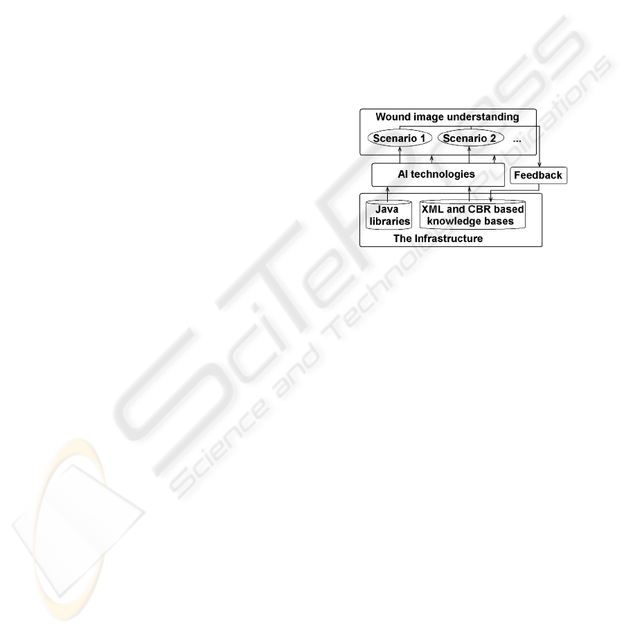
Figure 6: The generation of the e-learning scenarios.
Our aim is to create and implement in Java an
automatic method which can be used as a reference
standard for colour and texture wound analysis. In
collaboration with medical experts, we will create
e-learning scenarios by applying the method
illustrated in Figure 6 to large amounts of wound
image data stored in XML knowledge bases. By
estimating the percentages for the main colours of
red, yellow and black, it is possible to assess the
gravity of the wound. The image processing
program allows the user to interactively control the
process. The user can set the tolerance for each
colour, that is the width of the band of acceptable
colours. Based on colour analysis and statistical
methods, it is possible to analyse successive states of
a wound, assessing the wound healing evolution.
The final goal is develop a flexible and adaptable
system for wound image understanding, based on
new paradigms of Artificial Intelligence (such as
Bayesian Inference, Case Based Reasoning and
Intelligent Agents). The students create electronic
portfolios, consisting of wound images and reports
about wound healing, based on wound healing
simulation scenarios, allowing to assess the wound
image understanding (Rusu and Prodan, 2006).
WEBIST 2008 - International Conference on Web Information Systems and Technologies
392

The functionality of our system will aim to creating
new e-learning tools, to be used by the students in
medicine and pharmacy, at graduate, postgraduate
and residency levels, for developing appropriate
skills in wound management. Our effort is supported
by a continuous collaboration with physicians and
wound care experts from our university and from
health care and medical units. We are endowed with
a continuous access to actual medical records,
allowing us to have in view the wound evolution and
to verify the accuracy and the consistency of our
system. The observed and the estimated values of
the colour are all the time compared with each other.
Based on colour and texture analysis, it is possible to
identify the main barriers to wound healing, such as
tissue non-viable, infection, inflammation, moisture
imbalance, or edge non-advancing. These results are
used to implement algorithms for wound healing
simulation. The advantage of using Java for this
purpose is the integration without any difficulty with
other Web based facilities.
The methods presented in this paper should be
useful as an adjunct to traditional teaching and
learning resources. In a context of blended learning,
the teachers and learners may combine the colour
and texture based parameters with traditional
parameters, such as smell, venous and arterial status,
patient history, etc.
6 CONCLUSIONS
This paper presents a Java framework for analysing
and processing wound images, to be used in
teaching, learning and research activities. The colour
image processing methods have many advantages
over traditional human methods in assessment of
wounds. Computer based methods are objective,
repeatable and with a large potential of processing.
The analysis of a wound from a specific distance
involves procedures devoted to identify its
boundaries, to calculate its area and to estimate
proportions of the main colours red, yellow and
black. Generally, wounds have a non-uniform
mixture of yellow slough, red granulation tissue and
black necrotic tissue. To analyse the actual state of
the wound and the healing evolution, it is necessary
to determine the proportions of these main colours.
We create XML based databases containing
knowledge extracted from previous wound healing
experiences and from medical experts knowledge.
The students create electronic portfolios, consisting
of wound images and reports about wound healing,
based on wound healing simulation scenarios,
allowing to assess the wound image understanding.
Our experience demonstrated that electronic
portfolios may improve the teaching-learning
relation. As a future work, we have to implement
e-learning tools and e-learning scenarios enabling to
perform quantitative measurements of wound
evolution in time and to assess changes in wound
healing, i.e. the recovery or worse evolution. This is
our initial work towards a model of colour and
texture based simulation for the wound healing. We
intend to simulate wound healing based on various
treatments and to compare the results with
experimental observations.
REFERENCES
Baldwin, R. G., 2005. Processing Image Pixels using Java:
Controlling Contrast and Brightness. Available (Last
access at January 17, 2008) at address:
http://www.developer.com/java/other/article.php/3441
391
Garrison, D. R., Kanuka, H., 2004. Blended learning:
Uncovering its transformative potential in higher
education, The Internet and Higher Education, 7, 95-
105.
Leake, D. P., 1996. Case-Based Reasoning: Experiences,
Lessons, and Future Directions (Menlo Park: AAAI
Press / MIT Press).
Nwana, H. S., 1996. Software Agents: An Overview,
Knowledge Engineering Review, 11(3), 205-244.
Prodan, A., Câmpean, R., Rusu, M., Prodan, R., 2007. An
e-learning framework for wound image understanding.
In C. P. Constantinou et al. (Eds.), Proceedings of the
CBLIS 2007 (Computer Based Learning In Science),
Contemporary Perspectives on New Technologies in
Science and Education, pp. 225-235, ISBN 978-9963-
671-06-9, 30 June - 6 July 2007, Crete Island.
Prodan, A., Rusu, M., Campean, R. and Prodan R., 2006.
A Java framework for analyzing and processing
wound images for medical education. In Wolfgang
Borutzky et al. (Eds.), Proceedings of the ECMS 2006
(European Conference on Modelling and Simulation),
pp. 421-426, ISBN 0-9553018-0-7, 28-31 May 2006,
Bonn, Germany.
Prodan, A. and Prodan, M., 1997. Java Environment for
Internet, Editura Promedia-Plus, Cluj-Napoca,
Romania, ISBN 973-9275-07-09.
Rusu, M., Prodan, A., 2006. Electronic portfolios as
means of improving the teaching-learning relation. In
Ken Fernstrom et al. (Eds.), Proceedings of the
ICICTE 2006 (International Conference on
Information Communications Technologies in
Education), 17-21, ISBN: 1-895802-26-8, ISSN:1109-
2084, 6-8 July 2006, Rhodes Island.
E-LEARNING TOOLS FOR WOUND IMAGE UNDERSTANDING
393
