
UTERINE FIBROID SEGMENTATION ON MRI BASED ON
CHAN-VESE LEVEL SET METHOD AND SHAPE PRIOR
MODEL
Alireza Fallahi, Mohammad Pooyan
Biomedical Engineering Department, Shahed University, Tehran, Iran
Hassan Hashemi
1
, Hassan Khotanlou
2
, Mohammad Ali Oghabian
1
, Kavous Firuznia
1
1
Advanced Diagnostic and Interventional Radiology Research, Tehran, Iran
2
Computer Engineering Department, Bu-Ali Sina University, Hamedan, Iran
Keywords: Uterine Fibroid, MRI Images, Image Segmentation, Chan-vese Level Set, Prior Shape Model.
Abstract: Uterine fibroid the most common benign tumor of the female pelvic affected 20%- 50% of the women in the
world. The efficacy of medical treatment is gauged by shrinkage of the size of these tumors after surgery.
Complex fibroids anatomy, nonhomogeneity region and missing boundary in some cases are a challenging
task in the segmentation. In this paper, we present a method to robustly segment these fibroids on MRI and
measure the volume. Our method is based on combination of two step Chan-Vese level set method and
geometric shape prior model. With calculating an initial region inside the fibroid using Chan-Vese level sets
method, rough segmentation obtained followed by a prior shape model. We found the algorithm efficient
and that it has some good results.
1 INTRODUCTION
Magnetic resonance image (MRI) is widely used in
radiology diagnosis especially in soft tissues.
Different modalities like T1, T2 and FLAIR and the
fusion of information provided by them can be
useful in diagnosis. One of the recent applications of
MRI is to diagnosis uterine fibroid. As the uterine
fibroid is the most common benign tumors of the
female pelvic (VeKaut, 1993), MR imaging can be
very useful in follow-up the patient condition,
diagnosis and treatment process (Cura and Bugnone,
2006). Uterine segmentation and volume
measurement is one of the important tasks that
usually is a time consuming and inaccurate work
when is performed manually. By automated and
semi-automated segmentation techniques we can
assist physicians to have a more accurate result and a
fast process. In (Guyon et al.,2003) geodesic active
contours and fast marching level set have been used
for segmenting fibroid and rigid landmark
registration has been applied for tracking its
variation over time. In (Jianhua et al, 2006) fast
marching level set has been used for initial
segmentation and Laplacian level set has been
applied to refine the segmentation result. These
methods can perform only in homogeneous and
connected boundary regions.
Even with great advantages of level set based
methods, active contours that are based on image
gradients are highly sensitive to the presence of
noise, poor image contrast and missing boundaries.
These lead to bad segmentation results. To
overcome these problems, some methods have
proposed robust region-based evolution criteria into
active contour energy function, built from intensity
statistics and homogeneity requirements (Paragios
and Deriche, 2002; Chan andVese, 2001). Prior
knowledge is usually very helpful to segment or
localize an anatomical structure. Several methods
have been proposed to incorporate prior shape
information into boundary determination. (Cootes et
al, 1995) have proposed an active shape model to
construct a statistical shape model from a set of
training images for image segmentation. The model
is built by outlining the contours and finding point
correspondences across shapes. (Staib and Duncan,
51
Fallahi A., Pooyan M., Hashemi H., Khotanlou H., Ali Oghabian M. and Firuznia K. (2010).
UTERINE FIBROID SEGMENTATION ON MRI BASED ON CHAN-VESE LEVEL SET METHOD AND SHAPE PRIOR MODEL.
In Proceedings of the International Conference on Imaging Theor y and Applications and International Conference on Information Visualization Theory
and Applications, pages 51-55
Copyright
c
SciTePress

1992) incorporate global shape information into the
segmentation process by using an elliptic Fourier
decomposition of the boundary and placing a
Gaussian prior on the Fourier coefficients.
(Leventon, 2000) incorporate statistical shape
influence into the evolution process of geodesic
active contours (Caselles, 1997) by attracting the
evolving contour toward the shape priors. The
correspondence problem is solved in their approach
by embedding the prior shape as the zero level set of
a level set function map. (Chen et al, 2001) propose
a variational method that minimizes an energy
function defined by the image gradients and the
shapes of interest. (Bresson et al., 2006) propose a
method by combination of region, boundary and
shape features based on Mumford-Shah function
(Mumford and Shah, 1989), level set approach and
prior shape model that is based on PCA of training
data to robustly segmentation.
In this paper we propose a tow step method for
robustly segmentation of uterine fibroids. First raw
segmentation obtained using Chan-Vese level set
method based on Mumford-Shah function. Then
segmentation is refined by applying prior shape
model using Bresson et al, method. In the proposed
method, the previous training data is not required.
We generate training data from ellipses model of the
segmented region.
In section 2 we describe the first step prior
segmentation using Chan-Vese level set method.
Section 3 explains the segmentation refinement
based on Bresson et al. prior shape model. Section 4
presents the segmentation results obtained by the
proposed approach. Section 5 presents some
conclusions and future extensions to this approach.
2 INITIAL SEGMENTATION
BASED ON CHAN-VESE LEVEL
SET METHOD
All classical snakes and active contour models are
based on edge-function depending on the image
gradient. These models can detect only objects with
edges defined by gradient to stop the curve
evolution. In practice, the discrete gradients are
bounded and then the stopping function is never zero
on the edges and algorithm fail to segment region.
(Chan and Vese, 2001) proposed a different
active contour method, that is not based on the
gradient of the image for the stopping process. The
stopping term is based on Mumford–Shah
segmentation techniques (Mumford and Shah,
1989).
The Mumford–Shah function for segmentation
is:
C
MS
dxdyyxu
dxdyyxuyxu
CLengthcuF
\
2
2
0
),(|
|),(),(
)(.),(
(1)
Where
Ru :
0
is a given image,
and
are positive parameters. Their active contour model
is a particular case of the minimal partition problem,
in which they look for the best approximation
u
of
0
u
, as a function taking only two values, namely
and with one edge C, represented by the snake or the
active contour. This particular case of the minimal
partition problem can be formulated and solved
using the level set method (Osher and Sethian 1988).
outsideCuaverage
insideCuaverage
u
)(
)(
0
0
(2)
Associated Euler–Lagrange equation deduced
for
represented in equation 3 which parameterize
the descent direction by an artificial time
0t
.
The
equation
),,( yxt
(with
),(),,0(
0
yxyx
)
defining the initial contour is:
),0(0])(
)()()[(
2
202
2
101
incu
cuvdiv
t
e
(3)
inyxyx ),(),,0(
0
on
n
e
0
)(
In uterine fibroids that have a calcified and
infarcted region, edge based method fail to segment
whole region. We applied this method to the images
for initial segmentation. The segmentation is then
performed in whole image with initial manual region
in first slice. Figure.1 shows segmentation result in
some steps for a sample image.
IMAGAPP 2010 - International Conference on Imaging Theory and Applications
52
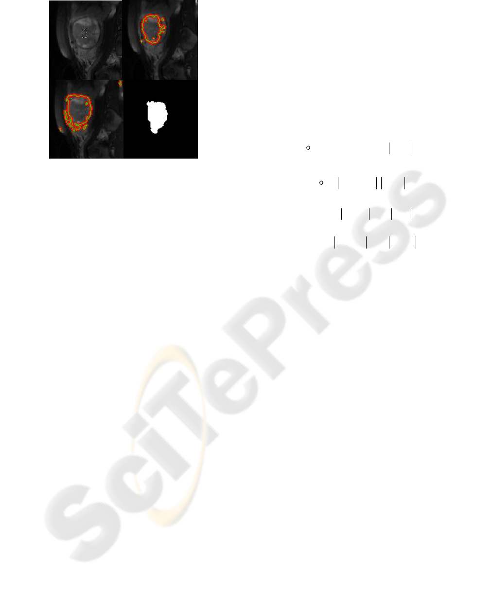
Figure 1: Result of applying Chan-Vese method, a: Initial
manual contour in the region, b and c: Evolving level set
to region boundary, d: Final segmentation result.
3 SEGMENTATION REFINMENT
USING ACTIVE PRIOR SHAPE
MODEL
The shape prior can be defined by different models
such as Fourier descriptors, medial axis or atlas-
based parametric models. Recently, the level set
representation of shapes has been employed as a
shape model (Leventon et al., 2000; Paragios et al.,
2003; Charpiat et al., 2003). This shape description
presents strong advantages since parameterization
free. It can represent shapes of any dimension such
as curves, surfaces and hyper-surfaces and basic
geometric properties such as the curvature. Finally,
this shape representation is also naturally consistent
with the level set framework of active contours. In
(Leventon et al, 2000), authors have used a level set
representation to model the shape prior. They have
defined a shape model of the object of interest by
computing principal components analysis (PCA) of
training shapes embedded in level set functions.
They have then integrated this shape model in an
evolution equation to globally drive the active
contour towards the prior shape. However, their
evolution equation is not expressed by a partial
differential equation (PDE) and there is no
variational formulation associated with his evolution
equation.
Recently Bresson et al, have proposed a
variational approach following the energy functional
model of (Chen et al. 2002) where integrate the
shape prior of (Leventon et al, 2002). They added a
region-based energy term based on the Mumford-
Shah function (Mumford and Shah, 1989) to
improve the robustness of segmentation model with
respect of noise, poor image contrast and initial
position of the contour.
They proposed the following energy functional
to address the problem of object segmentation using
a geometric shape prior with local and global image
information:
),,,(
)(),,(
outinTpcaregionr
boundarybTpcashapes
uuXXF
CFXXCFF
(4)
With
dqqCqChxF
T
xpcashape
)()))((,(
0
^
(5)
dqqCqCIgF
boundary
)()))(((
0
(6)
),(
),(
)(
)(
22
22
T
X
pca
Xout
T
X
pca
Xin
duuI
duuIF
ioutout
ininregion
(7)
where C is the active contour, φˆ is the shape
function of the object of interest given by the PCA
,x
pca
is the vector of PCA eigen coefficients, h
xT
is an
element of a group of geometric transformations
parameterized by x
T
(the vector of parameters), g is
an edge detecting function
in
and
out
are the
inside and outside regions of the zero level set of φˆ,
u
in
and u
out
are smooth approximations of the
original image I in
in
and
out
and β
b
, β
s
, β
r
are
arbitrary positive constants that balance the
contributions of the boundary, shape and region
terms .We chose β
b
, β
s
, β
r
, 1, 1/3 and 10
respectively.
3.1 Training Data for Computing PCA
The above method needs some initial training data
for calculating PCA eigen coefficients. Because of
explicit algebraic form we use ellipse model for
generating this training data based on region that
segmented from past stage. We calculate statistical
properties of the region: center, major axis, minor
axis and orientation. Then we generate ellipses
based on these parameters and some variations of
them for calculating PCA. Figure.2 shows generated
training data for segmented region.
We applied this method to refine segmented
region and consider active contour as a segmentation
result. Figure.3 shows the results.
b
c
d
a
UTERINE FIBROID SEGMENTATION ON MRI BASED ON CHAN-VESE LEVEL SET METHOD AND SHAPE
PRIOR MODEL
53
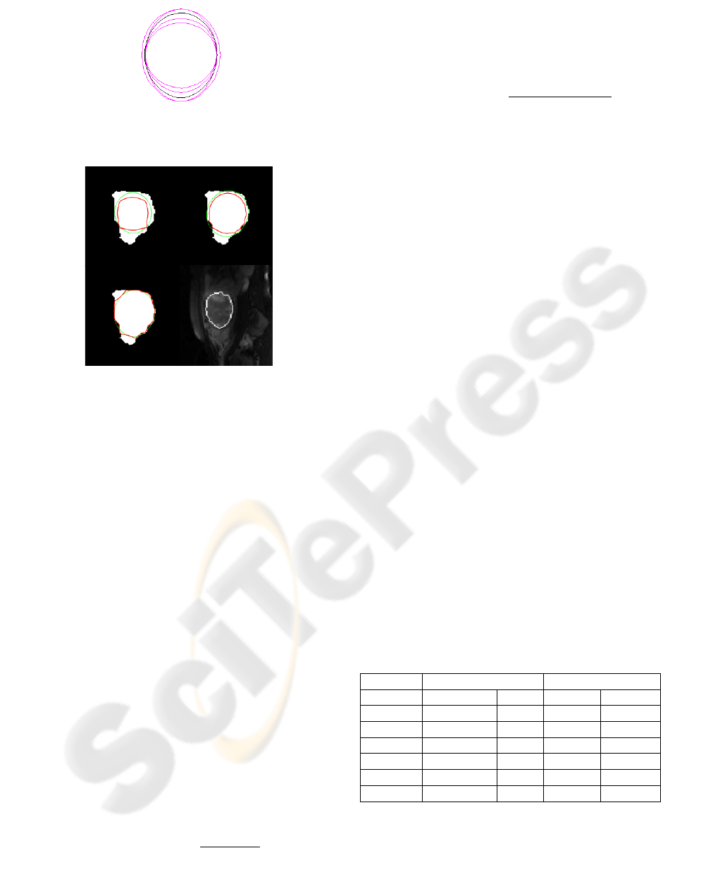
Figure 2: Training data generated from ellipse model of
segmented region.
Figure 3: Refined segmented region based on Bresson et
al, method and ellipses model; green contours denotes
shape model and red contours denote active contour.
4 RESULTS
We have used the above mentioned method on the
dataset containing the MR images of 5 patients that
were acquired at the Imam Khomeini hospital
Medical Imaging Centre. All the patients were
imaged on a 1.5T MR scanner used standard clinical
imaging protocol to obtain T2-weighted. Each MR
image has an in-plan resolution of 512×512 and slice
thickness of 5 mm with 15-20 slices.
Fig. 4 shows the segmentation results. With
manual initial region selection on first slice, other
slices were automatically segmented. The curves are
able to converge on the desired boundaries even
though some parts of the boundaries are too blurred
or missed to be detected by only gray level
information. To validate the segmentation results,
we compare obtained results with manual
segmentation performed by a senior radiologist. We
used four measures to evaluate the results which are
(M denotes the manually segmented area and an
automated segmented area):
• Similarity index:
%100*
2
AM
T
i
NN
N
S
p
Where
p
T
N
the number of true positive voxels and
M
N
is the cardinality of M and
A
N
is the
cardinality of A;
• Jaccard index:
%100*
p
p
TAM
T
i
NNN
N
J
• Hausdorff distance between A and M, defined
as HD = max (h (M, A), h (A, M)) where h (M, A) =
maxm∈M mina∈A d (m, a), and d (m, a) denotes the
Euclidean distance between m and a (m and a are
points of M and A, respectively).
•Average distance (MD) between the surfaces of
M and A.
As seen in Table 1 similarity index varies from
87.26% to 90.1% with a mean of 87.70%. The
Jaccard index varies from 74.65% to 82.62% with a
mean of 76.62% which shows a good accuracy of
segmentation. The average of Hausdorff distance is
3.42mm that smaller than a voxel size, and
constitutes good result. The mean value of the
average distance is 0.35mm that represented
accuracy of segmentation. Segmentation of fibroids
that have calcified and infarcted regions is a
challenging task due to global nonhomogenities.
Two published papers in uterine fibroid
segmentation used level set and active contour
methods. These methods are gradient based and are
not able to segment these types of tumors. Thus,
they only can be useful for complete infarcted
fibroids with clear boundaries. We have tried to
overcome these limitations by introducing a new
method that allows segmentation of these tumors by
region based method as initial segmentation. Then,
we applied combination of level set based and prior
shape model as final segmentation.
Table 1: Evaluation of the segmentation results of images
for which a manual segmentation was available.
Patient
Volume
metric (%)
Distance
metric(mm)
SI
JI
HD
MD
1
88.29
79.76
2.92
0.27
2
88.1
78.9
3.18
0.21
3
90.1
82.62
3.76
0.32
4
87.26
77.55
3.03
0.29
5
84.79
74.65
4.3
0.65
Average
87.70
78.62
3.42
0.35
IMAGAPP 2010 - International Conference on Imaging Theory and Applications
54
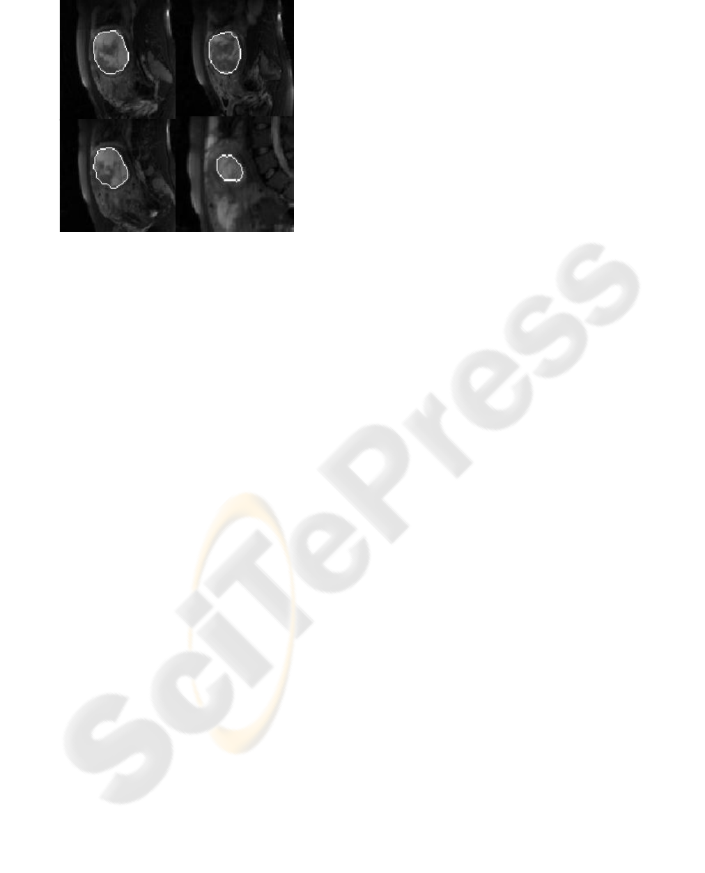
Figure 4: segmentation result using proposed method for
some slices of a patient.
5 CONCLUSIONS
This paper proposed an automatic method for the
segmentation of uterine fibroid in MR images. Using
Chan-Vese method initial segmentation obtained. In
second step segmentation refined by applying prior
shape model based on Bresson et al, method and
ellipses model. The quantitative results illustrate the
good performance of this method according to
nonhomogeneity region and missing boundary in
these types of fibroids. By uterine fibroid
segmentation in the future works we can analyze
fibroid properties like infarcted or calcified percent
region. This task has essential features in diagnosis
and treatment of uterine fibroids.
ACKNOWLEDGEMENTS
The authors would like to thank Dr A.Jalali and Dr
M.Shakiba of the Diagnostic and Interventional
Radiology Research Center (ADIR) for supplying all
patient images.
REFERENCES
Bresson, X., Vandercheynst, P., and Thiran, J.P., 2006, A
Variational Model for Object Segmentation Using
Boundary Information and Shape Prior Driven by the
Mumford-Shah Functional. International Journal of
Computer Vision, 68(2):145-162.
Caselles, V., Kimmel, R., and Sapiro, G., 1997, Geodesic
active contours, Int. J. Computer Vision, 22(1); 61–
79.
Chan, T.F. and Vese, L.A. 2001. Active contours without
edges. IEEE Transactions on Image Processing,
10(2):266–277.
Charpiat, G., Faugeras, O., and Keriven, R. 2003. Shape
metrics, warping and statistics. In IEEE International
Conference on Image Processing, 627–630.
Chen, S. Thiruvenkadam, H. D. Tagare, F. Huang, D.
Wilson, and E. A. Geiser, 2001, On the incorporation
of shape priors into geometric active contours, IEEE
Workshop on Variational and Level Set Methods in
Computer Vision, 145–152.
Cootes, T, F,. Taylor, C, J,. Cooper, D, H and Graham, J,
1995, Active shape models – their training and
application, Computer Vision Image Understand., 61
(1);38–59.
Cura M, Cura A , Bugnone A,. 2006, Role of Magnetic
Resonance Imaging in Patient Selection for Uterine
Artery Embolization. Acta Radiol ; 1105-1114.
Guyon J.P, Foskey M,Kim J, Firat Z, Davis Ylward B,.
2003, VETOT,Volume Estimation and Tracking Over
Time:Framework and Validation. Proceedings in
MICCAI; 142:149.
Jianhua Y , Chen D, Wenzhu L , Premkumar A., 2006,
Uterine fibroid segmentation and volume
measurement on MRI. Progress in biomedical optics
and imaging;(7)
Leventon, M. E,. Grimson, . W, E, L. and Faugeras,
2000, Statistical shape influence in geodesic active
contours, in Proc. IEEE Computer Society Conf.
Computer Vision and Pattern Recognition (CVPR),
;316–323.
Mumford, D. and Shah, J., 1989, Optimal approximations
of iecewise smooth functions and associated
variational problems. Communications on Pure and
Applied Mathematics, 42:577–685.
Osher, S. and Sethian, J.A. 1988. Fronts propagating with
curvaturedependent speed: Algorithms based on
Hamilton-Jacobi formulations. Journal of
Computational Physics, 79(1):2–49.
Paragios, N., Rousson, M., and Ramesh, V. 2003. Non-
rigid registr ation using distance functions . Journal of
Computer Vision and Image Understanding, 89(2–
3):142–165.
Staib,V and, Duncan, J,S, 1992, Boundary finding with
parametrically deformable models, IEEE Trans.
Pattern Anal. and Machine Intell., vol. 14, no. 11, pp.
1061–1075, 1992.
VeKaut, M, B,. 1993, Changing trends in treatment of
leiomyomata uteri. Curr Opin Obstet Gynecol 5:301.
UTERINE FIBROID SEGMENTATION ON MRI BASED ON CHAN-VESE LEVEL SET METHOD AND SHAPE
PRIOR MODEL
55
